Downloaded 52 times


![Artifacts & Interferences in EEG
Sources of Artifacts
Environmental factors (e.g. power noise,
sound/optical interference, EM-coupling from
earth, etc.)
Experiment factors (e.g. electrode position
altering, connecting wire movement, etc. due
to mainly subject motion )
Physiological factors (e.g. EOG, ECG, EMG,
etc.)
Problems with Artifacts
Can cause electronics saturation [1]
High dynamic range required (Higher ENOB in ADC) [2]
Increase false alarms in epileptic seizure
detection [ ]
Mistakes in BCI classifications
0 0.2 0.4 0.6 0.8 1 1.2 1.4 1.6 1.8 2
x 10
5
-2
-1.5
-1
-0.5
0
0.5
1
1.5
2
x 10
-3
Time Sample
Voltage,V
[1]
260 265 270 275 280 285 290 295
-15
-10
-5
0
5
x 10
-4
Time, Second
Voltage,Volt
[2]
[ ]](https://image.slidesharecdn.com/eegguestlectureiubeee541-170404063409/85/EEG-guest-lecture_iub_eee541-7-320.jpg)
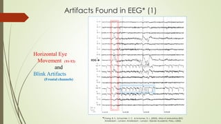
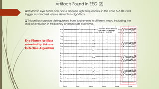
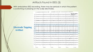
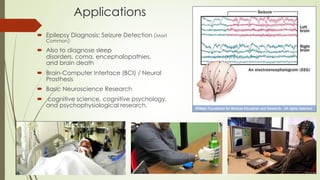
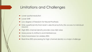

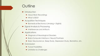
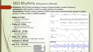
The document discusses electroencephalography (EEG), detailing its definition, acquisition techniques, signal analysis, and applications, particularly in diagnosing neurological disorders and brain-computer interfaces. It covers types of brain recordings (both invasive and non-invasive), artifacts and interferences affecting EEG signals, and various frequency bands associated with brain activity. Additionally, it emphasizes the limitations and challenges of EEG technology, including issues with spatial resolution and the prevalence of artifacts.


