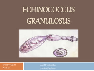
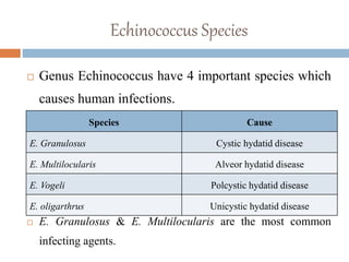
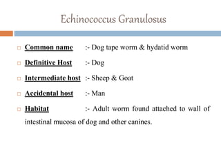
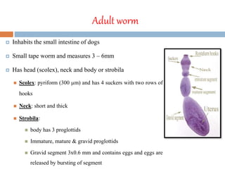
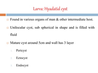
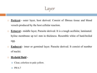
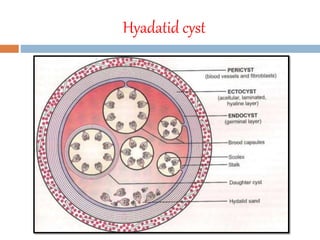
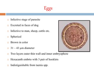
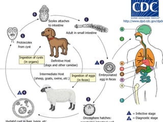
ECHINOCOCCUS
GRANULOSUS
ANKUr vashishtha
Assistant Professor
IIMT UNIVERSITY
MEERUT
Echinococcus Species
 Genus Echinococcus have 4 important species which
causes human infections.
 E. Granulosus & E. Multilocularis are the most common
infecting agents.
Species Cause
E. Granulosus Cystic hydatid disease
E. Multilocularis Alveor hydatid disease
E. Vogeli Polcystic hydatid disease
E. oligarthrus Unicystic hydatid disease
Echinococcus Granulosus
 Common name :- Dog tape worm & hydatid worm
 Definitive Host :- Dog
 Intermediate host :- Sheep & Goat
 Accidental host :- Man
 Habitat :- Adult worm found attached to wall of
intestinal mucosa of dog and other canines.
Morphology
 Three morphological form
1. Adult worm
2. Larva
3. Egg
Adult worm
 Inhabits the small intestine of dogs
 Small tape worm and measures 3 – 6mm
 Has head (scolex), neck and body or strobila
 Scolex: pyriform (300 µm) and has 4 suckers with two rows of
hooks
 Neck: short and thick
 Strobila:
 body has 3 proglottids
 Immature, mature & gravid proglottids
 Gravid segment 3x0.6 mm and contains eggs and eggs are
released by bursting of segment
Larva: Hyadatid cyst
 Found in various organs of man & other intermediate host.
 Unilocular cyst, sub spherical in shape and is filled with
fluid
 Mature cyst around 5cm and wall has 3 layer
1. Pericyst
2. Ectocyst
3. Endocyst
Layer
 Pericyst:- outer layer, host derived. Consist of fibrous tissue and blood
vessels produced by the host cellular reaction.
 Ectocyst:- middle layer, Parasite derived. It is a tough acellular, laminated.
hyline membrane up to1 mm in thickness. Resemble white of hard-boiled
egg.
 Endocyst:- inner or germinal layer. Parasite derived. It consist of number
of nuclei.
 Hydatid fluid:-
 Clear, colorless to pale yellow.
 PH 6.7
Hyadatid cyst
Eggs
 Infective stage of parasite
 Excreted in faces of dog
 Infective to man, sheep, cattle etc.
 Spherical
 Brown in color
 31 – 43 µm diameter
 Two layers outer thin wall and inner embryophore
 Hexacanth embrto with 3 pair of hooklets
 Indistiguishable from taenia spp.
Transmission
 Direct contact with infected dog
 Contaminated with egg Food & water
LIFE CYCLE
 E. Granulosus passes its life cycle in two host.
 Adult worm live attached to mucosa of small intestine of dog and
other canine animals.
 The egg are discharge in the faeces.
 Egg swallowed by intermediate host while grazing in the field.
 Man acquire infection by direct contact with infection dog or from
contaminated food or water.
 In duodenum embryo hatch out and penetrate the intestinal wall and
enter to portal vein and carried to liver lungs & other organs.
 Most of the embryo destroy in host immune response but few
escape which latter develop in to pericyst.
 Inside pericyst embryo developed in to hydatid cyst.
 Man is dead end of its life cycle.
Lab diagnosis
 Parasitic diagnosis
 Sample:- Diagnostic aspiration
 Hydatid fluid micrscopy
ļ‚§ LPCB:- Scolices and protosclices appear deep blue
ļ‚§ ZN staining:- scolices and protoscolices appear purple
or pink.
 Histopathological diagnosis:-
ļ‚§ PAS or HE stained FNAC or biopsy material.
Immuno-diagnosis
 Casonis skin test (type I Hypersentivity):-
 0.2 ml of sterile hydatid fluid injected intradermally in
one arm and 0.2 ml of saline on the other.
 Large wheal measuring 5cm or more within 30 min
 Low sensitivity 55% - 75%
Serological test
 Antibody detection:-
 Screening test:-
 IHA
 CIEP
 ELISA
 IFA
 DIGFA
 Confirmation test are western blot
CONT…..
 Antigen detection :-
 ELISA
 CIEP
 Latex Agglutination test
Molecular methods:-
PCR
Imaging methods
 X-ray
 USG
 CT Scan
 MRI
Treatment
 Surgery:- main stay of treatment
 Puncture-Aspiration-injection-Re-aspiration (PAIR)
 Chemotherapy
Echinococcus granulosus

Echinococcus granulosus

  • 1.
  • 2.
    Echinococcus Species  GenusEchinococcus have 4 important species which causes human infections.  E. Granulosus & E. Multilocularis are the most common infecting agents. Species Cause E. Granulosus Cystic hydatid disease E. Multilocularis Alveor hydatid disease E. Vogeli Polcystic hydatid disease E. oligarthrus Unicystic hydatid disease
  • 3.
    Echinococcus Granulosus  Commonname :- Dog tape worm & hydatid worm  Definitive Host :- Dog  Intermediate host :- Sheep & Goat  Accidental host :- Man  Habitat :- Adult worm found attached to wall of intestinal mucosa of dog and other canines.
  • 4.
    Morphology  Three morphologicalform 1. Adult worm 2. Larva 3. Egg
  • 5.
    Adult worm  Inhabitsthe small intestine of dogs  Small tape worm and measures 3 – 6mm  Has head (scolex), neck and body or strobila  Scolex: pyriform (300 µm) and has 4 suckers with two rows of hooks  Neck: short and thick  Strobila:  body has 3 proglottids  Immature, mature & gravid proglottids  Gravid segment 3x0.6 mm and contains eggs and eggs are released by bursting of segment
  • 6.
    Larva: Hyadatid cyst Found in various organs of man & other intermediate host.  Unilocular cyst, sub spherical in shape and is filled with fluid  Mature cyst around 5cm and wall has 3 layer 1. Pericyst 2. Ectocyst 3. Endocyst
  • 7.
    Layer  Pericyst:- outerlayer, host derived. Consist of fibrous tissue and blood vessels produced by the host cellular reaction.  Ectocyst:- middle layer, Parasite derived. It is a tough acellular, laminated. hyline membrane up to1 mm in thickness. Resemble white of hard-boiled egg.  Endocyst:- inner or germinal layer. Parasite derived. It consist of number of nuclei.  Hydatid fluid:-  Clear, colorless to pale yellow.  PH 6.7
  • 8.
  • 9.
    Eggs  Infective stageof parasite  Excreted in faces of dog  Infective to man, sheep, cattle etc.  Spherical  Brown in color  31 – 43 µm diameter  Two layers outer thin wall and inner embryophore  Hexacanth embrto with 3 pair of hooklets  Indistiguishable from taenia spp.
  • 10.
    Transmission  Direct contactwith infected dog  Contaminated with egg Food & water
  • 11.
    LIFE CYCLE  E.Granulosus passes its life cycle in two host.  Adult worm live attached to mucosa of small intestine of dog and other canine animals.  The egg are discharge in the faeces.  Egg swallowed by intermediate host while grazing in the field.  Man acquire infection by direct contact with infection dog or from contaminated food or water.  In duodenum embryo hatch out and penetrate the intestinal wall and enter to portal vein and carried to liver lungs & other organs.
  • 12.
     Most ofthe embryo destroy in host immune response but few escape which latter develop in to pericyst.  Inside pericyst embryo developed in to hydatid cyst.  Man is dead end of its life cycle.
  • 14.
    Lab diagnosis  Parasiticdiagnosis  Sample:- Diagnostic aspiration  Hydatid fluid micrscopy ļ‚§ LPCB:- Scolices and protosclices appear deep blue ļ‚§ ZN staining:- scolices and protoscolices appear purple or pink.  Histopathological diagnosis:- ļ‚§ PAS or HE stained FNAC or biopsy material.
  • 15.
    Immuno-diagnosis  Casonis skintest (type I Hypersentivity):-  0.2 ml of sterile hydatid fluid injected intradermally in one arm and 0.2 ml of saline on the other.  Large wheal measuring 5cm or more within 30 min  Low sensitivity 55% - 75%
  • 16.
    Serological test  Antibodydetection:-  Screening test:-  IHA  CIEP  ELISA  IFA  DIGFA  Confirmation test are western blot
  • 17.
    CONT…..  Antigen detection:-  ELISA  CIEP  Latex Agglutination test Molecular methods:- PCR
  • 18.
    Imaging methods  X-ray USG  CT Scan  MRI
  • 19.
    Treatment  Surgery:- mainstay of treatment  Puncture-Aspiration-injection-Re-aspiration (PAIR)  Chemotherapy

Editor's Notes