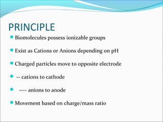
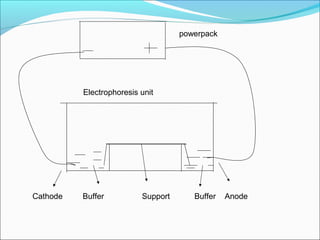
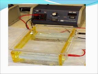
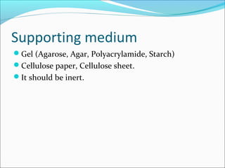
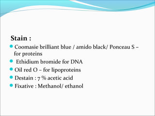
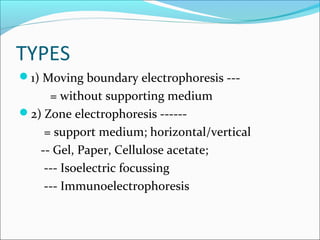
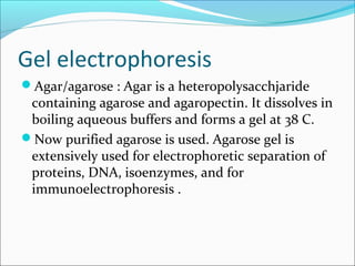
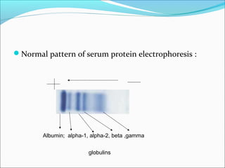
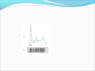
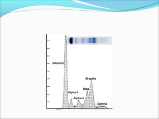
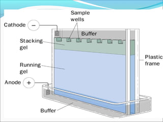
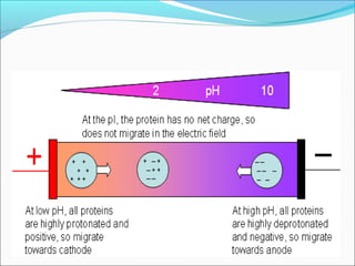
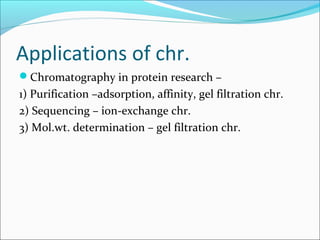
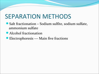
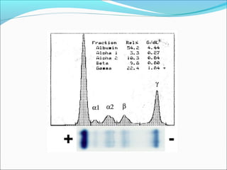
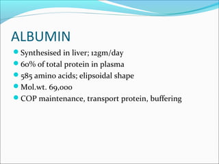
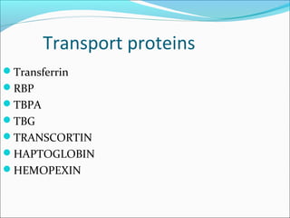
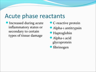
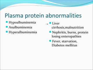
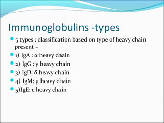
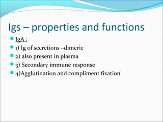
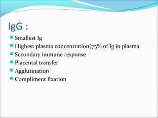
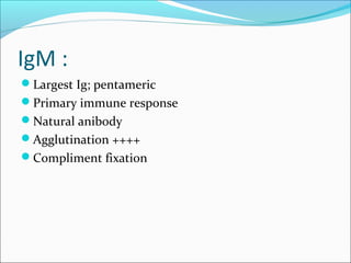
Prabin Shah MSc(Biochemistry)
DEFINITION
Electrophoresis is separative technique.
It is the migration of charged partcles in electric field.
The first electrophoretic technique to be widely used
was that of Tiselius in 1937.
PRINCIPLE
Biomolecules possess ionizable groups
Exist as Cations or Anions depending on pH
Charged particles move to opposite electrode
 -- cations to cathode
 ---- anions to anode
Movement based on charge/mass ratio
powerpack
Cathode Buffer Support Buffer Anode
Electrophoresis unit
Requirements
Electrophoresis chamber/unit
Powerpack
Buffer
Stain
Destaining solution
Fixative solution
Supporting medium
Buffer
Maintains the pH
pH of the buffer decides the ionic nature of the molecule.
It determines and stabilizes the charges in molecules to be
separated.
It also conducts current on the supporting medium.
Buffers in common use are
 barbitone buffer (pH 8.6, for serum protein electrophoresis)
 citrate buffer (pH 6.2 for hemoglobin electrophoresis )
 tris-EDTA-borate buffer (pH 8.6,for hemoglobin
electrophoresis).
Power supply
Constant current/constant voltage mode
Low voltage (100 to 500 V ) electrophoresis is
routinely used.
High voltage (500 V to 10000 V) electrophoretic
technique is employed in the separation of amino
acids and small peptides; it requires a cooling system
in the electrophoresis unit.
Supporting medium
Gel (Agarose, Agar, Polyacrylamide, Starch)
Cellulose paper, Cellulose sheet.
It should be inert.
Stain :
Coomasie brilliant blue / amido black/ Ponceau S –
for proteins
 Ethidium bromide for DNA
Oil red O – for lipoproteins
Destain : 7 % acetic acid
Fixative : Methanol/ ethanol
TYPES
1) Moving boundary electrophoresis ---
= without supporting medium
2) Zone electrophoresis ------
= support medium; horizontal/vertical
-- Gel, Paper, Cellulose acetate;
--- Isoelectric focussing
--- Immunoelectrophoresis
Gel electrophoresis
Agar/agarose : Agar is a heteropolysacchjaride
containing agarose and agaropectin. It dissolves in
boiling aqueous buffers and forms a gel at 38 C.
Now purified agarose is used. Agarose gel is
extensively used for electrophoretic separation of
proteins, DNA, isoenzymes, and for
immunoelectrophoresis .
Protocol of electrophoresis :
The sample is applied as a narrow line (2-5 microliters) on
the support medium, towards the designated cathode end
(in case of alkaline pH) , leaving about 1/3rd distance from
that end.
The support medium with the sample is kept on the solid
support inside the electrophoresis chamber. It is
connected to the buffer on both sides with filter paper
wicks.
• The electrodes are connected to power pack,
chamber is closed.
The power is switched on, power supply can be
adjusted in constant current ( 7 milliamperes /
slide) or constant voltage (100 volts) mode.
Allow the electrophoresis to run for fixed time
(alternatively, sample mixed with a dye may be
applied and the movement of the dye can be
tracked, till it reached the other end of the slide)
After the run, remove the support medium from
the chamber, immerse in a fixative(generally
methanol or alcohol for proteins) for 15 minutes,
the dry it.
For visualization of separated bands, stain with a
staining agent. For proteins, coomassie brilliant
blue, ponceau S or amido black can be used.
Destain with a washing solution (generally 3%
acetic acid), to make the background clear.
• Quantitation is possible by densitometry. The
slide/cellulose sheet with stained bands is
scanned through a densitometer, and a plot of
absorbance of each band against the distance
moved gives an electrophoretogram.
• Based on the height and area of each band/peak,
concentration is calculated.
Normal pattern of serum protein electrophoresis :
Albumin; alpha-1, alpha-2, beta ,gamma
globulins
Polyacrylamide Gel Electrophoresis
PAGE : Polyacrylamide gels are prepared immediately
before use from a number of highly toxic, synthetic
chemicals. Acrylamide monomer is copolymerized with a
cross-linking agent, usually N,N’- methylene bisacrylamide,
in the presence of a catalyst ammonium persulfate and
initiator TEMED (tetramenthylene diamine).
 Gels may prepared containing from 3% to 30% acrylamide,
corresponding to pore sizes of 0.5 nm and 0.2 nm
respectively. PAGE is most suitable for separation of
proteins.
SDS-PAGE is specialized form of PAGE in which a
detergent sodium dodecyl sulfate is included. This
particularly useful in determination of molecular weights of
proteins.
PAGE PATTERN
Cellulose acetate
 High purity cellulose strips are commercially
available, which have a micropore structure.
Hemoglobin electrophoresis at alkaline pH is
best carried on cellulose acetate strip, at a voltage
of 450V and the procedure gets completed in half
an hour,
Cellulose acetate is also suitable for
electrophoresis of glycoproteins and lipoproteins.
Isoelectric focussing
Across the gel layer, voltage and pH gradients are
established. Proteins get focused at their isoelectric
pH, as bands.
This technique is very useful in separation of
peptides, proteins, isoenzymes.
Immunoelectrophoresis
This technique combines together the specificity
of antigen-antibody binding with the
electrophoretic technique.
 In the quantitative form of
immunoelectrophoresis called as Laurell’s rocket
electrophoresis, the sample is applied wells cut in
agarose gel containing antiserum to the antigen to
be assayed .
After the completion of electrophoretic
migration, antigen-antibody complexes form as
rocket shapes. The area under rocket shape is
directly proportional to antigen concentration
Used in diagnosis of immune deificincies,
autoimmune disorders, chronic infections
-----detect Igs, viral/bacterial antigens.
Rocket electrophoresis
APPLICATIONS
In Clinical diagnosis – serum proteins,
lipoproteins, DNA, Hemoglobin, Enzymes
--- infections, immune disorders,
hemoglobinopathies, Lipid abnormalities.
In Protein Research --- Isolation, purification and
molecular weight determination
CHROMATOGRAPHY
Chromatography is collective term referring to a
group of separation processes whereby solutes in a
mixture are separated from one another by a
differential distribution of solutes between two
phases – stationary phase & mobile phase
CHROMATOGRAPHY
This technique received its name from the work of
Tswett in 1906.
 He adsorbed a mixture of plant pigments on to finely
divided charcoal and then separated the components
into a series of coloured bands by washing with
solvents.
 chromatography ,derived from greek word meaning
colour writing.
 now it is used for separation of many colourless
substances.
A variety of attractive forces between the solutes to
be separated and the stationary phase leads to the
selective retardation of the stationary phase relative
to the mobile phase.
Mobile phase carries the mixture of solutes through
the stationary phase.
Stationary phase : solid/liquid supported by glass
column/ gel layer/ cellulose paper
Mobile phase : Liquid/ Gas
CLASSIFICATION
 a) Classification based on principle/ mechanism of
separation :
i) Partition chromatography
ii) ion-exchange chromatography
iii)adsorption chromatography
Iv) Gel filtration chrromatography
V)Affinity chromatography
Classification based on type of physical
apparatus :
Paper, thin layer and column chromatography.
 Partition chromatography is carried out on
Cellulose sheet(paper), layer of gel (thin-layer) or
column .
Ion-exchange, affinity, gel filtration types are
carried in columns.
Adsorption chromatography is carried in thin-layer
and column modes.
PARTITION
CHROMATOGRAPHY
This is based on the separation of solutes by use
of differences in their distribution between two
immiscible phases. Separation is based on
solubility of solute (the polarity) in two phases.
In liquid-liquid partition chromatography, the
both stationary and mobile phases are liquids. In
gas-liquid chromatography, liquid is stationary
phase and gas is mobile phase.
 In liquid-liquid parttion chromatography
otherwise simply called as liquid
chromatography(LC), solutes are partitioned
between stationary and mobile phases, based on
distribution coefficient.
Distribution coefficient (kd)/ partition coefficient
= concentration of solute in solvent A
concentration of solute in solvent B
In normal phase LC, stationary phase is polar and
mobile phase is non-polar. water is the stationary
phase; hexane, benzene, chloroform or butanol
form the mobile phase.
In reverse phase LC, stationary phase is non-polar
(eg. octadecyl silane packing in a column) and
mobile phase is polar (solvents like methanol,
acetonitrile used in column mode of
chromatography).
PAPER chromatography
Partition principle
Ascending / Descending
Paper chr.--ascending
Paper chromatography.
Chr.chamber
Paper with
sample spot
solvent
Solvent for amino acid chr. : butanol:acetic acid:water 12/3/5
TLC(Thin layer
chromatography)
Silica / Alumina layered over a glass plate -----thin
layer
Procedure same as that for paper chr.
Better resolution than paper chr.
After the run, Paper/TLC plate
Solvent
front
Components
separated
as
spots/bands
Identification of sample
components in paper chr./TLC
Rf = Relative front ;
=solute front divided
by solvent front
• Run standards
parallel with samples
Chr.pattern
HPLC (High-performance liquid
chromatography )
 conducted in a column; use of high pressure and
smaller particle size when compared to routine LC
techniques, gives greater resolution.
HPLC can be run in normal as well as reverse
phase modes and any principle of
chromatography can be used in this
(partition/affinity/gel filtration/ion exchange).
It is the most versatile form of chromatography
used for analysis of wide variety of compounds.
GAS CHROMATOGRAPHY(GC)
Gas is the mobile phase and liquid/solid is the
stationary phase.
The sample is injected into a stream of inert gas
usually at an elevated temperature.
The vapourized sample is carried into a column
packed with the stationary phase.
ION-EXCHANGE
CHROMATOGRAPHY
Separates molecules
based on charge.
 Electrostatic attraction
Mobile phase
Generally liquid
Stationary phase
Electrostatically charged
ions bound to insoluble,
chemically inert martrix.
Elution of Molecules
 Ion-exchange resins H2
C
C
O
O-
carboxymethyl (CM)
cellulose
H2
C
C
H2
N
C
H2
CH3
H2C
CH3
H
diethylaminoethyl (DEAE)
cellulose
ADSORPTION CHROMATOGRAPHY
Adsorption is a process whereby one substance
adheres to another because of attractive forces
between surface atoms .
In adsorption chromatography, adsorbents are
used. Examples of adsorbents are charcoal, silica
gel and alumina.
This form of chromatography can be carried out
both in thin-layer and column modes.
Affinity chr.
This is based on strong
covalent binding between
a macromolecule and a
ligand.
Eg. of molecule and
ligand----
Enzyme-substrate
Hormone-receptor
Antigen-antibody
Gel filtration/ exclusion/molecular
sieve chr.
Separates molecules based
on size.
Large molecules exit first.
Mobile phase
Liquid
Stationary phase
Insoluble, porous
carbohydrate beads
Applications of chr.
• In clinical diagnosis : detection and estimation of of
amino acids, metabolites, sugars,
mucopolysaccharides in urine and blood –useful for
screening and diagnosis of inborn metabolic disorders
: Aminoacidurias, hemoglobinopathies,
mucopolysaccharidoses, etc.
In clinical diagnosis
Paper chr.,TLC –qualitative
HPLC, GC –For quantitation
In clinical diagnosis
Assay of Hormones, drugs, vitamins, metabolites
---HPLC and GC
Applications of chr.
Chromatography in protein research –
1) Purification –adsorption, affinity, gel filtration chr.
2) Sequencing – ion-exchange chr.
3) Mol.wt. determination – gel filtration chr.
PLASMA PROTEINS
ALBUMIN
GLOBULINS
FIBRINOGEN
SEPARATION METHODS
Salt fractionation – Sodium sulfite, sodium sulfate,
ammonium sulfate
Alcohol fractionation
Electrophoresis --- Main five fractions
Plasma proteins in each fraction
Alpha-1 globulins Alpha-1 antitrypsin
Alpha-1 acid
glycoprotein
Alpha lipoprotein
Alpha-2 globulins Ceruloplasmin
Alpha-2 macroglobulin
Haptoglobin
Beta-globulins Beta-lipoprotein
Transferrin
Hemopexin
Some Igs
FUNCTIONS
1)Maintenance of colloidal osmotic pressure(COP)—
80% by albumin
*In arterioles *In Venules
COP
25mm HgBP 35mm Hg
BP
15mmHg
functions
2) Transport
3) Nutritional
4) Buffering
5) Defense –clotting,
immunoglobulins
6) Viscosity
Albumin– free fatty
acids, steroid hormones,
bilirubin, copper
Transferrin –iron
Retinol-binding
protein : vitamin A
Lipoproteins : lipids
ALBUMIN
Synthesised in liver; 12gm/day
60% of total protein in plasma
585 amino acids; elipsoidal shape
Mol.wt. 69,000
COP maintenance, transport protein, buffering
Transport proteins
Transferrin
RBP
TBPA
TBG
TRANSCORTIN
HAPTOGLOBIN
HEMOPEXIN
Acute phase reactants
Increased during acute
inflammatory states or
secondary to certain
types of tissue damage
C-reactive protein
Alpha-1 antitrypsin
Haptoglobin
Alpha-1 acid
glycoprotein
fibrinogen
Plasma protein abnormalities
Hypoalbuminemia
Analbuminemia
Hyperalbuminemia
Liver
cirrhosis,malnutrition
Nephritis, burns, protein
losing enteropathies
Fever, starvation,
Diabetes mellitus
Plasma protein abnormalities
HYPER
GAMMAGLOBULINEMIAS
Hypogamaglobulinemia
Agammaglobulinemia
Chronic infections
Multiple myeloma
Electrophoretic patterns
1) Myeloma : M band
2) Nephrotic syndrome : increased alpha-2, decreased
albumin
3) Liver disease : decreased albumin, beta-gamma
bridging
4) Chronic infections : broad-based increase in
gamma globulins
IMMUNOGLOBULINS
4 polypeptide chains – 2 light and 2 heavy
Light chains – 2
kappa (k) or
2 lamda (λ)chains ;
Heavy chains – 2
chains of any of the five
types ---
α , γ, δ, ε, or µ type
Immunoglobulins -types
5 types : classification based on type of heavy chain
present –
1) IgA : α heavy chain
2) IgG : γ heavy chain
3) IgD: δ heavy chain
4) IgM: µ heavy chain
5)IgE: ε heavy chain
Igs – properties and functions
IgA :
1) Ig of secretions –dimeric
2) also present in plasma
3) Secondary immune response
4)Agglutination and compliment fixation
IgG :
Smallest Ig
Highest plasma concentration(75% of Ig in plasma
Secondary immune response
Placental transfer
Agglutination
Compliment fixation
IgM :
Largest Ig; pentameric
Primary immune response
Natural anibody
Agglutination ++++
Compliment fixation
IgE:
1)Fix On mast cells & basophils
2)Mediate anaphylaxis
3)Igs of allergic reactions and response to worm
infections
IgD : No well known fn., found on surface of B
lymphocytes
Multiple myeloma
Proliferation of plasma cells
Paraproteinemia
Abnormal Igs in plasma
Only heavy chain /only light chain
Immunodeficiency
Increased total protein, M band
Bence Jones Proteinuria
Plasma proteins

Plasma proteins

  • 1.
  • 2.
    DEFINITION Electrophoresis is separativetechnique. It is the migration of charged partcles in electric field. The first electrophoretic technique to be widely used was that of Tiselius in 1937.
  • 3.
    PRINCIPLE Biomolecules possess ionizablegroups Exist as Cations or Anions depending on pH Charged particles move to opposite electrode  -- cations to cathode  ---- anions to anode Movement based on charge/mass ratio
  • 4.
    powerpack Cathode Buffer SupportBuffer Anode Electrophoresis unit
  • 6.
  • 7.
    Buffer Maintains the pH pHof the buffer decides the ionic nature of the molecule. It determines and stabilizes the charges in molecules to be separated. It also conducts current on the supporting medium. Buffers in common use are  barbitone buffer (pH 8.6, for serum protein electrophoresis)  citrate buffer (pH 6.2 for hemoglobin electrophoresis )  tris-EDTA-borate buffer (pH 8.6,for hemoglobin electrophoresis).
  • 8.
    Power supply Constant current/constantvoltage mode Low voltage (100 to 500 V ) electrophoresis is routinely used. High voltage (500 V to 10000 V) electrophoretic technique is employed in the separation of amino acids and small peptides; it requires a cooling system in the electrophoresis unit.
  • 9.
    Supporting medium Gel (Agarose,Agar, Polyacrylamide, Starch) Cellulose paper, Cellulose sheet. It should be inert.
  • 10.
    Stain : Coomasie brilliantblue / amido black/ Ponceau S – for proteins  Ethidium bromide for DNA Oil red O – for lipoproteins Destain : 7 % acetic acid Fixative : Methanol/ ethanol
  • 11.
    TYPES 1) Moving boundaryelectrophoresis --- = without supporting medium 2) Zone electrophoresis ------ = support medium; horizontal/vertical -- Gel, Paper, Cellulose acetate; --- Isoelectric focussing --- Immunoelectrophoresis
  • 12.
    Gel electrophoresis Agar/agarose :Agar is a heteropolysacchjaride containing agarose and agaropectin. It dissolves in boiling aqueous buffers and forms a gel at 38 C. Now purified agarose is used. Agarose gel is extensively used for electrophoretic separation of proteins, DNA, isoenzymes, and for immunoelectrophoresis .
  • 13.
    Protocol of electrophoresis: The sample is applied as a narrow line (2-5 microliters) on the support medium, towards the designated cathode end (in case of alkaline pH) , leaving about 1/3rd distance from that end. The support medium with the sample is kept on the solid support inside the electrophoresis chamber. It is connected to the buffer on both sides with filter paper wicks.
  • 14.
    • The electrodesare connected to power pack, chamber is closed. The power is switched on, power supply can be adjusted in constant current ( 7 milliamperes / slide) or constant voltage (100 volts) mode. Allow the electrophoresis to run for fixed time (alternatively, sample mixed with a dye may be applied and the movement of the dye can be tracked, till it reached the other end of the slide)
  • 15.
    After the run,remove the support medium from the chamber, immerse in a fixative(generally methanol or alcohol for proteins) for 15 minutes, the dry it. For visualization of separated bands, stain with a staining agent. For proteins, coomassie brilliant blue, ponceau S or amido black can be used. Destain with a washing solution (generally 3% acetic acid), to make the background clear.
  • 16.
    • Quantitation ispossible by densitometry. The slide/cellulose sheet with stained bands is scanned through a densitometer, and a plot of absorbance of each band against the distance moved gives an electrophoretogram. • Based on the height and area of each band/peak, concentration is calculated.
  • 17.
    Normal pattern ofserum protein electrophoresis : Albumin; alpha-1, alpha-2, beta ,gamma globulins
  • 20.
    Polyacrylamide Gel Electrophoresis PAGE: Polyacrylamide gels are prepared immediately before use from a number of highly toxic, synthetic chemicals. Acrylamide monomer is copolymerized with a cross-linking agent, usually N,N’- methylene bisacrylamide, in the presence of a catalyst ammonium persulfate and initiator TEMED (tetramenthylene diamine).  Gels may prepared containing from 3% to 30% acrylamide, corresponding to pore sizes of 0.5 nm and 0.2 nm respectively. PAGE is most suitable for separation of proteins. SDS-PAGE is specialized form of PAGE in which a detergent sodium dodecyl sulfate is included. This particularly useful in determination of molecular weights of proteins.
  • 22.
  • 23.
    Cellulose acetate  Highpurity cellulose strips are commercially available, which have a micropore structure. Hemoglobin electrophoresis at alkaline pH is best carried on cellulose acetate strip, at a voltage of 450V and the procedure gets completed in half an hour, Cellulose acetate is also suitable for electrophoresis of glycoproteins and lipoproteins.
  • 24.
    Isoelectric focussing Across thegel layer, voltage and pH gradients are established. Proteins get focused at their isoelectric pH, as bands. This technique is very useful in separation of peptides, proteins, isoenzymes.
  • 26.
    Immunoelectrophoresis This technique combinestogether the specificity of antigen-antibody binding with the electrophoretic technique.  In the quantitative form of immunoelectrophoresis called as Laurell’s rocket electrophoresis, the sample is applied wells cut in agarose gel containing antiserum to the antigen to be assayed .
  • 27.
    After the completionof electrophoretic migration, antigen-antibody complexes form as rocket shapes. The area under rocket shape is directly proportional to antigen concentration Used in diagnosis of immune deificincies, autoimmune disorders, chronic infections -----detect Igs, viral/bacterial antigens.
  • 28.
  • 29.
    APPLICATIONS In Clinical diagnosis– serum proteins, lipoproteins, DNA, Hemoglobin, Enzymes --- infections, immune disorders, hemoglobinopathies, Lipid abnormalities. In Protein Research --- Isolation, purification and molecular weight determination
  • 30.
    CHROMATOGRAPHY Chromatography is collectiveterm referring to a group of separation processes whereby solutes in a mixture are separated from one another by a differential distribution of solutes between two phases – stationary phase & mobile phase
  • 31.
    CHROMATOGRAPHY This technique receivedits name from the work of Tswett in 1906.  He adsorbed a mixture of plant pigments on to finely divided charcoal and then separated the components into a series of coloured bands by washing with solvents.  chromatography ,derived from greek word meaning colour writing.  now it is used for separation of many colourless substances.
  • 32.
    A variety ofattractive forces between the solutes to be separated and the stationary phase leads to the selective retardation of the stationary phase relative to the mobile phase. Mobile phase carries the mixture of solutes through the stationary phase.
  • 33.
    Stationary phase :solid/liquid supported by glass column/ gel layer/ cellulose paper Mobile phase : Liquid/ Gas
  • 34.
    CLASSIFICATION  a) Classificationbased on principle/ mechanism of separation : i) Partition chromatography ii) ion-exchange chromatography iii)adsorption chromatography Iv) Gel filtration chrromatography V)Affinity chromatography
  • 35.
    Classification based ontype of physical apparatus : Paper, thin layer and column chromatography.  Partition chromatography is carried out on Cellulose sheet(paper), layer of gel (thin-layer) or column . Ion-exchange, affinity, gel filtration types are carried in columns. Adsorption chromatography is carried in thin-layer and column modes.
  • 36.
    PARTITION CHROMATOGRAPHY This is basedon the separation of solutes by use of differences in their distribution between two immiscible phases. Separation is based on solubility of solute (the polarity) in two phases. In liquid-liquid partition chromatography, the both stationary and mobile phases are liquids. In gas-liquid chromatography, liquid is stationary phase and gas is mobile phase.
  • 37.
     In liquid-liquidparttion chromatography otherwise simply called as liquid chromatography(LC), solutes are partitioned between stationary and mobile phases, based on distribution coefficient. Distribution coefficient (kd)/ partition coefficient = concentration of solute in solvent A concentration of solute in solvent B
  • 38.
    In normal phaseLC, stationary phase is polar and mobile phase is non-polar. water is the stationary phase; hexane, benzene, chloroform or butanol form the mobile phase. In reverse phase LC, stationary phase is non-polar (eg. octadecyl silane packing in a column) and mobile phase is polar (solvents like methanol, acetonitrile used in column mode of chromatography).
  • 39.
  • 40.
  • 41.
    Paper chromatography. Chr.chamber Paper with samplespot solvent Solvent for amino acid chr. : butanol:acetic acid:water 12/3/5
  • 42.
    TLC(Thin layer chromatography) Silica /Alumina layered over a glass plate -----thin layer Procedure same as that for paper chr. Better resolution than paper chr.
  • 43.
    After the run,Paper/TLC plate Solvent front Components separated as spots/bands
  • 44.
    Identification of sample componentsin paper chr./TLC Rf = Relative front ; =solute front divided by solvent front • Run standards parallel with samples
  • 45.
  • 46.
    HPLC (High-performance liquid chromatography)  conducted in a column; use of high pressure and smaller particle size when compared to routine LC techniques, gives greater resolution. HPLC can be run in normal as well as reverse phase modes and any principle of chromatography can be used in this (partition/affinity/gel filtration/ion exchange). It is the most versatile form of chromatography used for analysis of wide variety of compounds.
  • 48.
    GAS CHROMATOGRAPHY(GC) Gas isthe mobile phase and liquid/solid is the stationary phase. The sample is injected into a stream of inert gas usually at an elevated temperature. The vapourized sample is carried into a column packed with the stationary phase.
  • 49.
    ION-EXCHANGE CHROMATOGRAPHY Separates molecules based oncharge.  Electrostatic attraction Mobile phase Generally liquid Stationary phase Electrostatically charged ions bound to insoluble, chemically inert martrix. Elution of Molecules  Ion-exchange resins H2 C C O O- carboxymethyl (CM) cellulose H2 C C H2 N C H2 CH3 H2C CH3 H diethylaminoethyl (DEAE) cellulose
  • 50.
    ADSORPTION CHROMATOGRAPHY Adsorption isa process whereby one substance adheres to another because of attractive forces between surface atoms . In adsorption chromatography, adsorbents are used. Examples of adsorbents are charcoal, silica gel and alumina. This form of chromatography can be carried out both in thin-layer and column modes.
  • 51.
    Affinity chr. This isbased on strong covalent binding between a macromolecule and a ligand. Eg. of molecule and ligand---- Enzyme-substrate Hormone-receptor Antigen-antibody
  • 52.
    Gel filtration/ exclusion/molecular sievechr. Separates molecules based on size. Large molecules exit first. Mobile phase Liquid Stationary phase Insoluble, porous carbohydrate beads
  • 53.
    Applications of chr. •In clinical diagnosis : detection and estimation of of amino acids, metabolites, sugars, mucopolysaccharides in urine and blood –useful for screening and diagnosis of inborn metabolic disorders : Aminoacidurias, hemoglobinopathies, mucopolysaccharidoses, etc.
  • 54.
    In clinical diagnosis Paperchr.,TLC –qualitative HPLC, GC –For quantitation In clinical diagnosis Assay of Hormones, drugs, vitamins, metabolites ---HPLC and GC
  • 55.
    Applications of chr. Chromatographyin protein research – 1) Purification –adsorption, affinity, gel filtration chr. 2) Sequencing – ion-exchange chr. 3) Mol.wt. determination – gel filtration chr.
  • 56.
  • 57.
    SEPARATION METHODS Salt fractionation– Sodium sulfite, sodium sulfate, ammonium sulfate Alcohol fractionation Electrophoresis --- Main five fractions
  • 59.
    Plasma proteins ineach fraction Alpha-1 globulins Alpha-1 antitrypsin Alpha-1 acid glycoprotein Alpha lipoprotein
  • 60.
  • 61.
  • 62.
    FUNCTIONS 1)Maintenance of colloidalosmotic pressure(COP)— 80% by albumin *In arterioles *In Venules COP 25mm HgBP 35mm Hg BP 15mmHg
  • 63.
    functions 2) Transport 3) Nutritional 4)Buffering 5) Defense –clotting, immunoglobulins 6) Viscosity Albumin– free fatty acids, steroid hormones, bilirubin, copper Transferrin –iron Retinol-binding protein : vitamin A Lipoproteins : lipids
  • 64.
    ALBUMIN Synthesised in liver;12gm/day 60% of total protein in plasma 585 amino acids; elipsoidal shape Mol.wt. 69,000 COP maintenance, transport protein, buffering
  • 65.
  • 66.
    Acute phase reactants Increasedduring acute inflammatory states or secondary to certain types of tissue damage C-reactive protein Alpha-1 antitrypsin Haptoglobin Alpha-1 acid glycoprotein fibrinogen
  • 67.
  • 68.
  • 69.
    Electrophoretic patterns 1) Myeloma: M band 2) Nephrotic syndrome : increased alpha-2, decreased albumin 3) Liver disease : decreased albumin, beta-gamma bridging 4) Chronic infections : broad-based increase in gamma globulins
  • 70.
    IMMUNOGLOBULINS 4 polypeptide chains– 2 light and 2 heavy Light chains – 2 kappa (k) or 2 lamda (λ)chains ; Heavy chains – 2 chains of any of the five types --- α , γ, δ, ε, or µ type
  • 72.
    Immunoglobulins -types 5 types: classification based on type of heavy chain present – 1) IgA : α heavy chain 2) IgG : γ heavy chain 3) IgD: δ heavy chain 4) IgM: µ heavy chain 5)IgE: ε heavy chain
  • 73.
    Igs – propertiesand functions IgA : 1) Ig of secretions –dimeric 2) also present in plasma 3) Secondary immune response 4)Agglutination and compliment fixation
  • 74.
    IgG : Smallest Ig Highestplasma concentration(75% of Ig in plasma Secondary immune response Placental transfer Agglutination Compliment fixation
  • 75.
    IgM : Largest Ig;pentameric Primary immune response Natural anibody Agglutination ++++ Compliment fixation
  • 76.
    IgE: 1)Fix On mastcells & basophils 2)Mediate anaphylaxis 3)Igs of allergic reactions and response to worm infections IgD : No well known fn., found on surface of B lymphocytes
  • 77.
    Multiple myeloma Proliferation ofplasma cells Paraproteinemia Abnormal Igs in plasma Only heavy chain /only light chain Immunodeficiency Increased total protein, M band Bence Jones Proteinuria