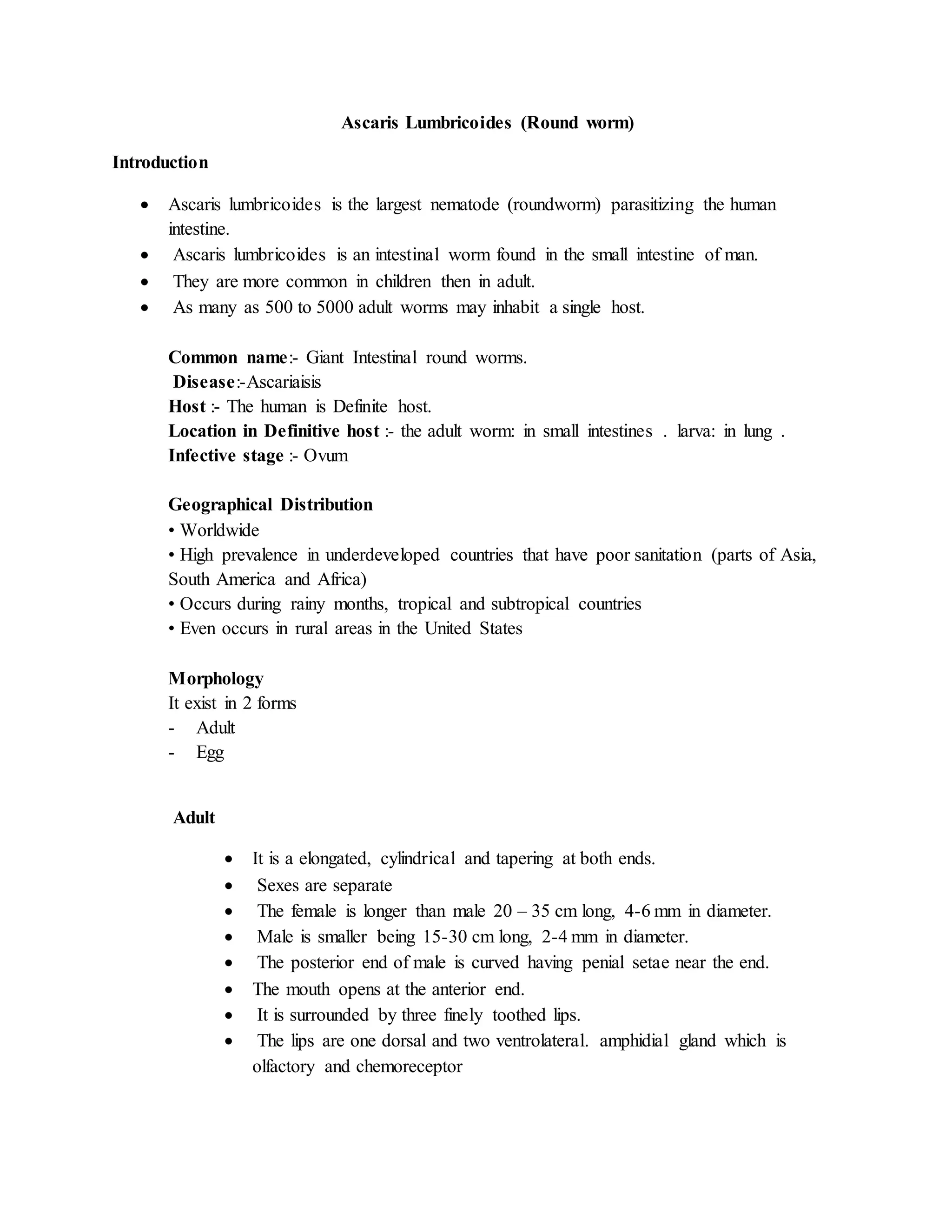
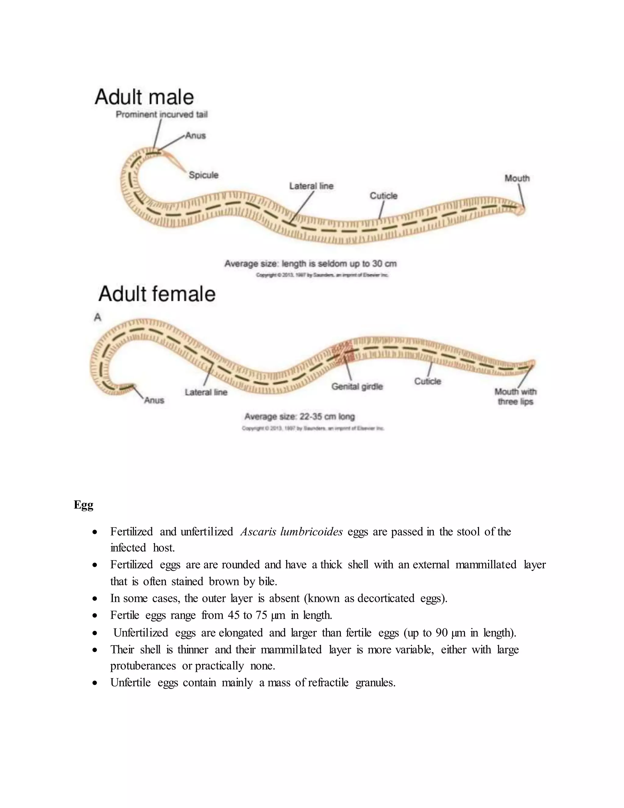
Ascaris Lumbricoides (Round worm)
Introduction
 Ascaris lumbricoides is the largest nematode (roundworm) parasitizing the human
intestine.
 Ascaris lumbricoides is an intestinal worm found in the small intestine of man.
 They are more common in children then in adult.
 As many as 500 to 5000 adult worms may inhabit a single host.
Common name:- Giant Intestinal round worms.
Disease:-Ascariaisis
Host :- The human is Definite host.
Location in Definitive host :- the adult worm: in small intestines . larva: in lung .
Infective stage :- Ovum
Geographical Distribution
• Worldwide
• High prevalence in underdeveloped countries that have poor sanitation (parts of Asia,
South America and Africa)
• Occurs during rainy months, tropical and subtropical countries
• Even occurs in rural areas in the United States
Morphology
It exist in 2 forms
- Adult
- Egg
Adult
 It is a elongated, cylindrical and tapering at both ends.
 Sexes are separate
 The female is longer than male 20 – 35 cm long, 4-6 mm in diameter.
 Male is smaller being 15-30 cm long, 2-4 mm in diameter.
 The posterior end of male is curved having penial setae near the end.
 The mouth opens at the anterior end.
 It is surrounded by three finely toothed lips.
 The lips are one dorsal and two ventrolateral. amphidial gland which is
olfactory and chemoreceptor
Egg
 Fertilized and unfertilized Ascaris lumbricoides eggs are passed in the stool of the
infected host.
 Fertilized eggs are are rounded and have a thick shell with an external mammillated layer
that is often stained brown by bile.
 In some cases, the outer layer is absent (known as decorticated eggs).
 Fertile eggs range from 45 to 75 µm in length.
 Unfertilized eggs are elongated and larger than fertile eggs (up to 90 µm in length).
 Their shell is thinner and their mammillated layer is more variable, either with large
protuberances or practically none.
 Unfertile eggs contain mainly a mass of refractile granules.
Life Cycle
 Site of inhabitation: small intestine
 Infective stage: embryonated eggs
 Route of infection: by mouth
 No intermediate and reservoir hosts
 Life span of the adult: about 1 year
Stage I: Eggs in faeces
 Sexually mature female produces as many as 200,000 eggs per day, which are shed along
with faeces in unembryonated form. They are non infective
Stage II: Development in soil
 Embryonation occurs in soil as optimum temperature of 20-25C with sufficient moisture
and O2
 Infective larva develops within egg in about 3-6 weeks
Stage III: Human infection and liberation of larvae
 Human get infection with ingestion of embryonated egg contaminated food and water
 Within embryonated state inside egg, first stage larvae develops into second stage larvae.
This second stage larvae is known as Rhabtitiform larvae
 Second stage larve is stimulated to hatch out by the presence of alkaline pH in small
intestine and solubilization of its outer layer by bile.
Stage IV: migration of larvae through
 Hatched out larvae penetrates the intestinal wall and carried to liver through portal
circulation
 It then travels via blood to heart and to lungs by pulmonary circulation within 4-7 days of
infection.
 The larvae in lungs molds twice, enlarge and breaks into alveoli.
Stage V: Re-entry to stomach and small intestine
 From alveoli, the Larvae then pass up through bronchi and into trachea and then
swallowed.
 The larvae passes down the oesophagus to the stomach and reached into small intestine
once again.
 Small intestine is the normal habitat of Ascaris and it colonises here.
 Within intestine parasite molds twice and mature into adult worm.
 Sexual maturation occurs with 6-10 weeks and the mature female discharges its eggs in
intestinal lumen and excreted along with faeces, continuing the life cycle.
 The life span of parasite is 12-18 months
Pathogenesis:
1. Mode of transmission:
 faeco-oral route, by contaminated vegetables or water.
2. Pathogenesis:
Infection of A. lumbricoides in man is known as Ascariasis. There are two phase in ascariasis.
Phase I: migrating larvae
 The migrating larvae causes pathological lesions. The severity of lesions depends upon
the sensitivity of host, nutritional status of host and number of migrating larvae.
 During migration and molding through lungs, larvae may causes pneumonia with low
grade fever, cough and other allergic symptoms.
Phase II: Adult worm
 Few worm in intestine produce no major symptoms and but some time give abdominal
pain especially in children.
 The adult worm produce trauma in host tissue and the wandering adults may block the
appendical lumen or common bile duct and even small intestine.
 Large number of adult worms affects the nutritional status of host by robbing the
nutrition leading to malnutrition and growth retardation in children.
 The metabolites of living or dead worm are toxic and immunogenic.
 lumbricoides also produces various allergic toxin, which manifests fever, conjunctivitis
and irritation
Clinical manifestation:
Most of the Ascaris infection is asymptomatic
1. Symptomatic ascariasis; two types
2. Intestinal Ascariasis
3. Pulmonary Ascariasis
1. Intestinal ascariasis;
 Nausea
 Vomiting
 Colicky abdominal pain
 Abdominal distention
 Weight loss and diarrhea
 Malbasorption of nutrition
 Growth retardation
 Heavy worm in children leads to intussusception and total obstruction
 Complications: Appendicitis, Biliary colic and perforation of bile duct, Hepatomegaly
2. Pulmonary ascariasis;
 Transient eosinophilic pneumonitis (loeffler’s disease); elevated IgE
 Bronchospasm
 Dyspnea and wheezing
 Fever
 Non-productive cough and chest pain
Lab diagnosis:
1. Specimen: stool, sputum
2. Microscopy: examination of stool by saline emulsion or concentration by floatation
methods employed to unembryonated egg
3. X-ray
4. Serodiagnosis: Indirect haemagglutination test, Immuno-fluorescence assay
5. Ultrasonography and CT scan
6. Other test: blood count shown peripheral eosinophilia
Treatment and prophylaxis:
 Mebendazole: drug of choice, (100mg twice a day for 3 days)
 Albendazole: 500mg single dose
 Pyrantel pamoate: single dose of 1omg/kg weight
 Piperazine citrate

Ascaris lumbricoides

  • 1.
    Ascaris Lumbricoides (Roundworm) Introduction  Ascaris lumbricoides is the largest nematode (roundworm) parasitizing the human intestine.  Ascaris lumbricoides is an intestinal worm found in the small intestine of man.  They are more common in children then in adult.  As many as 500 to 5000 adult worms may inhabit a single host. Common name:- Giant Intestinal round worms. Disease:-Ascariaisis Host :- The human is Definite host. Location in Definitive host :- the adult worm: in small intestines . larva: in lung . Infective stage :- Ovum Geographical Distribution • Worldwide • High prevalence in underdeveloped countries that have poor sanitation (parts of Asia, South America and Africa) • Occurs during rainy months, tropical and subtropical countries • Even occurs in rural areas in the United States Morphology It exist in 2 forms - Adult - Egg Adult  It is a elongated, cylindrical and tapering at both ends.  Sexes are separate  The female is longer than male 20 – 35 cm long, 4-6 mm in diameter.  Male is smaller being 15-30 cm long, 2-4 mm in diameter.  The posterior end of male is curved having penial setae near the end.  The mouth opens at the anterior end.  It is surrounded by three finely toothed lips.  The lips are one dorsal and two ventrolateral. amphidial gland which is olfactory and chemoreceptor
  • 2.
    Egg  Fertilized andunfertilized Ascaris lumbricoides eggs are passed in the stool of the infected host.  Fertilized eggs are are rounded and have a thick shell with an external mammillated layer that is often stained brown by bile.  In some cases, the outer layer is absent (known as decorticated eggs).  Fertile eggs range from 45 to 75 µm in length.  Unfertilized eggs are elongated and larger than fertile eggs (up to 90 µm in length).  Their shell is thinner and their mammillated layer is more variable, either with large protuberances or practically none.  Unfertile eggs contain mainly a mass of refractile granules.
  • 3.
    Life Cycle  Siteof inhabitation: small intestine  Infective stage: embryonated eggs  Route of infection: by mouth  No intermediate and reservoir hosts  Life span of the adult: about 1 year Stage I: Eggs in faeces  Sexually mature female produces as many as 200,000 eggs per day, which are shed along with faeces in unembryonated form. They are non infective Stage II: Development in soil  Embryonation occurs in soil as optimum temperature of 20-25C with sufficient moisture and O2  Infective larva develops within egg in about 3-6 weeks Stage III: Human infection and liberation of larvae  Human get infection with ingestion of embryonated egg contaminated food and water  Within embryonated state inside egg, first stage larvae develops into second stage larvae. This second stage larvae is known as Rhabtitiform larvae  Second stage larve is stimulated to hatch out by the presence of alkaline pH in small intestine and solubilization of its outer layer by bile. Stage IV: migration of larvae through
  • 4.
     Hatched outlarvae penetrates the intestinal wall and carried to liver through portal circulation  It then travels via blood to heart and to lungs by pulmonary circulation within 4-7 days of infection.  The larvae in lungs molds twice, enlarge and breaks into alveoli. Stage V: Re-entry to stomach and small intestine  From alveoli, the Larvae then pass up through bronchi and into trachea and then swallowed.  The larvae passes down the oesophagus to the stomach and reached into small intestine once again.  Small intestine is the normal habitat of Ascaris and it colonises here.  Within intestine parasite molds twice and mature into adult worm.  Sexual maturation occurs with 6-10 weeks and the mature female discharges its eggs in intestinal lumen and excreted along with faeces, continuing the life cycle.  The life span of parasite is 12-18 months
  • 6.
    Pathogenesis: 1. Mode oftransmission:  faeco-oral route, by contaminated vegetables or water. 2. Pathogenesis: Infection of A. lumbricoides in man is known as Ascariasis. There are two phase in ascariasis. Phase I: migrating larvae  The migrating larvae causes pathological lesions. The severity of lesions depends upon the sensitivity of host, nutritional status of host and number of migrating larvae.  During migration and molding through lungs, larvae may causes pneumonia with low grade fever, cough and other allergic symptoms. Phase II: Adult worm  Few worm in intestine produce no major symptoms and but some time give abdominal pain especially in children.  The adult worm produce trauma in host tissue and the wandering adults may block the appendical lumen or common bile duct and even small intestine.  Large number of adult worms affects the nutritional status of host by robbing the nutrition leading to malnutrition and growth retardation in children.  The metabolites of living or dead worm are toxic and immunogenic.  lumbricoides also produces various allergic toxin, which manifests fever, conjunctivitis and irritation Clinical manifestation: Most of the Ascaris infection is asymptomatic 1. Symptomatic ascariasis; two types 2. Intestinal Ascariasis 3. Pulmonary Ascariasis 1. Intestinal ascariasis;  Nausea  Vomiting  Colicky abdominal pain
  • 7.
     Abdominal distention Weight loss and diarrhea  Malbasorption of nutrition  Growth retardation  Heavy worm in children leads to intussusception and total obstruction  Complications: Appendicitis, Biliary colic and perforation of bile duct, Hepatomegaly 2. Pulmonary ascariasis;  Transient eosinophilic pneumonitis (loeffler’s disease); elevated IgE  Bronchospasm  Dyspnea and wheezing  Fever  Non-productive cough and chest pain Lab diagnosis: 1. Specimen: stool, sputum 2. Microscopy: examination of stool by saline emulsion or concentration by floatation methods employed to unembryonated egg 3. X-ray 4. Serodiagnosis: Indirect haemagglutination test, Immuno-fluorescence assay 5. Ultrasonography and CT scan 6. Other test: blood count shown peripheral eosinophilia Treatment and prophylaxis:  Mebendazole: drug of choice, (100mg twice a day for 3 days)  Albendazole: 500mg single dose  Pyrantel pamoate: single dose of 1omg/kg weight  Piperazine citrate