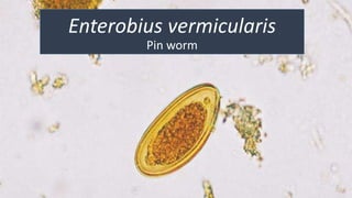
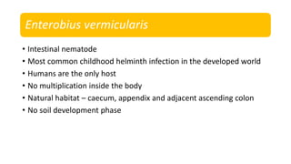
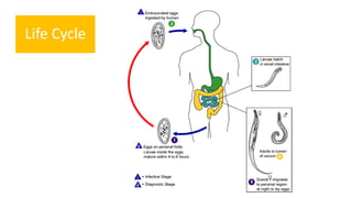
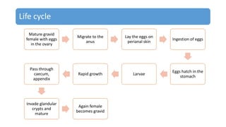
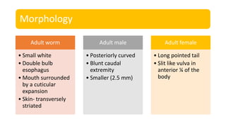
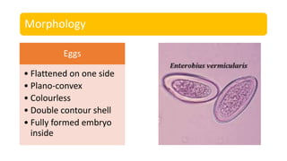
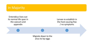
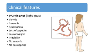
Enterobius vermicularis
Pin worm
Enterobius vermicularis
• Intestinal nematode
• Most common childhood helminth infection in the developed world
• Humans are the only host
• No multiplication inside the body
• Natural habitat – caecum, appendix and adjacent ascending colon
• No soil development phase
Life Cycle
Life cycle
Mature gravid
female with eggs
in the ovary
Migrate to the
anus
Lay the eggs on
perianal skin
Ingestion of eggs
Eggs hatch in the
stomach
LarvaeRapid growth
Pass through
caecum,
appendix
Invade glandular
crypts and
mature
Again female
becomes gravid
Morphology
Adult worm
• Small white
• Double bulb
esophagus
• Mouth surrounded
by a cuticular
expansion
• Skin- transversely
striated
Adult male
• Posteriorly curved
• Blunt caudal
extremity
• Smaller (2.5 mm)
Adult female
• Long pointed tail
• Slit like vulva in
anterior ¼ of the
body
Morphology
Eggs
• Flattened on one side
• Plano-convex
• Colourless
• Double contour shell
• Fully formed embryo
inside
Transmission
Contaminated food and water
Autoinfection
• Scratching of perianal skin leads to deposition of eggs in finger
nails
Retroinfection
• Eggs laid on the perianal skin immediately hatch and larvae
migrate into the colon and develop into worms
In Majority
Enterobius lives out
its normal life span in
the caecum and
appendix
Migrate down to the
anus to lay eggs
Larvae re-establish in
the host causing few
/ no symptoms
Clinical features
• Pruritis anus (itchy anus)
• Vulvitis
• Insomnia
• Restlessness
• Loss of appetite
• Loss of weight
• Irritability
• No anaemia
• No eosinophilia
Diagnosis
• Eggs in faeces
• Perianal scrapings / swabs
• Usually at night
Treatment
• Albendazole
• Mebendazole
• Pyrantel pamoate
• Piperazine
Thank you

Enterobius vermicularis

  • 1.
  • 2.
    Enterobius vermicularis • Intestinalnematode • Most common childhood helminth infection in the developed world • Humans are the only host • No multiplication inside the body • Natural habitat – caecum, appendix and adjacent ascending colon • No soil development phase
  • 3.
  • 5.
    Life cycle Mature gravid femalewith eggs in the ovary Migrate to the anus Lay the eggs on perianal skin Ingestion of eggs Eggs hatch in the stomach LarvaeRapid growth Pass through caecum, appendix Invade glandular crypts and mature Again female becomes gravid
  • 6.
    Morphology Adult worm • Smallwhite • Double bulb esophagus • Mouth surrounded by a cuticular expansion • Skin- transversely striated Adult male • Posteriorly curved • Blunt caudal extremity • Smaller (2.5 mm) Adult female • Long pointed tail • Slit like vulva in anterior ¼ of the body
  • 7.
    Morphology Eggs • Flattened onone side • Plano-convex • Colourless • Double contour shell • Fully formed embryo inside
  • 8.
    Transmission Contaminated food andwater Autoinfection • Scratching of perianal skin leads to deposition of eggs in finger nails Retroinfection • Eggs laid on the perianal skin immediately hatch and larvae migrate into the colon and develop into worms
  • 9.
    In Majority Enterobius livesout its normal life span in the caecum and appendix Migrate down to the anus to lay eggs Larvae re-establish in the host causing few / no symptoms
  • 10.
    Clinical features • Pruritisanus (itchy anus) • Vulvitis • Insomnia • Restlessness • Loss of appetite • Loss of weight • Irritability • No anaemia • No eosinophilia
  • 11.
    Diagnosis • Eggs infaeces • Perianal scrapings / swabs • Usually at night
  • 12.
    Treatment • Albendazole • Mebendazole •Pyrantel pamoate • Piperazine
  • 13.