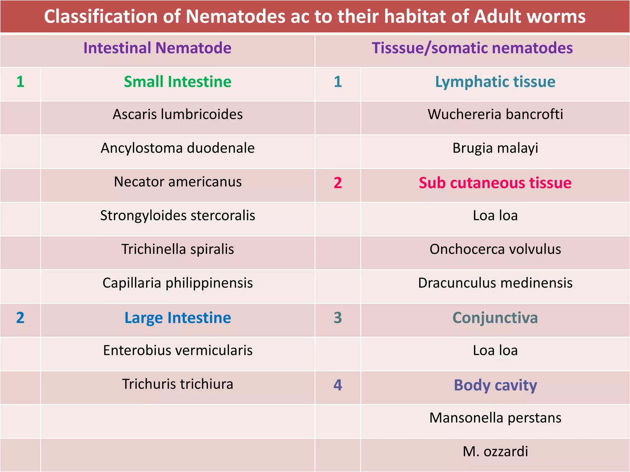
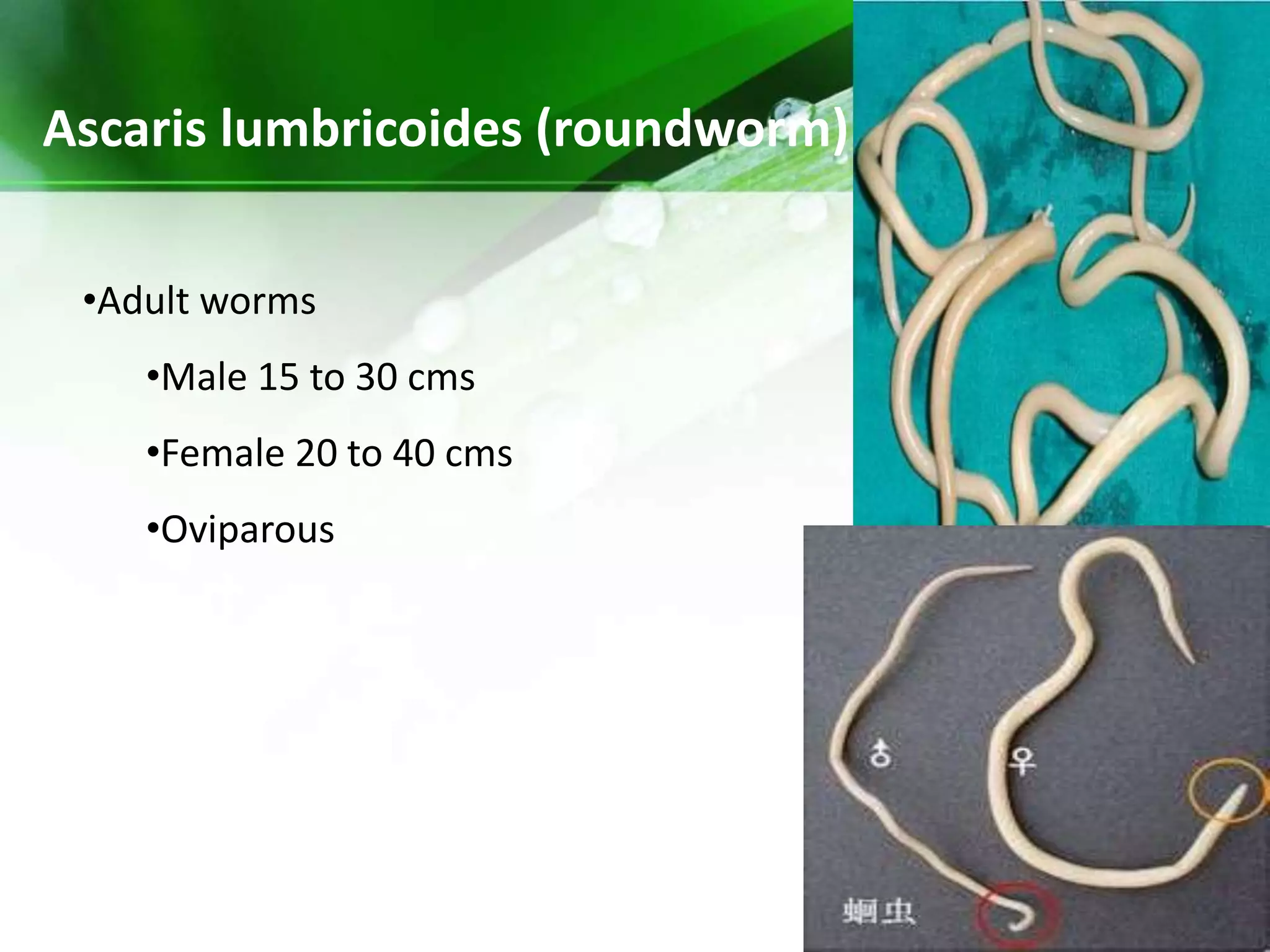
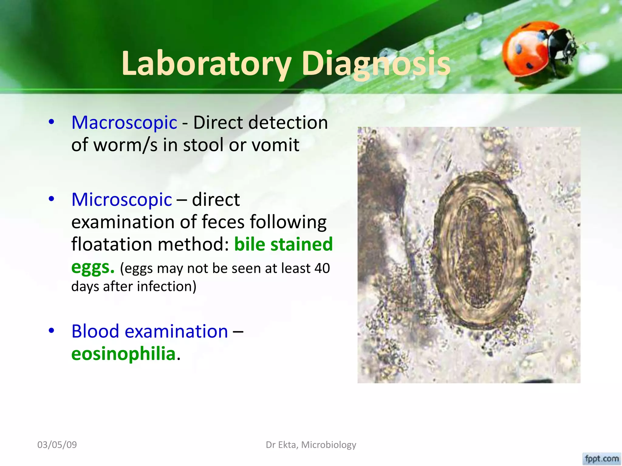
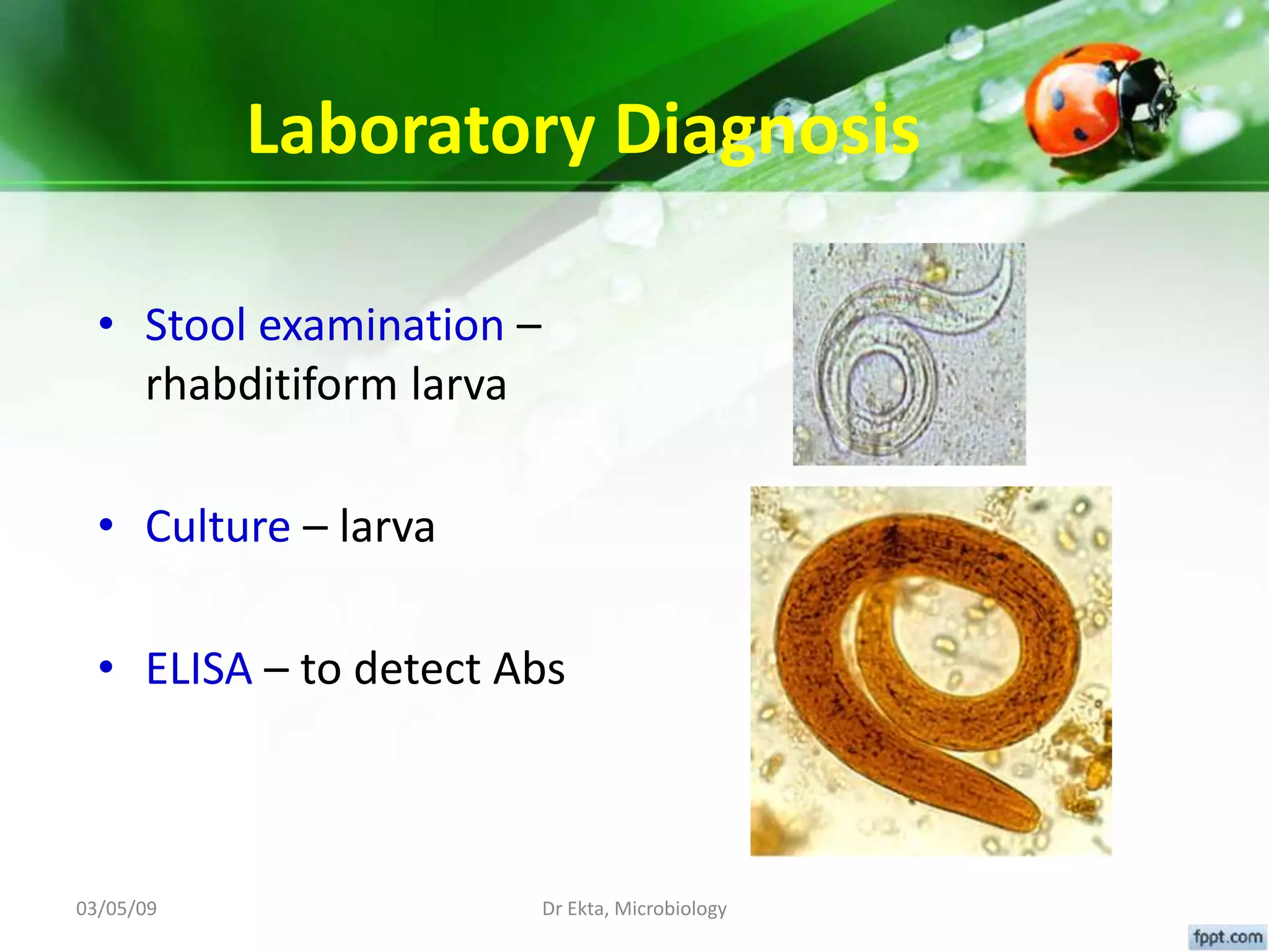
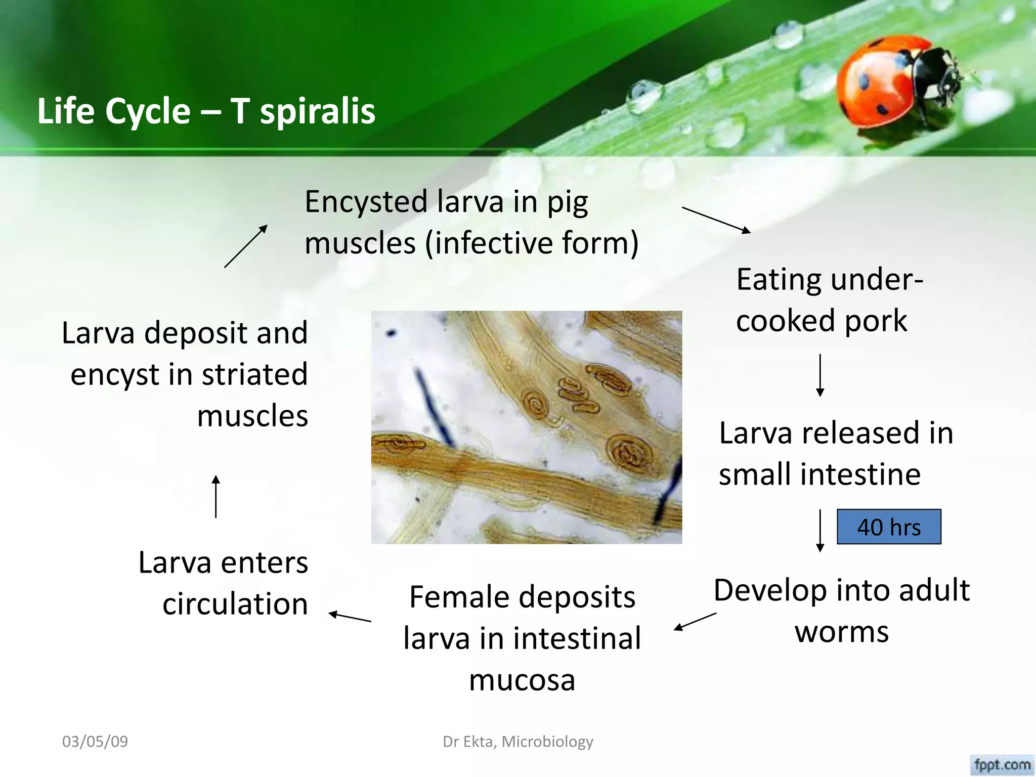
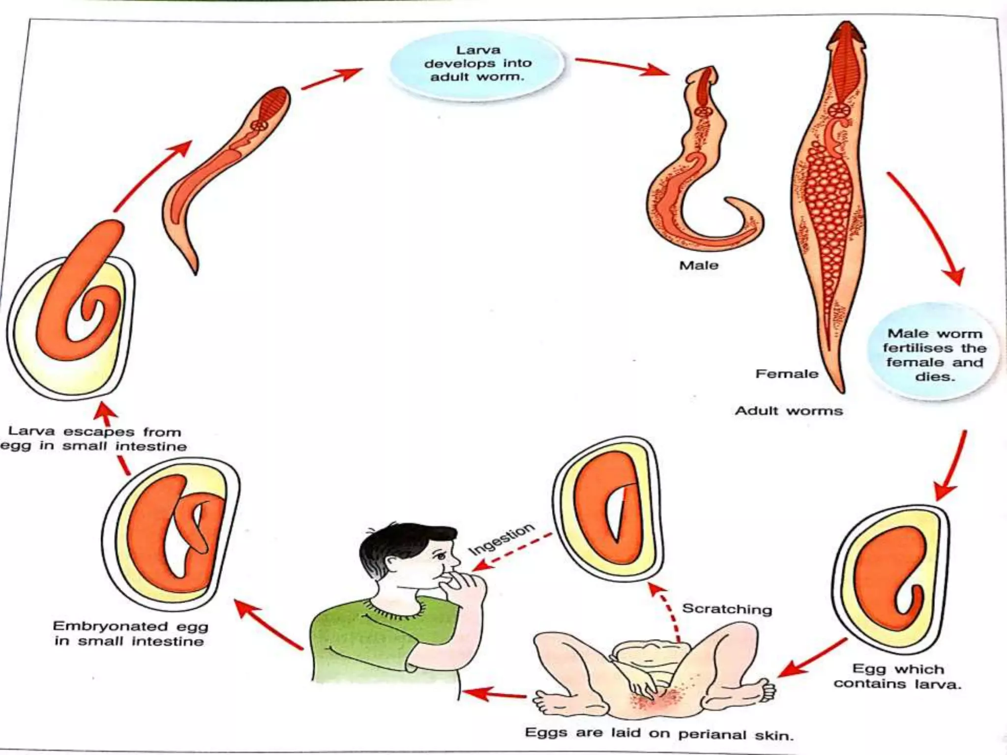
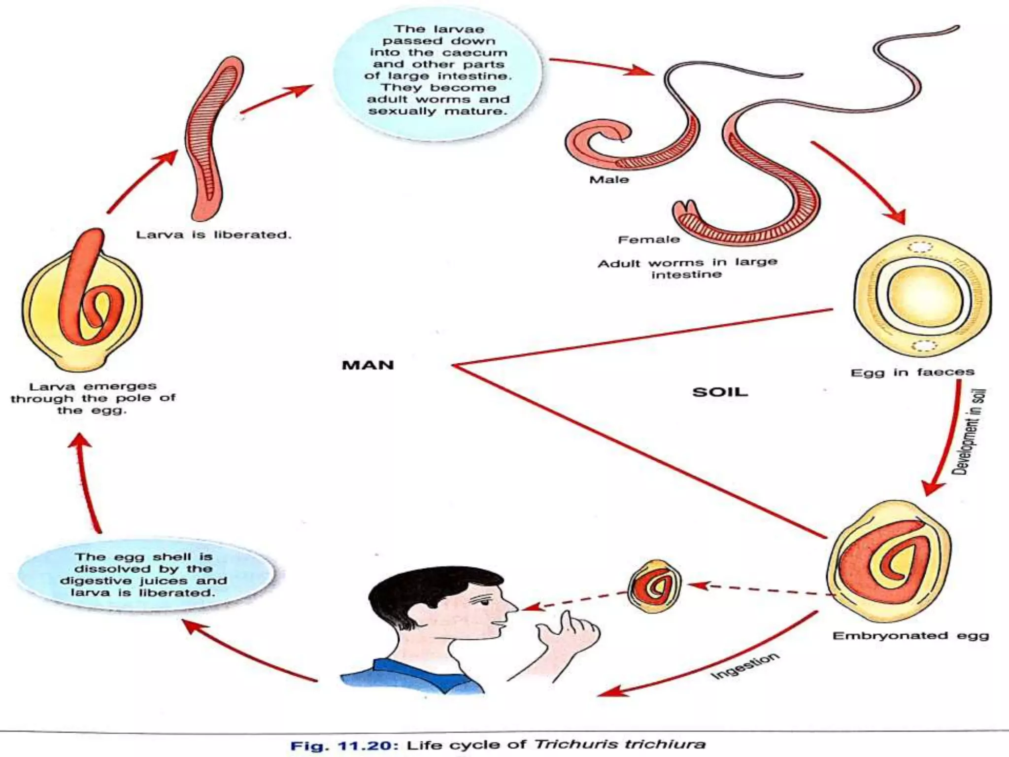
Nematodes
Dr. Rakesh Prasad Sah
Assistant Professor, Microbiology
Introduction
• “nematode”  thread like
• Are elongated bilaterally symmetrical, Non-segmented cylindrical worms
tapering at both ends
• Possess cuticle
• Sexes are separate (diecious), male is smaller than female & its posterior
end is curved ventrally
• Females are either Viviparous (produce larvae/ embryos) Oviparous (lay
eggs) or Ovo-viviparous (lay eggs which hatch immediately)
• Live in intestinal tract or tissues
Classification of Nematodes ac to their habitat of Adult worms
Intestinal Nematode Tisssue/somatic nematodes
1 Small Intestine 1 Lymphatic tissue
Ascaris lumbricoides Wuchereria bancrofti
Ancylostoma duodenale Brugia malayi
Necator americanus 2 Sub cutaneous tissue
Strongyloides stercoralis Loa loa
Trichinella spiralis Onchocerca volvulus
Capillaria philippinensis Dracunculus medinensis
2 Large Intestine 3 Conjunctiva
Enterobius vermicularis Loa loa
Trichuris trichiura 4 Body cavity
Mansonella perstans
M. ozzardi
Ascaris lumbricoides (roundworm)
•Adult worms
•Male 15 to 30 cms
•Female 20 to 40 cms
•Oviparous
Ascaris lumbricoides (roundworm)
•Eggs
•60 μ,
•bile stained
•Albuminous coat with unsegmented
ovum
•Infective form
•Embryonated eggs
•Mode of transmission
•Ingestion
•Site of localization
•Small intestine
Fertilised Egg Unfertilised egg
1 Round or oval (60-75 X 40-50) 1 Narrower & longer (80 X 55)
2 Bile stained & thus brown in colour 2 Bile stained & thus brown in
colour
3 Thick, traslucent shell with an
outer albuminous coat in the form
of rugosities.
3 Thin shell with an irregular
coating of albumin.
4 Outer coat sometimes lost
(decorticated egg)
4
5 Contains large unsegmented ovum
with clear crescentric area at each
pole
5 Contains a small atrophied ovum.
6 Floats in saturated solution of
common salts.
6. Does not float in saturated
solution of common salts
(heaviest of all helminthic eggs)
Adult worms in the
intestine
Unembryonated egg (stoll)
Embryonated eggs in 2-3 weeks
in soil (eggs containg
rhabditiform larvae)
Ingestion of eggs
Rhabditiform larva
hatches &
Penetrate intestine, reach
liver
Right heart, lung, bronchi,
trachea, larynx, pharynx
Larvae swallowed and reach
intestine
03/05/09 Dr Ekta, Microbiology
Pathogenicity & Clinical Features
• Ascariasis – infection of A.lumbricoides
• Majority of infections are asymptomatic
• Clinical disease is largely restricted to individuals
with a high worm load
• Symptoms divided into two groups: those
produced by
1. Migrating larvae
2. Adult worms
03/05/09 Dr Ekta, Microbiology
Symptoms & Complications
• Symptoms produced by Migrating larvae
1. Pneumonia (loeffler’s syndrome) –
• fever,
• cough,
• dyspnoea,
• blood tinged sputum that may contain larva,
• urticarial rash & eosinophilia
2. Visceral larva migrans – if larvae enter systemic circulation
(from pulmonary capillaries) to reach other organs like
brain, spinal cord, heart, kidney; usually seen in massive
infection
Symptoms & Complications
• Symptoms produced by Adult worms
1. Abdominal discomfort, anorexia, nausea &
diarrhoea.
2. PEM, Vit. A deficiency (night blindness)
3. Intestinal obstruction (particularly in
children 1-5 years), intussusception &
volvulus
4. Penetration through intestinal ulcer
(perforation) – peritonitis
5. Hypersensitivity reactions to worm Ags
(toxic body fluids) – urticaria, edema of
face, conjunctivitis, irritation of URT
Pathogenicity & clinical features
Symptoms produced by Adult worms
6. Ectopic Ascariasis – due to
migration of worm up into the
stomach. It may
• be vomited out,
• pass up through the oesophagus
at night & comes out through
mouth or nose,
• enter larynx to cause asphyxia.
• migrate to other organs and
cause
– appendicitis,
– cholecystitis,
– biliary colic,
– Cholangitis,
– pancreatitis
03/05/09 Dr Ekta, Microbiology
Laboratory Diagnosis
• Macroscopic - Direct detection
of worm/s in stool or vomit
• Microscopic – direct
examination of feces following
floatation method: bile stained
eggs. (eggs may not be seen at least 40
days after infection)
• Blood examination –
eosinophilia.
03/05/09 Dr Ekta, Microbiology
Other modes of diagnosis
• Imaging – large collections of worms in abdomen
• USG - to diagnose hepatobiliary or pancreatic
ascariasis
• Serology (Ab detection) – mainly reserved for
epidemiological studies.
03/05/09 Dr Ekta, Microbiology
Adult worms Male 8 -11mm
Female 10-13 mm, oviparous
Eggs 60 µ, non bile stained (colorless)
Segmented, 4 blastomeres
Infective form 3rd stage filariform larva
Mode of infection Penetration into skin
Site of localization Small intestine
Ancylostoma duodenale (hook worm)
Differences between Male and Female Ancylostoma duodenale
Characteristic Male Female
Size Smaller (5-11mm X 0.4 X 0.5mm) Longer than male (9-13mm X
0.6mm)
Posterior end Expanded in an umbrella like
fashion, named as copulatory
bursa
Tapering and there is no
expanded bursa
Genital opening Posteriorly and opens with cloaca Present at the junction of
posterior and middle third of
the body
• Egg
– Oval, (L -65 µm & B-40 µm)
– Not bile stained
– Surrounded by a thin transparent
hyaline shell-membrane
– Contains segmented ovum with four
blastomeres.
– Clear space between egg shell and the
segmented ovum
– Floats in S.S.S.
Sites of skin penetration
• Most common sites are:
1. Thin skin between toes
2. Dorsum of the feet
3. Inner side of the soles
• Gardeners & miners – skin of hands
03/05/09 Dr Ekta, Microbiology
48 hours
6 to 8 days
6 to 8 weeks
8 to 10 days
Life cycle of
hookworm
Adult worms in the small
intestine
Eggs in faeces
In soil (eggs containg
rhabditiform larvae hatch out)
Filariform larva
Penetrates skin &
reach in circulation
Lungs, trachea,larynx
Pharynx  Swallowed
Larvae settles down in the small
intestine
03/05/09 Dr Ekta, Microbiology
Hook worms in the intestine
03/05/09 Dr Ekta, Microbiology
Pathogenicity & Clinical Features
• Ancylostomiasis or hookworm disease,
characterised by iron deficiency anaemia
• Majority of infections are asymptomatic
• Symptoms develop in heavy infections and divided
into two groups: those produced by
1. Migrating larvae
2. Adult worms
03/05/09 Dr Ekta, Microbiology
Symptoms produced by larvae
• Lesions in the skin:
1. Ancylostome dermatitis or Ground itch – occurs at the
site of entry (more common in necator), lasts for 2 to 4
weeks.
Intense itching and burning followed by erythema and
oedema of the area.
1. Creeping eruption – reddish itchy papule
along the path traversed by filariform
larvae (larva migrans)
• Lesions in the lungs – bronchitis &
bronchopneumonia.
03/05/09 Dr Ekta, Microbiology
Symptoms produced by adult worm
• Epigastric pain, diarrhoea & vomiting during early
phase of infection.
• Microcytic hypochromic (Iron deficiency) anaemia –
due to chronic blood loss:
– a single adult hookworm sucks 0.2ml of blood/
day
– Hemorrhages from punctured sites
Clinical features of hookworm anemia
• Extreme pallor
• Abnormal appetite
• Epigastric tenderness with dyspepsia
• Constipation
• Puffy face with swelling of lower eyelids
• Pedal edema
• Growth retardation
03/05/09 Dr Ekta, Microbiology
Laboratory Diagnosis
• Stool examination –
microscopy: non bile
stained egg, segmented
• Occult blood in stool –
positive
• Blood examination –
anaemia, eosinophilia
Necator americanus
• Similar as A. duodenale.
Differentiation features of A. duodenale and N. americanus
Feature A. Duodenale N. Americanus
Size Larger & thicker Smaller & more slender
Anterior end Bends in the same direction as the body
curvature
Bend in the opposite direction to
the body curvature
Buccal capsule Six teeth  4 hook on V.S. and 2 Knob
like on D.S.
4 chitinous plate  2 on each V.S.
& D.S.
Copulatory
bursa
13 rays present 14 rays present
Posterior end of
female
Spine present Spine absent
Vulval opening Present behind the middle of the body Present in front of the middle of
the body
Pathogenicity More pathogenic Less pathogenic
03/05/09 Dr Ekta, Microbiology
Strongyloides stercoralis
Adult worms 2 - 2.5mm, ovoviviparous,
eggs laid in the tissues
Free living worms
Infective form Filariform larvae
Mode of transmission Penetration / autoinfection
Site of localization Wall of Small intestine, mainly
duodenum & jejunum
Moist soil
Strongyloides stercoralis
03/05/09 Dr Ekta, Microbiology
Life cycle – S. stercoralis
Adult worms in
intestine
Eggs in intestine
Rhabditiform larvae hatch
Autoinfection
Larvae in feces - soil
Direct Indirect
Larvae into adults
Eggs produced
Rhabditiform larvae hatch
Filariform larvae
(infective form)
Penetrate
skin
Larvae in blood
Larvae pass
through similar
to Ascaris
Larvae reach
intestine
Rhabditiform to
filariform
Larvae penetrate
intestine
03/05/09 Dr Ekta, Microbiology
Pathogenicity
1. Skin lesions (2 types) – “larva currens”
– At the site of entry – itching & erythema, pruritus & urticaria rash
– In the perianal region – linear, erythematous urticarial wheal
2. Pulmonary lesions – due to migrating larva
– Alveolar hemorrhages
– Bronchopneumonia
3. Intestinal lesions - “burrowing lesions”
– Epigastric pain
– Diarrhoea with blood & mucus
– Nausea
– Weight loss
03/05/09 Dr Ekta, Microbiology
Important terms to know
• Autoinfection – filariform larva
1. In the Intestinal lumen
2. Perineal & perianal skin penetration
• Hyperinfection – can result in autoinfection
1. Steroids or Immunosuppressive therapy
2. Malignancy
3. Malnutrition
4. Pregnancy
5. AIDS
• Persistence of infection – due to autoinfection
03/05/09 Dr Ekta, Microbiology
Laboratory Diagnosis
• Stool examination –
rhabditiform larva
• Culture – larva
• ELISA – to detect Abs
03/05/09 Dr Ekta, Microbiology
Trichinella spiralis (Trichina Worm)
Adult worms (smallest
nematode infecting man)
Male 1.4 – 1.6 mm
Female 3 - 4 mm, viviparous
Infective form Encysted larvae (100µ) in
striated muscles of pig
Mode of transmission Ingestion of improperly cooked
pork
Site of localization Small intestine
Commonly involved
muscles
Diaphragm, Intercostals, Deltoid,
Pectoralis major, Biceps
03/05/09 Dr Ekta, Microbiology
Encysted larva in pig
muscles (infective form)
Eating under-
cooked pork
Larva released in
small intestine
Develop into adult
worms
Female deposits
larva in intestinal
mucosa
Larva enters
circulation
Larva deposit and
encyst in striated
muscles
Life Cycle – T spiralis
40 hrs
03/05/09 Dr Ekta, Microbiology
Pathogenicity
• Trichinelliasis / Trichinosis – clinical features depends on
the stage:
1. Stage of intestinal invasion: 5-7 days, pain in abdomen, nausea,
vomiting, diarrhoea
2. Stage of larval migration: fever, urticarial rash, splinter
hemorrhages, periorbital & facial edema
3. Stage of encystation: asymptomatic in light infections; myalgia,
weakness in heavy infections
• Complications – during migration:
– myocarditis, encephalitis
03/05/09 Dr Ekta, Microbiology
Laboratory Diagnosis
• Muscle biopsy – encysted larva
• Blood – eosinophilia between 2nd &
4th week
• Serology – ELISA
• Intradermal Test – named Bachman
test 0.1ml (1in 1000 dil of larval Ag)
 erythematous wheal within 15-
20min
• Radiological examination – Calcified
cysts
03/05/09 Dr Ekta, Microbiology
Enterobius vermicularis
Adult worms Male 2 - 5 mm
Female 8 -13 mm, oviparous
Eggs 60 µ, non bile stained
Plano-convex with coiled embryo
Infective form Embryonated egg
Mode of transmission Ingestion,
Autoinfection
Site of localization Large intestine –
caecum & appendix
(Pin Worm)
03/05/09 Dr Ekta, Microbiology
Clinical features
• Due to migration of worm - Perianal, perineal &
vaginal itching (pruritis) worsens at night.
• Insomnia and restlessness
• Nocturnal enuresis
• In girls migrating female worms  vagina & urethra 
Vaginitis & Urethritis.
03/05/09
Laboratory Diagnosis & Treatment
• Detection of adult worms in-
– Feces
– Perianal region
• NIH swab – scrapings from
perianal region
• Microscopy – non bile stained
eggs
• Mebendazole, pyrantel
pamoate
03/05/09 Dr Ekta, Microbiology
Trichuris trichiura (Whip Worm)
Adult worm 30 – 50 mm
Eggs 60 µ, bile stained
Barrel-shaped with Mucus plug at
each pole
Unsegmented ovum
Infective form Mature embryonated eggs
Mode of transmission Ingestion
Site of localization Large intestine -
caecum
03/05/09 Dr Ekta, Microbiology
Clinical features
• Infection – Trichuriasis
• Symptoms depend on worm burden
– Less than 10 worms – asymptomatic
– Heavier infections –
1. chronic profuse mucus and bloody diarrhea with
abdominal pains and edematous rectum
2. malnutrition, weight loss and anemia
03/05/09 Dr Ekta, Microbiology
Laboratory diagnosis & Treatment
• Stool examination – bile stained
eggs with bipolar mucus plugs
• Treatment – albendazole /
mebendazole
• Prevention –
– Proper disposal of night soil
– Prevention of consumption of
uncooked vegetables & fruits .
03/05/09 Dr Ekta, Microbiology
Key to the diagnosis of Intestinal Nematodes
Intestinal
Nematodes
Larvae in Stool
S. stercoralis
Eggs in stool
Eggs on
Perianal Skin
Colored
(Bile Stained)
A. lumbricoides
T. trichiura
Colorless
(Non Bile Stained)
E. vermicularis
Colorless
(Non Bile Stained)
A. duodenale
N. americanus
E. vermicularis
Nematodes by Dr. Rakesh Prasad Sah

Nematodes by Dr. Rakesh Prasad Sah

  • 1.
    Nematodes Dr. Rakesh PrasadSah Assistant Professor, Microbiology
  • 2.
    Introduction • “nematode” thread like • Are elongated bilaterally symmetrical, Non-segmented cylindrical worms tapering at both ends • Possess cuticle • Sexes are separate (diecious), male is smaller than female & its posterior end is curved ventrally • Females are either Viviparous (produce larvae/ embryos) Oviparous (lay eggs) or Ovo-viviparous (lay eggs which hatch immediately) • Live in intestinal tract or tissues
  • 3.
    Classification of Nematodesac to their habitat of Adult worms Intestinal Nematode Tisssue/somatic nematodes 1 Small Intestine 1 Lymphatic tissue Ascaris lumbricoides Wuchereria bancrofti Ancylostoma duodenale Brugia malayi Necator americanus 2 Sub cutaneous tissue Strongyloides stercoralis Loa loa Trichinella spiralis Onchocerca volvulus Capillaria philippinensis Dracunculus medinensis 2 Large Intestine 3 Conjunctiva Enterobius vermicularis Loa loa Trichuris trichiura 4 Body cavity Mansonella perstans M. ozzardi
  • 4.
    Ascaris lumbricoides (roundworm) •Adultworms •Male 15 to 30 cms •Female 20 to 40 cms •Oviparous
  • 5.
    Ascaris lumbricoides (roundworm) •Eggs •60μ, •bile stained •Albuminous coat with unsegmented ovum •Infective form •Embryonated eggs •Mode of transmission •Ingestion •Site of localization •Small intestine
  • 6.
    Fertilised Egg Unfertilisedegg 1 Round or oval (60-75 X 40-50) 1 Narrower & longer (80 X 55) 2 Bile stained & thus brown in colour 2 Bile stained & thus brown in colour 3 Thick, traslucent shell with an outer albuminous coat in the form of rugosities. 3 Thin shell with an irregular coating of albumin. 4 Outer coat sometimes lost (decorticated egg) 4 5 Contains large unsegmented ovum with clear crescentric area at each pole 5 Contains a small atrophied ovum. 6 Floats in saturated solution of common salts. 6. Does not float in saturated solution of common salts (heaviest of all helminthic eggs)
  • 7.
    Adult worms inthe intestine Unembryonated egg (stoll) Embryonated eggs in 2-3 weeks in soil (eggs containg rhabditiform larvae) Ingestion of eggs Rhabditiform larva hatches & Penetrate intestine, reach liver Right heart, lung, bronchi, trachea, larynx, pharynx Larvae swallowed and reach intestine
  • 8.
    03/05/09 Dr Ekta,Microbiology Pathogenicity & Clinical Features • Ascariasis – infection of A.lumbricoides • Majority of infections are asymptomatic • Clinical disease is largely restricted to individuals with a high worm load • Symptoms divided into two groups: those produced by 1. Migrating larvae 2. Adult worms
  • 9.
    03/05/09 Dr Ekta,Microbiology Symptoms & Complications • Symptoms produced by Migrating larvae 1. Pneumonia (loeffler’s syndrome) – • fever, • cough, • dyspnoea, • blood tinged sputum that may contain larva, • urticarial rash & eosinophilia 2. Visceral larva migrans – if larvae enter systemic circulation (from pulmonary capillaries) to reach other organs like brain, spinal cord, heart, kidney; usually seen in massive infection
  • 10.
    Symptoms & Complications •Symptoms produced by Adult worms 1. Abdominal discomfort, anorexia, nausea & diarrhoea. 2. PEM, Vit. A deficiency (night blindness) 3. Intestinal obstruction (particularly in children 1-5 years), intussusception & volvulus 4. Penetration through intestinal ulcer (perforation) – peritonitis 5. Hypersensitivity reactions to worm Ags (toxic body fluids) – urticaria, edema of face, conjunctivitis, irritation of URT
  • 11.
    Pathogenicity & clinicalfeatures Symptoms produced by Adult worms 6. Ectopic Ascariasis – due to migration of worm up into the stomach. It may • be vomited out, • pass up through the oesophagus at night & comes out through mouth or nose, • enter larynx to cause asphyxia. • migrate to other organs and cause – appendicitis, – cholecystitis, – biliary colic, – Cholangitis, – pancreatitis
  • 12.
    03/05/09 Dr Ekta,Microbiology Laboratory Diagnosis • Macroscopic - Direct detection of worm/s in stool or vomit • Microscopic – direct examination of feces following floatation method: bile stained eggs. (eggs may not be seen at least 40 days after infection) • Blood examination – eosinophilia.
  • 13.
    03/05/09 Dr Ekta,Microbiology Other modes of diagnosis • Imaging – large collections of worms in abdomen • USG - to diagnose hepatobiliary or pancreatic ascariasis • Serology (Ab detection) – mainly reserved for epidemiological studies.
  • 14.
    03/05/09 Dr Ekta,Microbiology Adult worms Male 8 -11mm Female 10-13 mm, oviparous Eggs 60 µ, non bile stained (colorless) Segmented, 4 blastomeres Infective form 3rd stage filariform larva Mode of infection Penetration into skin Site of localization Small intestine Ancylostoma duodenale (hook worm)
  • 15.
    Differences between Maleand Female Ancylostoma duodenale Characteristic Male Female Size Smaller (5-11mm X 0.4 X 0.5mm) Longer than male (9-13mm X 0.6mm) Posterior end Expanded in an umbrella like fashion, named as copulatory bursa Tapering and there is no expanded bursa Genital opening Posteriorly and opens with cloaca Present at the junction of posterior and middle third of the body
  • 16.
    • Egg – Oval,(L -65 µm & B-40 µm) – Not bile stained – Surrounded by a thin transparent hyaline shell-membrane – Contains segmented ovum with four blastomeres. – Clear space between egg shell and the segmented ovum – Floats in S.S.S.
  • 17.
    Sites of skinpenetration • Most common sites are: 1. Thin skin between toes 2. Dorsum of the feet 3. Inner side of the soles • Gardeners & miners – skin of hands
  • 18.
    03/05/09 Dr Ekta,Microbiology 48 hours 6 to 8 days 6 to 8 weeks 8 to 10 days Life cycle of hookworm
  • 19.
    Adult worms inthe small intestine Eggs in faeces In soil (eggs containg rhabditiform larvae hatch out) Filariform larva Penetrates skin & reach in circulation Lungs, trachea,larynx Pharynx  Swallowed Larvae settles down in the small intestine
  • 20.
    03/05/09 Dr Ekta,Microbiology Hook worms in the intestine
  • 21.
    03/05/09 Dr Ekta,Microbiology Pathogenicity & Clinical Features • Ancylostomiasis or hookworm disease, characterised by iron deficiency anaemia • Majority of infections are asymptomatic • Symptoms develop in heavy infections and divided into two groups: those produced by 1. Migrating larvae 2. Adult worms
  • 22.
    03/05/09 Dr Ekta,Microbiology Symptoms produced by larvae • Lesions in the skin: 1. Ancylostome dermatitis or Ground itch – occurs at the site of entry (more common in necator), lasts for 2 to 4 weeks. Intense itching and burning followed by erythema and oedema of the area. 1. Creeping eruption – reddish itchy papule along the path traversed by filariform larvae (larva migrans) • Lesions in the lungs – bronchitis & bronchopneumonia.
  • 23.
    03/05/09 Dr Ekta,Microbiology Symptoms produced by adult worm • Epigastric pain, diarrhoea & vomiting during early phase of infection. • Microcytic hypochromic (Iron deficiency) anaemia – due to chronic blood loss: – a single adult hookworm sucks 0.2ml of blood/ day – Hemorrhages from punctured sites
  • 24.
    Clinical features ofhookworm anemia • Extreme pallor • Abnormal appetite • Epigastric tenderness with dyspepsia • Constipation • Puffy face with swelling of lower eyelids • Pedal edema • Growth retardation
  • 25.
    03/05/09 Dr Ekta,Microbiology Laboratory Diagnosis • Stool examination – microscopy: non bile stained egg, segmented • Occult blood in stool – positive • Blood examination – anaemia, eosinophilia
  • 26.
    Necator americanus • Similaras A. duodenale. Differentiation features of A. duodenale and N. americanus Feature A. Duodenale N. Americanus Size Larger & thicker Smaller & more slender Anterior end Bends in the same direction as the body curvature Bend in the opposite direction to the body curvature Buccal capsule Six teeth  4 hook on V.S. and 2 Knob like on D.S. 4 chitinous plate  2 on each V.S. & D.S. Copulatory bursa 13 rays present 14 rays present Posterior end of female Spine present Spine absent Vulval opening Present behind the middle of the body Present in front of the middle of the body Pathogenicity More pathogenic Less pathogenic
  • 27.
    03/05/09 Dr Ekta,Microbiology Strongyloides stercoralis Adult worms 2 - 2.5mm, ovoviviparous, eggs laid in the tissues Free living worms Infective form Filariform larvae Mode of transmission Penetration / autoinfection Site of localization Wall of Small intestine, mainly duodenum & jejunum Moist soil
  • 28.
  • 29.
    03/05/09 Dr Ekta,Microbiology Life cycle – S. stercoralis Adult worms in intestine Eggs in intestine Rhabditiform larvae hatch Autoinfection Larvae in feces - soil Direct Indirect Larvae into adults Eggs produced Rhabditiform larvae hatch Filariform larvae (infective form) Penetrate skin Larvae in blood Larvae pass through similar to Ascaris Larvae reach intestine Rhabditiform to filariform Larvae penetrate intestine
  • 30.
    03/05/09 Dr Ekta,Microbiology Pathogenicity 1. Skin lesions (2 types) – “larva currens” – At the site of entry – itching & erythema, pruritus & urticaria rash – In the perianal region – linear, erythematous urticarial wheal 2. Pulmonary lesions – due to migrating larva – Alveolar hemorrhages – Bronchopneumonia 3. Intestinal lesions - “burrowing lesions” – Epigastric pain – Diarrhoea with blood & mucus – Nausea – Weight loss
  • 31.
    03/05/09 Dr Ekta,Microbiology Important terms to know • Autoinfection – filariform larva 1. In the Intestinal lumen 2. Perineal & perianal skin penetration • Hyperinfection – can result in autoinfection 1. Steroids or Immunosuppressive therapy 2. Malignancy 3. Malnutrition 4. Pregnancy 5. AIDS • Persistence of infection – due to autoinfection
  • 32.
    03/05/09 Dr Ekta,Microbiology Laboratory Diagnosis • Stool examination – rhabditiform larva • Culture – larva • ELISA – to detect Abs
  • 34.
    03/05/09 Dr Ekta,Microbiology Trichinella spiralis (Trichina Worm) Adult worms (smallest nematode infecting man) Male 1.4 – 1.6 mm Female 3 - 4 mm, viviparous Infective form Encysted larvae (100µ) in striated muscles of pig Mode of transmission Ingestion of improperly cooked pork Site of localization Small intestine Commonly involved muscles Diaphragm, Intercostals, Deltoid, Pectoralis major, Biceps
  • 35.
    03/05/09 Dr Ekta,Microbiology Encysted larva in pig muscles (infective form) Eating under- cooked pork Larva released in small intestine Develop into adult worms Female deposits larva in intestinal mucosa Larva enters circulation Larva deposit and encyst in striated muscles Life Cycle – T spiralis 40 hrs
  • 36.
    03/05/09 Dr Ekta,Microbiology Pathogenicity • Trichinelliasis / Trichinosis – clinical features depends on the stage: 1. Stage of intestinal invasion: 5-7 days, pain in abdomen, nausea, vomiting, diarrhoea 2. Stage of larval migration: fever, urticarial rash, splinter hemorrhages, periorbital & facial edema 3. Stage of encystation: asymptomatic in light infections; myalgia, weakness in heavy infections • Complications – during migration: – myocarditis, encephalitis
  • 37.
    03/05/09 Dr Ekta,Microbiology Laboratory Diagnosis • Muscle biopsy – encysted larva • Blood – eosinophilia between 2nd & 4th week • Serology – ELISA • Intradermal Test – named Bachman test 0.1ml (1in 1000 dil of larval Ag)  erythematous wheal within 15- 20min • Radiological examination – Calcified cysts
  • 38.
    03/05/09 Dr Ekta,Microbiology Enterobius vermicularis Adult worms Male 2 - 5 mm Female 8 -13 mm, oviparous Eggs 60 µ, non bile stained Plano-convex with coiled embryo Infective form Embryonated egg Mode of transmission Ingestion, Autoinfection Site of localization Large intestine – caecum & appendix (Pin Worm)
  • 41.
    03/05/09 Dr Ekta,Microbiology Clinical features • Due to migration of worm - Perianal, perineal & vaginal itching (pruritis) worsens at night. • Insomnia and restlessness • Nocturnal enuresis • In girls migrating female worms  vagina & urethra  Vaginitis & Urethritis.
  • 42.
    03/05/09 Laboratory Diagnosis &Treatment • Detection of adult worms in- – Feces – Perianal region • NIH swab – scrapings from perianal region • Microscopy – non bile stained eggs • Mebendazole, pyrantel pamoate
  • 44.
    03/05/09 Dr Ekta,Microbiology Trichuris trichiura (Whip Worm) Adult worm 30 – 50 mm Eggs 60 µ, bile stained Barrel-shaped with Mucus plug at each pole Unsegmented ovum Infective form Mature embryonated eggs Mode of transmission Ingestion Site of localization Large intestine - caecum
  • 47.
    03/05/09 Dr Ekta,Microbiology Clinical features • Infection – Trichuriasis • Symptoms depend on worm burden – Less than 10 worms – asymptomatic – Heavier infections – 1. chronic profuse mucus and bloody diarrhea with abdominal pains and edematous rectum 2. malnutrition, weight loss and anemia
  • 48.
    03/05/09 Dr Ekta,Microbiology Laboratory diagnosis & Treatment • Stool examination – bile stained eggs with bipolar mucus plugs • Treatment – albendazole / mebendazole • Prevention – – Proper disposal of night soil – Prevention of consumption of uncooked vegetables & fruits .
  • 50.
    03/05/09 Dr Ekta,Microbiology Key to the diagnosis of Intestinal Nematodes Intestinal Nematodes Larvae in Stool S. stercoralis Eggs in stool Eggs on Perianal Skin Colored (Bile Stained) A. lumbricoides T. trichiura Colorless (Non Bile Stained) E. vermicularis Colorless (Non Bile Stained) A. duodenale N. americanus E. vermicularis