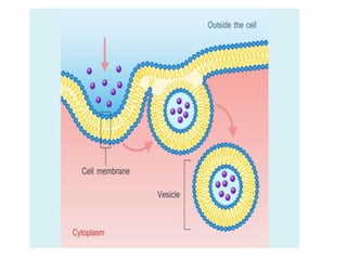
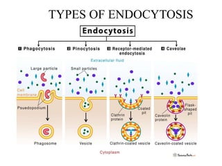
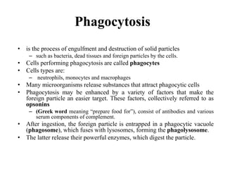
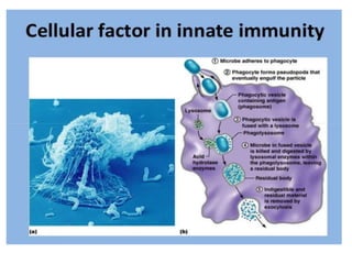
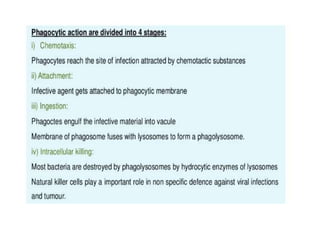
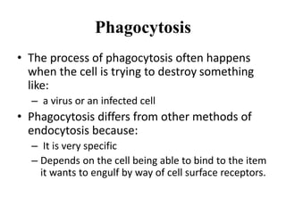
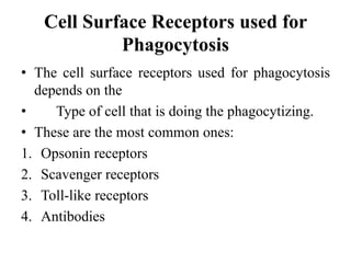
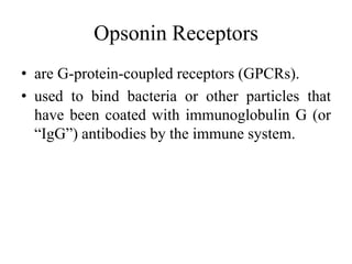
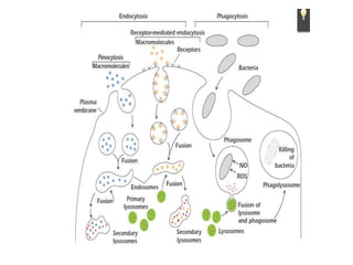
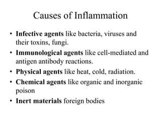
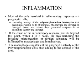
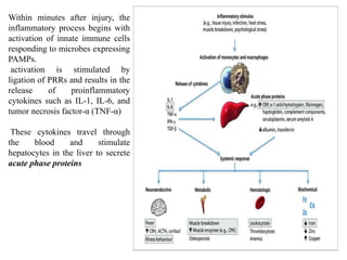
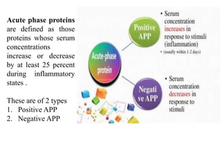
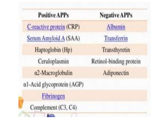
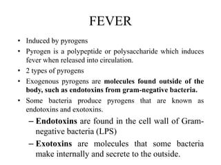
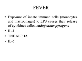
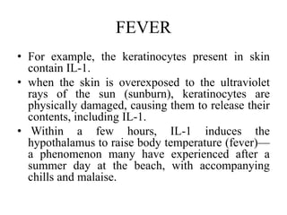
ENDOCYTOSIS & INFLAMMATION
LECTURE 8
BY: DR SADAF MOEEZ
Intracellular and Extracellular
Killing of Microorganisms
• Another hallmark of innate immunity is
– The ability of certain innate immune cells to sample
their environment by engulfing macroparticles
(endocytosis).
– Endocytosis: the process by which a cell moves
large amounts of material, or non-dissolved particles
into its cytoplasm from the outside environment.
– The word ‘endocytosis’ is derived from the Greek
words: ‘endon’= meaning ‘within’
– ‘kytos’= meaning ‘cell’,
– ‘-osis’= meaning ‘process’.
Endocytosis
• Ingestion of the foreign macromolecules generates
endocytic vesicles filled with the foreign material.
• Which then fuse with acidic compartments called
endosomes.
• Edosomes then fuse with lysosomes to form
endolysosomes.
– containing degradative enzymes (e.g., nucleases, lipases,
proteases) to reduce the ingested macromolecules to small
breakdown products, including nucleotides, sugars, and peptides.
– Nucleases: enzyme that cleaves nucleic acids
– Lipases: are a family of enzymes that break down triglycerides
into free fatty acids and glycerol.
– Proteases: enzymes that break down protein
Endocytosis
The 4 types of endocytosis are:
1. Phagocytosis (cellular eating)
2. Pinocytosis (cellular drinking)
3. Receptor mediated endocytosis
4. Caveolae
TYPES OF ENDOCYTOSIS
Phagocytosis
• is the process of engulfment and destruction of solid particles
– such as bacteria, dead tissues and foreign particles by the cells.
• Cells performing phagocytosis are called phagocytes
• Cells types are:
– neutrophils, monocytes and macrophages
• Many microorganisms release substances that attract phagocytic cells
• Phagocytosis may be enhanced by a variety of factors that make the
foreign particle an easier target. These factors, collectively referred to as
opsonins
– (Greek word meaning “prepare food for”), consist of antibodies and various
serum components of complement.
• After ingestion, the foreign particle is entrapped in a phagocytic vacuole
(phagosome), which fuses with lysosomes, forming the phagolysosome.
• The latter release their powerful enzymes, which digest the particle.
Phagocytosis
• The process of phagocytosis often happens
when the cell is trying to destroy something
like:
– a virus or an infected cell
• Phagocytosis differs from other methods of
endocytosis because:
– It is very specific
– Depends on the cell being able to bind to the item
it wants to engulf by way of cell surface receptors.
Cell Surface Receptors used for
Phagocytosis
• The cell surface receptors used for phagocytosis
depends on the
• Type of cell that is doing the phagocytizing.
• These are the most common ones:
1. Opsonin receptors
2. Scavenger receptors
3. Toll-like receptors
4. Antibodies
Opsonin Receptors
• are G-protein-coupled receptors (GPCRs).
• used to bind bacteria or other particles that
have been coated with immunoglobulin G (or
“IgG”) antibodies by the immune system.
Scavenger receptors
• heterogeneous family of surface receptors
• bind to molecules that are produced by bacteria.
• Most bacteria and other cellular species produce a
matrix of proteins surrounding themselves (called
an “extracellular matrix”).
– This matrix is a perfect way for the immune system to
identify foreign species in the body, because human
cells do not produce the same protein matrix.
Toll-like receptor
• bind to specific molecules produced by
bacteria.
• key part of the innate immune system because,
– once bound to a bacterial pathogen, they recognize
the specific bacteria and activate the immune
response
Antibodies
• immune cells make antibodies that can bind to
specific antigens.
• This is a process similar to how toll-like
receptors recognize and identify what type of
bacteria is infecting the host.
Phagocytosis
• Phagocytes can also damage invading
pathogens through the generation of toxic
products in a process known as the
respiratory burst.
• Production of these toxic metabolites is
induced during phagocytosis of pathogens
such as bacteria and catalyzed by a set of
interrelated enzyme pathways.
Toxic Metabolites
1. Oxygen Radicals: are highly reactive molecules
that react with proteins, lipids and other
biological molecules.
1. During physiological stress, the amount of oxygen
radicals in a cell can increase dramatically, causing
oxidative stress, which can destroy cell structures.
2. Nitric Oxide: is a reactive substance, similar to
oxygen radicals.
1. Reacts with superoxide to create further molecules
that damage various biological molecules.
Toxic Metabolites
3. Antimicrobial Proteins: are proteins that
specifically damage or kill bacteria.
1. Examples of antimicrobial proteins include:
1. Proteases, which kill various bacteria by destroying
essential proteins
2. Lysozyme, which attacks the cell walls of gram positive
bacteria.
4. Antimicrobial Peptides: similar to antimicrobial
proteins
1. Attack and kill bacteria, like defensins, attack
bacterial cell membranes.
Pinocytosis (cellular drinking)
• Steps of the Process
• The basic steps of pinocytosis are described below:
• Step 1 – Initiation: In the initial stage, an inducer molecule, such as sugar, protein,
or ion, comes in contact with the plasma membrane. As a result, an ionic interaction
occurs between the positively charged inducer and the negatively charged cell
membrane. This phenomenon triggers the binding of the molecule to the membrane.
• Step 2 – Folding of the Membrane: Following binding, the cell membrane gets
stimulated to fold inwards and form a small open-ended pocket, or invagination,
around the fluid containing the molecule.
• Step 3 – Invagination and Engulfment of the Fluid: The cell membrane continues
to fold inwards with the fluid and dissolved solutes in small pockets. Later, they start
to reconnect at the open end of the invagination to enclose the fluid and solutes.
• Step 4 – Detachment of the Pocket: This is the final stage when both ends of the
invagination meet. It gives rise to a vesicular structure called a pinosome that
contains extracellular fluid and dissolved solutes. Afterward, this pouch gets
detached or pinches off from the cell membrane. The molecules present inside the
vesicle are eventually released to be used by other parts of the cell, thus completing
the process.
Receptor mediated endocytosis
• Receptor-mediated endocytosis (RME), also
called clathrin-mediated endocytosis.
• Is a process by which cells absorb
– metabolites, hormones, proteins – and in some
cases viruses – by the inward budding of the plasma
membrane (invagination).
• This process forms vesicles containing the
absorbed substances and is strictly mediated
by receptors on the surface of the cell.
• Only the receptor-specific substances can enter
the cell through this process.
INFLAMMATION
• The word “inflammation” comes from the Latin
inflammare (to set on fire).
• Function of phagocytic cells is their participation in
inflammatory reactions.
• As a physiologic process, inflammation
• is typically initiated:
– by tissue damage caused by endogenous factors (such as
tissue necrosis or bone fracture)
– by exogenous factors.
– The latter includes various types of damage,
• such as mechanical injury (e.g., cuts), physical injury (e.g., burns),
chemical injury (e.g., exposure to corrosive chemicals),
immunologic injury (e.g., hypersensitivity reactions
Causes of Inflammation
• Infective agents like bacteria, viruses and
their toxins, fungi.
• Immunological agents like cell-mediated and
antigen antibody reactions.
• Physical agents like heat, cold, radiation.
• Chemical agents like organic and inorganic
poison
• Inert materials foreign bodies
INFLAMMATION
• Most of the cells involved in inflammatory responses are
phagocytic cells,
– consisting mainly of the polymorphonuclear leukocytes that
accumulate within 30 to 60 minutes, phagocytize the intruder or
damaged tissue, and release their lysosomal enzymes in an
attempt to destroy the intruder.
• If the cause of the inflammatory response persists beyond
this point, within 4 to 6 hours, the area harboring the
invading microorganism or foreign substance will be
infiltrated by macrophages and lymphocytes.
• The macrophages supplement the phagocytic activity of the
Polymorphonuclear cells, thus adding to the defense of the
area.
Within minutes after injury, the
inflammatory process begins with
activation of innate immune cells
responding to microbes expressing
PAMPs.
activation is stimulated by
ligation of PRRs and results in the
release of proinflammatory
cytokines such as IL-1, IL-6, and
tumor necrosis factor-α (TNF-α)
These cytokines travel through
the blood and stimulate
hepatocytes in the liver to secrete
acute phase proteins
Acute phase proteins
are defined as those
proteins whose serum
concentrations
increase or decrease
by at least 25 percent
during inflammatory
states .
These are of 2 types
1. Positive APP
2. Negative APP
FEVER
• Induced by pyrogens
• Pyrogen is a polypeptide or polysaccharide which induces
fever when released into circulation.
• 2 types of pyrogens
• Exogenous pyrogens are molecules found outside of the
body, such as endotoxins from gram-negative bacteria.
• Some bacteria produce pyrogens that are known as
endotoxins and exotoxins.
– Endotoxins are found in the cell wall of Gram-
negative bacteria (LPS)
– Exotoxins are molecules that some bacteria
make internally and secrete to the outside.
FEVER
• Exposure of innate immune cells (monocytes
and macrophages) to LPS causes their release
of cytokines called endogenous pyrogens
• IL-1
• TNF ALPHA
• IL-6
FEVER
• For example, the keratinocytes present in skin
contain IL-1.
• when the skin is overexposed to the ultraviolet
rays of the sun (sunburn), keratinocytes are
physically damaged, causing them to release their
contents, including IL-1.
• Within a few hours, IL-1 induces the
hypothalamus to raise body temperature (fever)—
a phenomenon many have experienced after a
summer day at the beach, with accompanying
chills and malaise.

ENDOCYTOSIS & INFLAMMATION-1.pptx

  • 1.
  • 2.
    Intracellular and Extracellular Killingof Microorganisms • Another hallmark of innate immunity is – The ability of certain innate immune cells to sample their environment by engulfing macroparticles (endocytosis). – Endocytosis: the process by which a cell moves large amounts of material, or non-dissolved particles into its cytoplasm from the outside environment. – The word ‘endocytosis’ is derived from the Greek words: ‘endon’= meaning ‘within’ – ‘kytos’= meaning ‘cell’, – ‘-osis’= meaning ‘process’.
  • 3.
    Endocytosis • Ingestion ofthe foreign macromolecules generates endocytic vesicles filled with the foreign material. • Which then fuse with acidic compartments called endosomes. • Edosomes then fuse with lysosomes to form endolysosomes. – containing degradative enzymes (e.g., nucleases, lipases, proteases) to reduce the ingested macromolecules to small breakdown products, including nucleotides, sugars, and peptides. – Nucleases: enzyme that cleaves nucleic acids – Lipases: are a family of enzymes that break down triglycerides into free fatty acids and glycerol. – Proteases: enzymes that break down protein
  • 5.
    Endocytosis The 4 typesof endocytosis are: 1. Phagocytosis (cellular eating) 2. Pinocytosis (cellular drinking) 3. Receptor mediated endocytosis 4. Caveolae
  • 6.
  • 7.
    Phagocytosis • is theprocess of engulfment and destruction of solid particles – such as bacteria, dead tissues and foreign particles by the cells. • Cells performing phagocytosis are called phagocytes • Cells types are: – neutrophils, monocytes and macrophages • Many microorganisms release substances that attract phagocytic cells • Phagocytosis may be enhanced by a variety of factors that make the foreign particle an easier target. These factors, collectively referred to as opsonins – (Greek word meaning “prepare food for”), consist of antibodies and various serum components of complement. • After ingestion, the foreign particle is entrapped in a phagocytic vacuole (phagosome), which fuses with lysosomes, forming the phagolysosome. • The latter release their powerful enzymes, which digest the particle.
  • 10.
    Phagocytosis • The processof phagocytosis often happens when the cell is trying to destroy something like: – a virus or an infected cell • Phagocytosis differs from other methods of endocytosis because: – It is very specific – Depends on the cell being able to bind to the item it wants to engulf by way of cell surface receptors.
  • 11.
    Cell Surface Receptorsused for Phagocytosis • The cell surface receptors used for phagocytosis depends on the • Type of cell that is doing the phagocytizing. • These are the most common ones: 1. Opsonin receptors 2. Scavenger receptors 3. Toll-like receptors 4. Antibodies
  • 12.
    Opsonin Receptors • areG-protein-coupled receptors (GPCRs). • used to bind bacteria or other particles that have been coated with immunoglobulin G (or “IgG”) antibodies by the immune system.
  • 13.
    Scavenger receptors • heterogeneousfamily of surface receptors • bind to molecules that are produced by bacteria. • Most bacteria and other cellular species produce a matrix of proteins surrounding themselves (called an “extracellular matrix”). – This matrix is a perfect way for the immune system to identify foreign species in the body, because human cells do not produce the same protein matrix.
  • 14.
    Toll-like receptor • bindto specific molecules produced by bacteria. • key part of the innate immune system because, – once bound to a bacterial pathogen, they recognize the specific bacteria and activate the immune response
  • 15.
    Antibodies • immune cellsmake antibodies that can bind to specific antigens. • This is a process similar to how toll-like receptors recognize and identify what type of bacteria is infecting the host.
  • 16.
    Phagocytosis • Phagocytes canalso damage invading pathogens through the generation of toxic products in a process known as the respiratory burst. • Production of these toxic metabolites is induced during phagocytosis of pathogens such as bacteria and catalyzed by a set of interrelated enzyme pathways.
  • 17.
    Toxic Metabolites 1. OxygenRadicals: are highly reactive molecules that react with proteins, lipids and other biological molecules. 1. During physiological stress, the amount of oxygen radicals in a cell can increase dramatically, causing oxidative stress, which can destroy cell structures. 2. Nitric Oxide: is a reactive substance, similar to oxygen radicals. 1. Reacts with superoxide to create further molecules that damage various biological molecules.
  • 18.
    Toxic Metabolites 3. AntimicrobialProteins: are proteins that specifically damage or kill bacteria. 1. Examples of antimicrobial proteins include: 1. Proteases, which kill various bacteria by destroying essential proteins 2. Lysozyme, which attacks the cell walls of gram positive bacteria. 4. Antimicrobial Peptides: similar to antimicrobial proteins 1. Attack and kill bacteria, like defensins, attack bacterial cell membranes.
  • 19.
    Pinocytosis (cellular drinking) •Steps of the Process • The basic steps of pinocytosis are described below: • Step 1 – Initiation: In the initial stage, an inducer molecule, such as sugar, protein, or ion, comes in contact with the plasma membrane. As a result, an ionic interaction occurs between the positively charged inducer and the negatively charged cell membrane. This phenomenon triggers the binding of the molecule to the membrane. • Step 2 – Folding of the Membrane: Following binding, the cell membrane gets stimulated to fold inwards and form a small open-ended pocket, or invagination, around the fluid containing the molecule. • Step 3 – Invagination and Engulfment of the Fluid: The cell membrane continues to fold inwards with the fluid and dissolved solutes in small pockets. Later, they start to reconnect at the open end of the invagination to enclose the fluid and solutes. • Step 4 – Detachment of the Pocket: This is the final stage when both ends of the invagination meet. It gives rise to a vesicular structure called a pinosome that contains extracellular fluid and dissolved solutes. Afterward, this pouch gets detached or pinches off from the cell membrane. The molecules present inside the vesicle are eventually released to be used by other parts of the cell, thus completing the process.
  • 21.
    Receptor mediated endocytosis •Receptor-mediated endocytosis (RME), also called clathrin-mediated endocytosis. • Is a process by which cells absorb – metabolites, hormones, proteins – and in some cases viruses – by the inward budding of the plasma membrane (invagination). • This process forms vesicles containing the absorbed substances and is strictly mediated by receptors on the surface of the cell. • Only the receptor-specific substances can enter the cell through this process.
  • 24.
    INFLAMMATION • The word“inflammation” comes from the Latin inflammare (to set on fire). • Function of phagocytic cells is their participation in inflammatory reactions. • As a physiologic process, inflammation • is typically initiated: – by tissue damage caused by endogenous factors (such as tissue necrosis or bone fracture) – by exogenous factors. – The latter includes various types of damage, • such as mechanical injury (e.g., cuts), physical injury (e.g., burns), chemical injury (e.g., exposure to corrosive chemicals), immunologic injury (e.g., hypersensitivity reactions
  • 25.
    Causes of Inflammation •Infective agents like bacteria, viruses and their toxins, fungi. • Immunological agents like cell-mediated and antigen antibody reactions. • Physical agents like heat, cold, radiation. • Chemical agents like organic and inorganic poison • Inert materials foreign bodies
  • 26.
    INFLAMMATION • Most ofthe cells involved in inflammatory responses are phagocytic cells, – consisting mainly of the polymorphonuclear leukocytes that accumulate within 30 to 60 minutes, phagocytize the intruder or damaged tissue, and release their lysosomal enzymes in an attempt to destroy the intruder. • If the cause of the inflammatory response persists beyond this point, within 4 to 6 hours, the area harboring the invading microorganism or foreign substance will be infiltrated by macrophages and lymphocytes. • The macrophages supplement the phagocytic activity of the Polymorphonuclear cells, thus adding to the defense of the area.
  • 27.
    Within minutes afterinjury, the inflammatory process begins with activation of innate immune cells responding to microbes expressing PAMPs. activation is stimulated by ligation of PRRs and results in the release of proinflammatory cytokines such as IL-1, IL-6, and tumor necrosis factor-α (TNF-α) These cytokines travel through the blood and stimulate hepatocytes in the liver to secrete acute phase proteins
  • 28.
    Acute phase proteins aredefined as those proteins whose serum concentrations increase or decrease by at least 25 percent during inflammatory states . These are of 2 types 1. Positive APP 2. Negative APP
  • 32.
    FEVER • Induced bypyrogens • Pyrogen is a polypeptide or polysaccharide which induces fever when released into circulation. • 2 types of pyrogens • Exogenous pyrogens are molecules found outside of the body, such as endotoxins from gram-negative bacteria. • Some bacteria produce pyrogens that are known as endotoxins and exotoxins. – Endotoxins are found in the cell wall of Gram- negative bacteria (LPS) – Exotoxins are molecules that some bacteria make internally and secrete to the outside.
  • 33.
    FEVER • Exposure ofinnate immune cells (monocytes and macrophages) to LPS causes their release of cytokines called endogenous pyrogens • IL-1 • TNF ALPHA • IL-6
  • 34.
    FEVER • For example,the keratinocytes present in skin contain IL-1. • when the skin is overexposed to the ultraviolet rays of the sun (sunburn), keratinocytes are physically damaged, causing them to release their contents, including IL-1. • Within a few hours, IL-1 induces the hypothalamus to raise body temperature (fever)— a phenomenon many have experienced after a summer day at the beach, with accompanying chills and malaise.