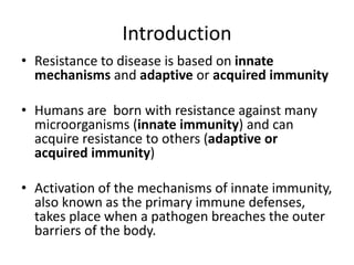
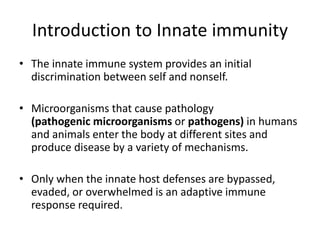
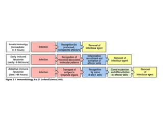
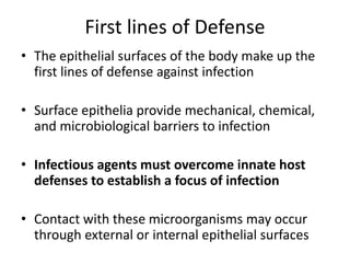
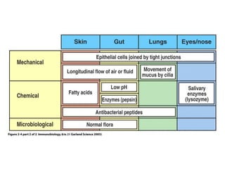
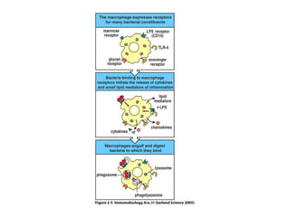
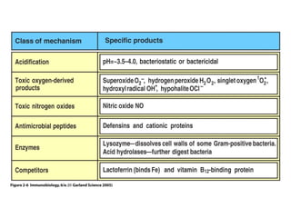
Innate Immunity
Hope C. Nkamba
10th March, 2022
Introduction
• Resistance to disease is based on innate
mechanisms and adaptive or acquired immunity
• Humans are born with resistance against many
microorganisms (innate immunity) and can
acquire resistance to others (adaptive or
acquired immunity)
• Activation of the mechanisms of innate immunity,
also known as the primary immune defenses,
takes place when a pathogen breaches the outer
barriers of the body.
Introduction to Innate immunity
• The innate immune system provides an initial
discrimination between self and nonself.
• Microorganisms that cause pathology
(pathogenic microorganisms or pathogens) in humans
and animals enter the body at different sites and
produce disease by a variety of mechanisms.
• Only when the innate host defenses are bypassed,
evaded, or overwhelmed is an adaptive immune
response required.
Figure 2-1
Routes of Infection for Pathogens
• The respiratory tract mucosa provides a route of entry
for airborne microorganisms
• The gastrointestinal mucosa for microorganisms in food
and water
• Insect bites and wounds allow microorganisms to
penetrate the skin
• Direct contact between individuals offers opportunities
for infection of the skin, the gut, and the mucosa of the
reproductive tract
Figure 2-2 part 1 of 2
Figure 2-2 part 2 of 2
First lines of Defense
• The epithelial surfaces of the body make up the
first lines of defense against infection
• Surface epithelia provide mechanical, chemical,
and microbiological barriers to infection
• Infectious agents must overcome innate host
defenses to establish a focus of infection
• Contact with these microorganisms may occur
through external or internal epithelial surfaces
First lines of Defense
• The internal epithelia are known as mucosal
epithelia because they secrete a viscous fluid
called mucus, which contains many glycoproteins
called mucins.
• Microorganisms coated in mucus may be
prevented from adhering to the epithelium and
in mucosal epithelia such as that of the
respiratory tract
• Microorganisms can be expelled in the flow of
mucus driven by the beating of epithelial cilia
First lines of Defense
• Infectious disease occurs when a microorganism
succeeds in evading or overwhelming innate host
defenses to establish a local site of infection and
replication that allows its further transmission.
• Pathogen spread is often countered by an
inflammatory response that recruits more
effector molecules and cells of the innate
immune system from local blood vessels
• The epithelial surfaces of the body are the first
defenses against infection
Figure 2-4 part 1 of 2
Barriers to infection
• Our body surfaces are defended by epithelia, which provide a
physical barrier between the internal milieu and the external world
that contains pathogens
• Epithelial cells are held together by tight junctions, which effectively
form a seal against the external environment. Epithelia comprise
the skin and the linings of the body’s tubular structures—the
gastrointestinal, respiratory, and urinogenital tracts
• Infections occur only when the pathogen can colonize or cross
through these barriers, and since the dry, protective layers of the
skin present a more formidable barrier, pathogen entry most often
occurs through the internal epithelial surfaces
• In the absence of wounding or disruption, pathogens normally cross
epithelial barriers by binding to molecules on internal epithelial
surfaces, or establish an infection by adhering to and colonizing
these surfaces
Barriers to infection
• This specific attachment allows the pathogen to infect the
epithelial cell, or to damage it so that the epithelium can be
crossed, or, in the case of colonizing pathogens, to avoid
being dislodged by the flow of air or fluid across the
epithelial surface
• The internal epithelia are known as mucosal epithelia
because they secrete a viscous fluid called mucus, which
contains many glycoproteins called mucins.
• Microorganisms coated in mucus may be prevented from
adhering to the epithelium, and in mucosal epithelia such
as that of the respiratory tract, microorganisms can be
expelled in the flow of mucus driven by the beating of
epithelial cilia.
• The efficacy of mucus flow in clearing infection is illustrated
by people with defective mucus secretion or inhibition of
ciliary movement; they frequently develop lung infections
caused by bacteria that colonize the epithelial surface
Barriers to infection
• In the gut, peristalsis is an important mechanism for keeping both
food and infectious agents moving through. Failure of peristalsis is
typically accompanied by overgrowth of bacteria within the
intestinal lumen.
• Our surface epithelia are more than mere physical barriers to
infection; they also produce chemical substances that are
microbicidal or inhibit microbial growth
• For example, the antibacterial enzyme lysozyme is secreted in tears
and saliva. The acid pH of the stomach and the digestive enzymes of
the upper gastrointestinal tract create a substantial chemical barrier
to infection.
• Further down the intestinal tract, antibacterial and antifungal
peptides called cryptidins or α-defensins are made by Paneth cells,
which are resident in the base of the crypts in the small intestine
beneath the epithelial stem cells.
Barriers to infection
• Related antimicrobial peptides, the β-defensins, are made by other
epithelia, primarily in the skin and respiratory tract.
• Antimicrobial peptides play a role in the immune defense of many
organisms. They are cationic peptides that are thought to kill
bacteria by damaging the bacterial cell membrane.
• Another type of antimicrobial protein is secreted into the fluid that
bathes the epithelial surfaces of the lung. This fluid contains two
proteins—surfactant proteins A and D—that bind to and coat the
surfaces of pathogens so that they are more easily phagocytosed by
macrophages that have left the subepithelial tissues to enter the
alveoli of the lung.
• Coating of a particle with proteins that facilitate its phagocytosis is
known as opsonization
Barriers to infection
• Surface epithelia provide mechanical, chemical, and microbiological
barriers to infection.
• In addition to these defenses, most epithelial surfaces are
associated with a normal flora of nonpathogenic bacteria that
compete with pathogenic microorganisms for nutrients and for
attachment sites on cells.
• The normal flora can also produce antimicrobial substances, such as
the colicins (anti-bacterial proteins made by Escherichia coli) that
prevent colonization by other bacteria
• When the nonpathogenic bacteria are killed by antibiotic
treatment, pathogenic microorganisms frequently replace them and
cause disease.
Figure 2-4 part 2 of 2
Innate Immunity- Phagocytes
• Phagocytes are the cells that protect the body
by ingesting (phagocytosing) harmful foreign
particles, bacteria, and dead or dying cells.
• Professional phagocytes include cells called
neutrophils, monocytes, macrophages,
dendritic cells, and mast cells.
Innate Immunity- Phagocytes
• If a microorganism crosses an epithelial barrier and begins
to replicate in the tissues of the host, it is recognized by the
mononuclear phagocytes, or macrophages.
• Macrophages mature continuously from monocytes which
leave the circulation to migrate into tissues throughout the
body
• They are found in especially large numbers in connective
tissue, in the submucosal layer of the gastrointestinal tract,
in the lung, along certain blood vessels in the liver and
throughout the spleen
• Macrophages are called different names, depending on the
tissue: Kupffer cells in the liver, histiocytes in connective
tissue, and alveolar macrophages in the lungs.
Innate Immunity- Phagocytes
• The second major family of phagocytes- the
neutrophils are short lived cells that are
abundant in the blood, but they are not
present in normal heath tissues.
• Both macrophages and neutrophils have a key
role in innate immunity because the can
recognize, ingest, and destroy many
pathogens without the aid of an adaptive
immune response
Innate Immunity- Phagocytes
• Macrophages located in the submucosal tissues,
are the first cells to encounter pathogens, but
they are soon reinforced by the recruitment of
large numbers of neutrophils to the site of
infection.
• Macrophages and neutrophils recognize
pathogens by means of their cell surface
receptors that can discriminate between the
surface molecules displayed by pathogens and
those of the host.
Innate Immunity- Phagocytes
• These receptors include the macrophage
mannose receptor, scavenger receptors, which
bind many negatively charged ligands such as
lipoteichoic acids, which are cell-wall
components of Gram- positive bacteria
• CD14 a receptor predominately found on
monocytes and macrophages is a receptor for
bacterial lipopolysaccharide (LPS).
Phagocytosis
• Phagocytosis is an active process, in which the bound
pathogen is first surrounded by the phagocyte
membrane and then internalized in a membrane-
bound vesicle known as a phagosome or endocytic
vacuole.
• The phagosome then becomes acidified, killing most
pathogens
• In addition to being phagocytic macrophages and
neutrophils have membrane-bounded granules, called
lysosomes.
Phagocytosis
• Lysosomes contain enzymes, proteins and
peptides that can mediate an intracellular
antimicrobial response.
• The phagosome fuses with one or more
lysosomes to generate a phagolysosome in
which the lysosomal contents are released to
destroy the pathogen.
Figure 2-5
Phagocytosis
• Upon phagocytosis, macrophages and
neutrophils also produce a variety of other toxic
products that help kill engulfed microorganisms
• They are generated by lysosomal NADPH oxidases
and other enzymes in a process known as the
respiratory burst
• The most important of these are nitric oxide
(NO), the superoxide anion(O2
-) and hydrogen
peroxide (H2O2), which are directly toxic to
bacteria.
Phagocytosis
• Chemical and enzymatic reactions further
produce a range of toxic chemicals from H2O2 ,
these include hydroxyl radical(OH.),
hypochlorite (OCl-) and hypobromite(OBr-)
ions.
Figure 2-6
Innate Immunity
• Neutrophils are short lived cells, dying soon
after they have accomplished a round of
phagocytosis.
• Dead and dying neutrophils are a major
component of pus that forms in some
infections; bacteria that gives raise to such
infections are thus known as pyogenic or pus-
forming bacteria.
Innate Immunity
• In contrast to neutrophils, macrophages are long
lived and continue to generate new lysosomes.
• Patients with chronic granulomatous disease
have a genetic deficiency of NADPH oxidase,
which means that their phagocytes do not
produce the toxic oxygen derivatives
characteristic of the respiratory burst, and so less
able to kill ingested microorganisms and clear
infection.
Innate Immunity
• People with a genetic deficiency of NADPH
oxidase are usually susceptible to bacterial
and fungal infections, especially in infancy.
• Invertebrates such as sea star rely entirely on
innate immunity for their defense against
infection.
Pathogens Avoid Destruction
• Pathogens have developed a variety of strategies
to avoid immediate destruction by macrophages.
• Many extracellular pathogenic bacteria coat
themselves with a thick polysaccharide capsule
that is not recognized by any phagocyte receptor.
• Other pathogens, for example mycobateria, have
evolved ways to grow inside macrophage
phagosomes by inhibiting their acidification and
fusion with lysosomes.
• When innate immunity fails, stimulatory
molecules which are induced and expressed
on macrophages and on dendritic cells,
enable these APCs to initiate an adaptive
immune response

Innate Immunity 2022.pptx

  • 1.
    Innate Immunity Hope C.Nkamba 10th March, 2022
  • 2.
    Introduction • Resistance todisease is based on innate mechanisms and adaptive or acquired immunity • Humans are born with resistance against many microorganisms (innate immunity) and can acquire resistance to others (adaptive or acquired immunity) • Activation of the mechanisms of innate immunity, also known as the primary immune defenses, takes place when a pathogen breaches the outer barriers of the body.
  • 3.
    Introduction to Innateimmunity • The innate immune system provides an initial discrimination between self and nonself. • Microorganisms that cause pathology (pathogenic microorganisms or pathogens) in humans and animals enter the body at different sites and produce disease by a variety of mechanisms. • Only when the innate host defenses are bypassed, evaded, or overwhelmed is an adaptive immune response required.
  • 4.
  • 5.
    Routes of Infectionfor Pathogens • The respiratory tract mucosa provides a route of entry for airborne microorganisms • The gastrointestinal mucosa for microorganisms in food and water • Insect bites and wounds allow microorganisms to penetrate the skin • Direct contact between individuals offers opportunities for infection of the skin, the gut, and the mucosa of the reproductive tract
  • 6.
  • 7.
  • 8.
    First lines ofDefense • The epithelial surfaces of the body make up the first lines of defense against infection • Surface epithelia provide mechanical, chemical, and microbiological barriers to infection • Infectious agents must overcome innate host defenses to establish a focus of infection • Contact with these microorganisms may occur through external or internal epithelial surfaces
  • 9.
    First lines ofDefense • The internal epithelia are known as mucosal epithelia because they secrete a viscous fluid called mucus, which contains many glycoproteins called mucins. • Microorganisms coated in mucus may be prevented from adhering to the epithelium and in mucosal epithelia such as that of the respiratory tract • Microorganisms can be expelled in the flow of mucus driven by the beating of epithelial cilia
  • 10.
    First lines ofDefense • Infectious disease occurs when a microorganism succeeds in evading or overwhelming innate host defenses to establish a local site of infection and replication that allows its further transmission. • Pathogen spread is often countered by an inflammatory response that recruits more effector molecules and cells of the innate immune system from local blood vessels • The epithelial surfaces of the body are the first defenses against infection
  • 11.
  • 12.
    Barriers to infection •Our body surfaces are defended by epithelia, which provide a physical barrier between the internal milieu and the external world that contains pathogens • Epithelial cells are held together by tight junctions, which effectively form a seal against the external environment. Epithelia comprise the skin and the linings of the body’s tubular structures—the gastrointestinal, respiratory, and urinogenital tracts • Infections occur only when the pathogen can colonize or cross through these barriers, and since the dry, protective layers of the skin present a more formidable barrier, pathogen entry most often occurs through the internal epithelial surfaces • In the absence of wounding or disruption, pathogens normally cross epithelial barriers by binding to molecules on internal epithelial surfaces, or establish an infection by adhering to and colonizing these surfaces
  • 13.
    Barriers to infection •This specific attachment allows the pathogen to infect the epithelial cell, or to damage it so that the epithelium can be crossed, or, in the case of colonizing pathogens, to avoid being dislodged by the flow of air or fluid across the epithelial surface • The internal epithelia are known as mucosal epithelia because they secrete a viscous fluid called mucus, which contains many glycoproteins called mucins. • Microorganisms coated in mucus may be prevented from adhering to the epithelium, and in mucosal epithelia such as that of the respiratory tract, microorganisms can be expelled in the flow of mucus driven by the beating of epithelial cilia. • The efficacy of mucus flow in clearing infection is illustrated by people with defective mucus secretion or inhibition of ciliary movement; they frequently develop lung infections caused by bacteria that colonize the epithelial surface
  • 14.
    Barriers to infection •In the gut, peristalsis is an important mechanism for keeping both food and infectious agents moving through. Failure of peristalsis is typically accompanied by overgrowth of bacteria within the intestinal lumen. • Our surface epithelia are more than mere physical barriers to infection; they also produce chemical substances that are microbicidal or inhibit microbial growth • For example, the antibacterial enzyme lysozyme is secreted in tears and saliva. The acid pH of the stomach and the digestive enzymes of the upper gastrointestinal tract create a substantial chemical barrier to infection. • Further down the intestinal tract, antibacterial and antifungal peptides called cryptidins or α-defensins are made by Paneth cells, which are resident in the base of the crypts in the small intestine beneath the epithelial stem cells.
  • 15.
    Barriers to infection •Related antimicrobial peptides, the β-defensins, are made by other epithelia, primarily in the skin and respiratory tract. • Antimicrobial peptides play a role in the immune defense of many organisms. They are cationic peptides that are thought to kill bacteria by damaging the bacterial cell membrane. • Another type of antimicrobial protein is secreted into the fluid that bathes the epithelial surfaces of the lung. This fluid contains two proteins—surfactant proteins A and D—that bind to and coat the surfaces of pathogens so that they are more easily phagocytosed by macrophages that have left the subepithelial tissues to enter the alveoli of the lung. • Coating of a particle with proteins that facilitate its phagocytosis is known as opsonization
  • 16.
    Barriers to infection •Surface epithelia provide mechanical, chemical, and microbiological barriers to infection. • In addition to these defenses, most epithelial surfaces are associated with a normal flora of nonpathogenic bacteria that compete with pathogenic microorganisms for nutrients and for attachment sites on cells. • The normal flora can also produce antimicrobial substances, such as the colicins (anti-bacterial proteins made by Escherichia coli) that prevent colonization by other bacteria • When the nonpathogenic bacteria are killed by antibiotic treatment, pathogenic microorganisms frequently replace them and cause disease.
  • 17.
  • 18.
    Innate Immunity- Phagocytes •Phagocytes are the cells that protect the body by ingesting (phagocytosing) harmful foreign particles, bacteria, and dead or dying cells. • Professional phagocytes include cells called neutrophils, monocytes, macrophages, dendritic cells, and mast cells.
  • 19.
    Innate Immunity- Phagocytes •If a microorganism crosses an epithelial barrier and begins to replicate in the tissues of the host, it is recognized by the mononuclear phagocytes, or macrophages. • Macrophages mature continuously from monocytes which leave the circulation to migrate into tissues throughout the body • They are found in especially large numbers in connective tissue, in the submucosal layer of the gastrointestinal tract, in the lung, along certain blood vessels in the liver and throughout the spleen • Macrophages are called different names, depending on the tissue: Kupffer cells in the liver, histiocytes in connective tissue, and alveolar macrophages in the lungs.
  • 20.
    Innate Immunity- Phagocytes •The second major family of phagocytes- the neutrophils are short lived cells that are abundant in the blood, but they are not present in normal heath tissues. • Both macrophages and neutrophils have a key role in innate immunity because the can recognize, ingest, and destroy many pathogens without the aid of an adaptive immune response
  • 21.
    Innate Immunity- Phagocytes •Macrophages located in the submucosal tissues, are the first cells to encounter pathogens, but they are soon reinforced by the recruitment of large numbers of neutrophils to the site of infection. • Macrophages and neutrophils recognize pathogens by means of their cell surface receptors that can discriminate between the surface molecules displayed by pathogens and those of the host.
  • 22.
    Innate Immunity- Phagocytes •These receptors include the macrophage mannose receptor, scavenger receptors, which bind many negatively charged ligands such as lipoteichoic acids, which are cell-wall components of Gram- positive bacteria • CD14 a receptor predominately found on monocytes and macrophages is a receptor for bacterial lipopolysaccharide (LPS).
  • 23.
    Phagocytosis • Phagocytosis isan active process, in which the bound pathogen is first surrounded by the phagocyte membrane and then internalized in a membrane- bound vesicle known as a phagosome or endocytic vacuole. • The phagosome then becomes acidified, killing most pathogens • In addition to being phagocytic macrophages and neutrophils have membrane-bounded granules, called lysosomes.
  • 24.
    Phagocytosis • Lysosomes containenzymes, proteins and peptides that can mediate an intracellular antimicrobial response. • The phagosome fuses with one or more lysosomes to generate a phagolysosome in which the lysosomal contents are released to destroy the pathogen.
  • 25.
  • 26.
    Phagocytosis • Upon phagocytosis,macrophages and neutrophils also produce a variety of other toxic products that help kill engulfed microorganisms • They are generated by lysosomal NADPH oxidases and other enzymes in a process known as the respiratory burst • The most important of these are nitric oxide (NO), the superoxide anion(O2 -) and hydrogen peroxide (H2O2), which are directly toxic to bacteria.
  • 27.
    Phagocytosis • Chemical andenzymatic reactions further produce a range of toxic chemicals from H2O2 , these include hydroxyl radical(OH.), hypochlorite (OCl-) and hypobromite(OBr-) ions.
  • 28.
  • 29.
    Innate Immunity • Neutrophilsare short lived cells, dying soon after they have accomplished a round of phagocytosis. • Dead and dying neutrophils are a major component of pus that forms in some infections; bacteria that gives raise to such infections are thus known as pyogenic or pus- forming bacteria.
  • 30.
    Innate Immunity • Incontrast to neutrophils, macrophages are long lived and continue to generate new lysosomes. • Patients with chronic granulomatous disease have a genetic deficiency of NADPH oxidase, which means that their phagocytes do not produce the toxic oxygen derivatives characteristic of the respiratory burst, and so less able to kill ingested microorganisms and clear infection.
  • 31.
    Innate Immunity • Peoplewith a genetic deficiency of NADPH oxidase are usually susceptible to bacterial and fungal infections, especially in infancy. • Invertebrates such as sea star rely entirely on innate immunity for their defense against infection.
  • 32.
    Pathogens Avoid Destruction •Pathogens have developed a variety of strategies to avoid immediate destruction by macrophages. • Many extracellular pathogenic bacteria coat themselves with a thick polysaccharide capsule that is not recognized by any phagocyte receptor. • Other pathogens, for example mycobateria, have evolved ways to grow inside macrophage phagosomes by inhibiting their acidification and fusion with lysosomes.
  • 33.
    • When innateimmunity fails, stimulatory molecules which are induced and expressed on macrophages and on dendritic cells, enable these APCs to initiate an adaptive immune response