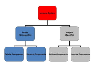
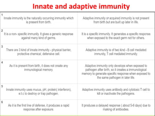
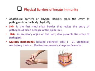
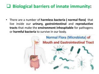
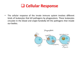
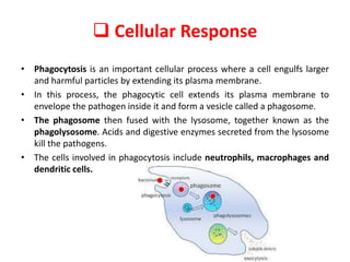
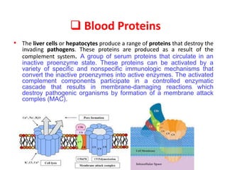
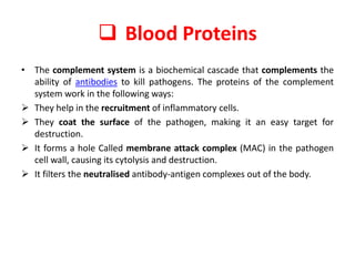
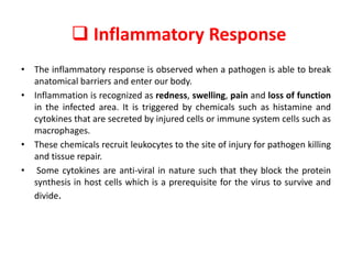
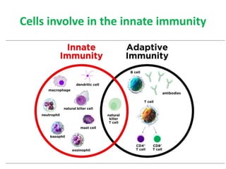
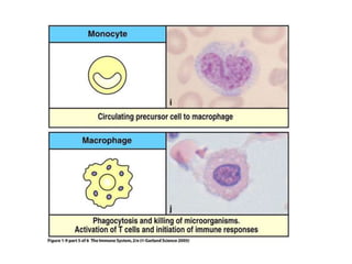
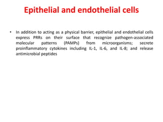
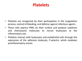
Immune System and Innate
immunity
Dr.Sherzad M Taher
Medical Immunology
College Of Pharmacy
UoD
September 12,2023
• Immunology
Immunology deals with physiological functioning of the immune system in
states of both health and disease as well as malfunctions of the immune
system in immunological disorders like allergies, hypersensitivities, immune
deficiency, transplant rejection and autoimmune disorders.
• Immune system
- includes all parts of the body that help in the recognition and destruction
of foreign materials. White blood cells, phagocytes and lymphocytes, bone
marrow, lymphnodes, tonsils, thymus, and your spleen are all part of the
immune system.
Innate and adaptive immunity
Components of the Innate Immune
System
 Physical( Anatomical ) barriers of innate immunity
 Chemical barriers of innate immunity
 Biological barriers of innate immunity
 Physiological barrier of innate immunity
 Cellular Response
 Blood Proteins
 Inflammatory Response
 Physical Barriers of Innate Immunity
• Anatomical barriers or physical barriers block the entry of
pathogens into the body physically.
• Skin is the first mechanical barrier that makes the entry of
pathogens difficult because of the epidermis.
• Hair, an accessory organ on the skin, also prevents the entry of
pathogens.
• Mucous membranes (ciliated epithelial cells; ) - GI, urogenital,
respiratory tracts - collectively represents a huge surface area.
• Chemical barriers block entry of pathogens at body openings
and inner body surfaces. Examples of chemical barriers
include sweat, breastmilk, mucus, saliva, tears and semen.
• Vaginal secretion is acidic in nature and is not endured by
pathogens.
• Semen has zinc which is intolerable by pathogens.
• The digestive juices (pepsin) produced in the stomach also kill
foreign invaders.
 Chemical barriers of innate immunity:
• Fatty acids, lactic acid
• Lysozyme -hydrolytic enzyme found in mucous secretions - able to
cleave the peptidoglycan layer of the bacterial cell wall
• Anti-microbial substances which directly destroy microorganims:
cryptidins and defensins
a- cryptidins (produced in base of crypts of small intestine - damage cell
membranes)
b-defensins (produced within skin, respiratory tract - also damages cell
membranes)
• surfactant proteins A & D (present in lungs - function as opsonins
which enhance the efficiency of phagocytosis)
 Chemical barriers of innate immunity:
• Interferons - group of proteins produced by cells following viral
infection. Secreted by the cells, and then binds to nearby cells and
induces mechanisms which inhibit viral replication.
• Acute phase proteins
– proteins in the plasma that increase during infection and
inflammation ex: C-reactive protein (CRP)
– can be used diagnostically in many diseases to give an
indication of acute inflammation
 Biological barriers of innate immunity:
• There are a number of harmless bacteria ( normal flora) that
live inside our urinary, gastrointestinal and reproductive
tracts that make the environment inhospitable for pathogens
or harmful bacteria to survive in our body.
 Physiological barrier of innate immunity
• Temperature - normal body temperature inhibits growth of many
microorganisms.
• Elevated body temperature (fever) can have a direct effect on
pathogenic microorganisms
 Cellular Response
• The cellular response of the innate immune system involves different
kinds of leukocytes that kill pathogens by phagocytosis. These leukocytes
circulate in the blood and single-handedly kill the pathogens that invade
our bodies.
 Cellular Response
• Phagocytosis is an important cellular process where a cell engulfs larger
and harmful particles by extending its plasma membrane.
• In this process, the phagocytic cell extends its plasma membrane to
envelope the pathogen inside it and form a vesicle called a phagosome.
• The phagosome then fused with the lysosome, together known as the
phagolysosome. Acids and digestive enzymes secreted from the lysosome
kill the pathogens.
• The cells involved in phagocytosis include neutrophils, macrophages and
dendritic cells.
 Blood Proteins
• The liver cells or hepatocytes produce a range of proteins that destroy the
invading pathogens. These proteins are produced as a result of the
complement system. A group of serum proteins that circulate in an
inactive proenzyme state. These proteins can be activated by a
variety of specific and nonspecific immunologic mechanisms that
convert the inactive proenzymes into active enzymes. The activated
complement components participate in a controlled enzymatic
cascade that results in membrane-damaging reactions which
destroy pathogenic organisms by formation of a membrane attack
comples (MAC).
 Blood Proteins
• The complement system is a biochemical cascade that complements the
ability of antibodies to kill pathogens. The proteins of the complement
system work in the following ways:
 They help in the recruitment of inflammatory cells.
 They coat the surface of the pathogen, making it an easy target for
destruction.
 It forms a hole Called membrane attack complex (MAC) in the pathogen
cell wall, causing its cytolysis and destruction.
 It filters the neutralised antibody-antigen complexes out of the body.
 Inflammatory Response
• The inflammatory response is observed when a pathogen is able to break
anatomical barriers and enter our body.
• Inflammation is recognized as redness, swelling, pain and loss of function
in the infected area. It is triggered by chemicals such as histamine and
cytokines that are secreted by injured cells or immune system cells such as
macrophages.
• These chemicals recruit leukocytes to the site of injury for pathogen killing
and tissue repair.
• Some cytokines are anti-viral in nature such that they block the protein
synthesis in host cells which is a prerequisite for the virus to survive and
divide.
Cells involve in the innate immunity
Neutrophils
• These cells are most abundant and effective during the inflammation and
phagocytosis processes. Neutrophils (PMN) are characterized as being the
first cell line that is recruited at the inflammation site after chemotactic
stimuli.
Eosinophils
• These granulocytes are present in the respiratory, gastrointestinal, and
urinary tract, and they are less abundant than neutrophils. Their effector
function is mediated by degranulation and release of histamine, cationic
proteins, major basic protein, and chemotactic factors such as
leukotrienes and prostaglandins
Basophils and mast cells
• These cells are not phagocytic in nature and have several receptors
including IgE receptors. The proportion of basophils in circulation is lower
than the proportion of other granulocytes. Mast cells are located in
tissues, mainly in mucosa, and their granules contain heparin, serotonin,
and histamine.
Monocyte and macrophages
• are considered important actors in both innate and adaptive immunity.
• Monocytes circulate in peripheral blood and have the ability to not only
migrate to the inflammatory site but also exhibit the plasticity to
transform themselves into tissue macrophages .
• Thus, macrophages take different names according to their tissue location,
such as osteoclasts (bone), alveolar macrophages (lung), microglial cells
(brain), histiocytes (connective tissue), Kupffer cells (liver), Langerhans
cells (LC) (skin), etc
Monocyte and macrophages
• macrophages functions:
• i) they are phagocytic and exhibit a microbicidal mechanism
through oxygen -dependent and-independent mechanism;
• ii) they are able to present antigens and activate lymphocytes;
• iii) once activated, they release and stimulate cytokine secretion;
• iv) they modulate the immune response;
• v) they participate in tissue reorganization after the inflammation
process has ceased through production of extracellular matrix
proteins (i.e., collagen and elastase) and matrix
metalloproteinases;
• vi) they produce cytotoxic factors involved in the immunity against
tumors
Dendritic cells (DCs)
• DCs are considered to be professional antigen-presenting cells (APC). They
reside in and patrol the skin and mucosal surfaces, thus playing an
important role in the innate immune system with subsequent activation of
T cell responses to provide a cell-mediated immunity against microbial
pathogens.
Natural killer (NK) cells
• NK cells exhibit an immunomodulatory role in the cell-mediated immune
responses due their cytotoxic activity. They are also involved in
antimicrobial defense and in the immunological surveillance by controlling
tumoral growth and maintaining the immunological homeostasis.
Epithelial and endothelial cells
• In addition to acting as a physical barrier, epithelial and endothelial cells
express PRRs on their surface that recognize pathogen-associated
molecular patterns (PAMPs) from microorganisms; secrete
proinflammatory cytokines including IL-1, IL-6, and IL-8; and release
antimicrobial peptides
Platelets
• Platelets are recognized by their participation in the coagulation
process, control of bleeding, and defense against infectious agents .
• These cells express PRRs on their surface and produce cytokines
and chemotactic molecules to recruit leukocytes at the
inflammatory site.
• Platelets interact with leukocytes and endothelial cells through the
expression of the adhesion molecule, P-selectin, which mediates
proinflammatory events
Innate Immunity ppt..pptx

Innate Immunity ppt..pptx

  • 1.
    Immune System andInnate immunity Dr.Sherzad M Taher Medical Immunology College Of Pharmacy UoD September 12,2023
  • 2.
    • Immunology Immunology dealswith physiological functioning of the immune system in states of both health and disease as well as malfunctions of the immune system in immunological disorders like allergies, hypersensitivities, immune deficiency, transplant rejection and autoimmune disorders. • Immune system - includes all parts of the body that help in the recognition and destruction of foreign materials. White blood cells, phagocytes and lymphocytes, bone marrow, lymphnodes, tonsils, thymus, and your spleen are all part of the immune system.
  • 4.
  • 5.
    Components of theInnate Immune System  Physical( Anatomical ) barriers of innate immunity  Chemical barriers of innate immunity  Biological barriers of innate immunity  Physiological barrier of innate immunity  Cellular Response  Blood Proteins  Inflammatory Response
  • 6.
     Physical Barriersof Innate Immunity • Anatomical barriers or physical barriers block the entry of pathogens into the body physically. • Skin is the first mechanical barrier that makes the entry of pathogens difficult because of the epidermis. • Hair, an accessory organ on the skin, also prevents the entry of pathogens. • Mucous membranes (ciliated epithelial cells; ) - GI, urogenital, respiratory tracts - collectively represents a huge surface area.
  • 7.
    • Chemical barriersblock entry of pathogens at body openings and inner body surfaces. Examples of chemical barriers include sweat, breastmilk, mucus, saliva, tears and semen. • Vaginal secretion is acidic in nature and is not endured by pathogens. • Semen has zinc which is intolerable by pathogens. • The digestive juices (pepsin) produced in the stomach also kill foreign invaders.  Chemical barriers of innate immunity:
  • 8.
     Chemical barriersof innate immunity: • Fatty acids, lactic acid • Lysozyme -hydrolytic enzyme found in mucous secretions - able to cleave the peptidoglycan layer of the bacterial cell wall • Anti-microbial substances which directly destroy microorganims: cryptidins and defensins a- cryptidins (produced in base of crypts of small intestine - damage cell membranes) b-defensins (produced within skin, respiratory tract - also damages cell membranes) • surfactant proteins A & D (present in lungs - function as opsonins which enhance the efficiency of phagocytosis)
  • 9.
     Chemical barriersof innate immunity: • Interferons - group of proteins produced by cells following viral infection. Secreted by the cells, and then binds to nearby cells and induces mechanisms which inhibit viral replication. • Acute phase proteins – proteins in the plasma that increase during infection and inflammation ex: C-reactive protein (CRP) – can be used diagnostically in many diseases to give an indication of acute inflammation
  • 10.
     Biological barriersof innate immunity: • There are a number of harmless bacteria ( normal flora) that live inside our urinary, gastrointestinal and reproductive tracts that make the environment inhospitable for pathogens or harmful bacteria to survive in our body.
  • 11.
     Physiological barrierof innate immunity • Temperature - normal body temperature inhibits growth of many microorganisms. • Elevated body temperature (fever) can have a direct effect on pathogenic microorganisms
  • 12.
     Cellular Response •The cellular response of the innate immune system involves different kinds of leukocytes that kill pathogens by phagocytosis. These leukocytes circulate in the blood and single-handedly kill the pathogens that invade our bodies.
  • 13.
     Cellular Response •Phagocytosis is an important cellular process where a cell engulfs larger and harmful particles by extending its plasma membrane. • In this process, the phagocytic cell extends its plasma membrane to envelope the pathogen inside it and form a vesicle called a phagosome. • The phagosome then fused with the lysosome, together known as the phagolysosome. Acids and digestive enzymes secreted from the lysosome kill the pathogens. • The cells involved in phagocytosis include neutrophils, macrophages and dendritic cells.
  • 14.
     Blood Proteins •The liver cells or hepatocytes produce a range of proteins that destroy the invading pathogens. These proteins are produced as a result of the complement system. A group of serum proteins that circulate in an inactive proenzyme state. These proteins can be activated by a variety of specific and nonspecific immunologic mechanisms that convert the inactive proenzymes into active enzymes. The activated complement components participate in a controlled enzymatic cascade that results in membrane-damaging reactions which destroy pathogenic organisms by formation of a membrane attack comples (MAC).
  • 15.
     Blood Proteins •The complement system is a biochemical cascade that complements the ability of antibodies to kill pathogens. The proteins of the complement system work in the following ways:  They help in the recruitment of inflammatory cells.  They coat the surface of the pathogen, making it an easy target for destruction.  It forms a hole Called membrane attack complex (MAC) in the pathogen cell wall, causing its cytolysis and destruction.  It filters the neutralised antibody-antigen complexes out of the body.
  • 16.
     Inflammatory Response •The inflammatory response is observed when a pathogen is able to break anatomical barriers and enter our body. • Inflammation is recognized as redness, swelling, pain and loss of function in the infected area. It is triggered by chemicals such as histamine and cytokines that are secreted by injured cells or immune system cells such as macrophages. • These chemicals recruit leukocytes to the site of injury for pathogen killing and tissue repair. • Some cytokines are anti-viral in nature such that they block the protein synthesis in host cells which is a prerequisite for the virus to survive and divide.
  • 17.
    Cells involve inthe innate immunity
  • 18.
    Neutrophils • These cellsare most abundant and effective during the inflammation and phagocytosis processes. Neutrophils (PMN) are characterized as being the first cell line that is recruited at the inflammation site after chemotactic stimuli.
  • 19.
    Eosinophils • These granulocytesare present in the respiratory, gastrointestinal, and urinary tract, and they are less abundant than neutrophils. Their effector function is mediated by degranulation and release of histamine, cationic proteins, major basic protein, and chemotactic factors such as leukotrienes and prostaglandins
  • 20.
    Basophils and mastcells • These cells are not phagocytic in nature and have several receptors including IgE receptors. The proportion of basophils in circulation is lower than the proportion of other granulocytes. Mast cells are located in tissues, mainly in mucosa, and their granules contain heparin, serotonin, and histamine.
  • 21.
    Monocyte and macrophages •are considered important actors in both innate and adaptive immunity. • Monocytes circulate in peripheral blood and have the ability to not only migrate to the inflammatory site but also exhibit the plasticity to transform themselves into tissue macrophages . • Thus, macrophages take different names according to their tissue location, such as osteoclasts (bone), alveolar macrophages (lung), microglial cells (brain), histiocytes (connective tissue), Kupffer cells (liver), Langerhans cells (LC) (skin), etc
  • 23.
    Monocyte and macrophages •macrophages functions: • i) they are phagocytic and exhibit a microbicidal mechanism through oxygen -dependent and-independent mechanism; • ii) they are able to present antigens and activate lymphocytes; • iii) once activated, they release and stimulate cytokine secretion; • iv) they modulate the immune response; • v) they participate in tissue reorganization after the inflammation process has ceased through production of extracellular matrix proteins (i.e., collagen and elastase) and matrix metalloproteinases; • vi) they produce cytotoxic factors involved in the immunity against tumors
  • 24.
    Dendritic cells (DCs) •DCs are considered to be professional antigen-presenting cells (APC). They reside in and patrol the skin and mucosal surfaces, thus playing an important role in the innate immune system with subsequent activation of T cell responses to provide a cell-mediated immunity against microbial pathogens.
  • 25.
    Natural killer (NK)cells • NK cells exhibit an immunomodulatory role in the cell-mediated immune responses due their cytotoxic activity. They are also involved in antimicrobial defense and in the immunological surveillance by controlling tumoral growth and maintaining the immunological homeostasis.
  • 26.
    Epithelial and endothelialcells • In addition to acting as a physical barrier, epithelial and endothelial cells express PRRs on their surface that recognize pathogen-associated molecular patterns (PAMPs) from microorganisms; secrete proinflammatory cytokines including IL-1, IL-6, and IL-8; and release antimicrobial peptides
  • 27.
    Platelets • Platelets arerecognized by their participation in the coagulation process, control of bleeding, and defense against infectious agents . • These cells express PRRs on their surface and produce cytokines and chemotactic molecules to recruit leukocytes at the inflammatory site. • Platelets interact with leukocytes and endothelial cells through the expression of the adhesion molecule, P-selectin, which mediates proinflammatory events