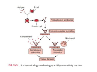
Hypersensitivity
BY: DR SADAF MOEEZ
Hypersensitivity
• It refers to undesirable reactions produced
by the normal immune system, including
allergies and autoimmunity.
• They are usually referred to as an over-reaction
of the immune system.
• These reactions may be damaging and
uncomfortable.
Caused by:
Complex set of
interactions between a
medication, your own
immune system, and
viruses in your body,
especially herpes viruses.
Signs of hypersensitivity:
• A pink or red rash with or without
pus-filled bumps or blisters.
• Scaly, flaky skin.
• Fever.
• Facial swelling.
• Swollen or tender lymph nodes.
• Swollen saliva glands.
• Dry mouth
• Abnormalities in your white
blood cell count
Types of hypersensitivity:
Types of Hypersensitivity
• Type I, II and III are immunoglobulin-
mediated (immediate) hypersensitivity
reactions while type IV reaction is lymphoid
cell-mediated or simply cell mediated
hypersensitivity (delayed-type).
TYPE I
MECHANISM:
• Allergen-specific lgE
antibodies bind to
mast cells via their fc
receptor. When the
specific allergen binds
to the lgE. Cross-
linking of IgE Induces
degranulation of mast
cells.
EXAMPLES
• local and systemic anaphylaxis
• seasonal hay fever
• food allergies
• drug allergies
Type II HYPERSENSITIVITY REACTION,
ADCC (CYTOTOXIC REACTION)
Definition
• In type II, the target is fixed in tissue or on the cell surface.
This is mediated by IgG or IgM binding to the specific cell
or tissue. The damage will be limited to the tissue or the
cells where this reaction will take place.
• Antigen: It is present or is a part of the cell membrane. It
may be:
1. Exogenous Ag: Microbes, parasites, drugs.
2. Intrinsic Ag: Autoimmune diseases, and these are self-Ag.
• Antibody: This is mainly due to IgG and occasionally IgM.
Rarely IgA can give this reaction.
• The effector cells are macrophages, neutrophils,
eosinophils, and NK (natural killer) cells.
Mechanism
1. Complement activation:
The antibody attaches to antigen on the surface of cells
and activates the complement system, which leads to
lysis.
2. Antibody-dependent cellular toxicity (ADCC):
Sometimes Antibody is attached to Antigen on the cell
surface will bring this complex near to NK cells or other
phagocytic cells possessing the Fc-Receptor and leads to
antibody dependant cellular cytotoxicity (ADCC).
3. Opsonization and phagocytosis:
The antibody binds to an antigen and makes it a target
for phagocytosis, and this process is called opsonization.
1. Complement activation:
2. Antibody-dependent cellular toxicity
(ADCC):
3. Opsonization and phagocytosis:
Type III Hypersensitivity Reaction
• Type III reaction is mediated by antigen–antibody
immune complexes, which induce an
inflammatory reaction in tissues.
• Activation of the complement results in the
formation of complement components, such as
C3a and 5a.
• The C5a attracts neutrophils to the site, but these
neutrophils fail to phagocytose large aggregated
mass of immune complexes, and instead release
lysosomal enzymes and lytic substances that
damage host tissue.
Type IV Hypersensitivity Reaction
• Type IV hypersensitivity occurs 24 hours after
contact with an antigen, usually starting at 2 or
3 days and often last for many days.
• For this reason, type IV hypersensitivity
reaction is termed as “delayed
hypersensitivity”.
• In the sensitization phase, the primary contact
with the antigen is established. During this
period, specific Th cells are sensitized and are
clonally expanded.
• In the effector phase, a subsequent exposure
to the same antigen induces the delayed type
hypersensitivity response.
activated macrophages
produce hydrolytic enzymes

hypersensitivity.pptx

  • 1.
  • 2.
    Hypersensitivity • It refersto undesirable reactions produced by the normal immune system, including allergies and autoimmunity. • They are usually referred to as an over-reaction of the immune system. • These reactions may be damaging and uncomfortable.
  • 3.
    Caused by: Complex setof interactions between a medication, your own immune system, and viruses in your body, especially herpes viruses.
  • 4.
    Signs of hypersensitivity: •A pink or red rash with or without pus-filled bumps or blisters. • Scaly, flaky skin. • Fever. • Facial swelling. • Swollen or tender lymph nodes. • Swollen saliva glands. • Dry mouth • Abnormalities in your white blood cell count
  • 5.
  • 6.
    Types of Hypersensitivity •Type I, II and III are immunoglobulin- mediated (immediate) hypersensitivity reactions while type IV reaction is lymphoid cell-mediated or simply cell mediated hypersensitivity (delayed-type).
  • 7.
    TYPE I MECHANISM: • Allergen-specificlgE antibodies bind to mast cells via their fc receptor. When the specific allergen binds to the lgE. Cross- linking of IgE Induces degranulation of mast cells.
  • 10.
    EXAMPLES • local andsystemic anaphylaxis • seasonal hay fever • food allergies • drug allergies
  • 11.
    Type II HYPERSENSITIVITYREACTION, ADCC (CYTOTOXIC REACTION) Definition • In type II, the target is fixed in tissue or on the cell surface. This is mediated by IgG or IgM binding to the specific cell or tissue. The damage will be limited to the tissue or the cells where this reaction will take place. • Antigen: It is present or is a part of the cell membrane. It may be: 1. Exogenous Ag: Microbes, parasites, drugs. 2. Intrinsic Ag: Autoimmune diseases, and these are self-Ag. • Antibody: This is mainly due to IgG and occasionally IgM. Rarely IgA can give this reaction. • The effector cells are macrophages, neutrophils, eosinophils, and NK (natural killer) cells.
  • 12.
    Mechanism 1. Complement activation: Theantibody attaches to antigen on the surface of cells and activates the complement system, which leads to lysis. 2. Antibody-dependent cellular toxicity (ADCC): Sometimes Antibody is attached to Antigen on the cell surface will bring this complex near to NK cells or other phagocytic cells possessing the Fc-Receptor and leads to antibody dependant cellular cytotoxicity (ADCC). 3. Opsonization and phagocytosis: The antibody binds to an antigen and makes it a target for phagocytosis, and this process is called opsonization.
  • 13.
  • 14.
  • 15.
    3. Opsonization andphagocytosis:
  • 16.
    Type III HypersensitivityReaction • Type III reaction is mediated by antigen–antibody immune complexes, which induce an inflammatory reaction in tissues. • Activation of the complement results in the formation of complement components, such as C3a and 5a. • The C5a attracts neutrophils to the site, but these neutrophils fail to phagocytose large aggregated mass of immune complexes, and instead release lysosomal enzymes and lytic substances that damage host tissue.
  • 18.
    Type IV HypersensitivityReaction • Type IV hypersensitivity occurs 24 hours after contact with an antigen, usually starting at 2 or 3 days and often last for many days. • For this reason, type IV hypersensitivity reaction is termed as “delayed hypersensitivity”.
  • 19.
    • In thesensitization phase, the primary contact with the antigen is established. During this period, specific Th cells are sensitized and are clonally expanded. • In the effector phase, a subsequent exposure to the same antigen induces the delayed type hypersensitivity response.
  • 20.