The document outlines a standard examination protocol for contact lens fitting, focusing on patient screening, anterior segment examination, ocular measurements, tear assessment, and spectacle refraction. It emphasizes the importance of identifying suitable candidates for contact lenses to prevent complications and ensure optimal fit and comfort. Key components include assessing ocular health, measuring corneal and pupil dimensions, and evaluating tear production.
Why a StandardProtocol for Contact
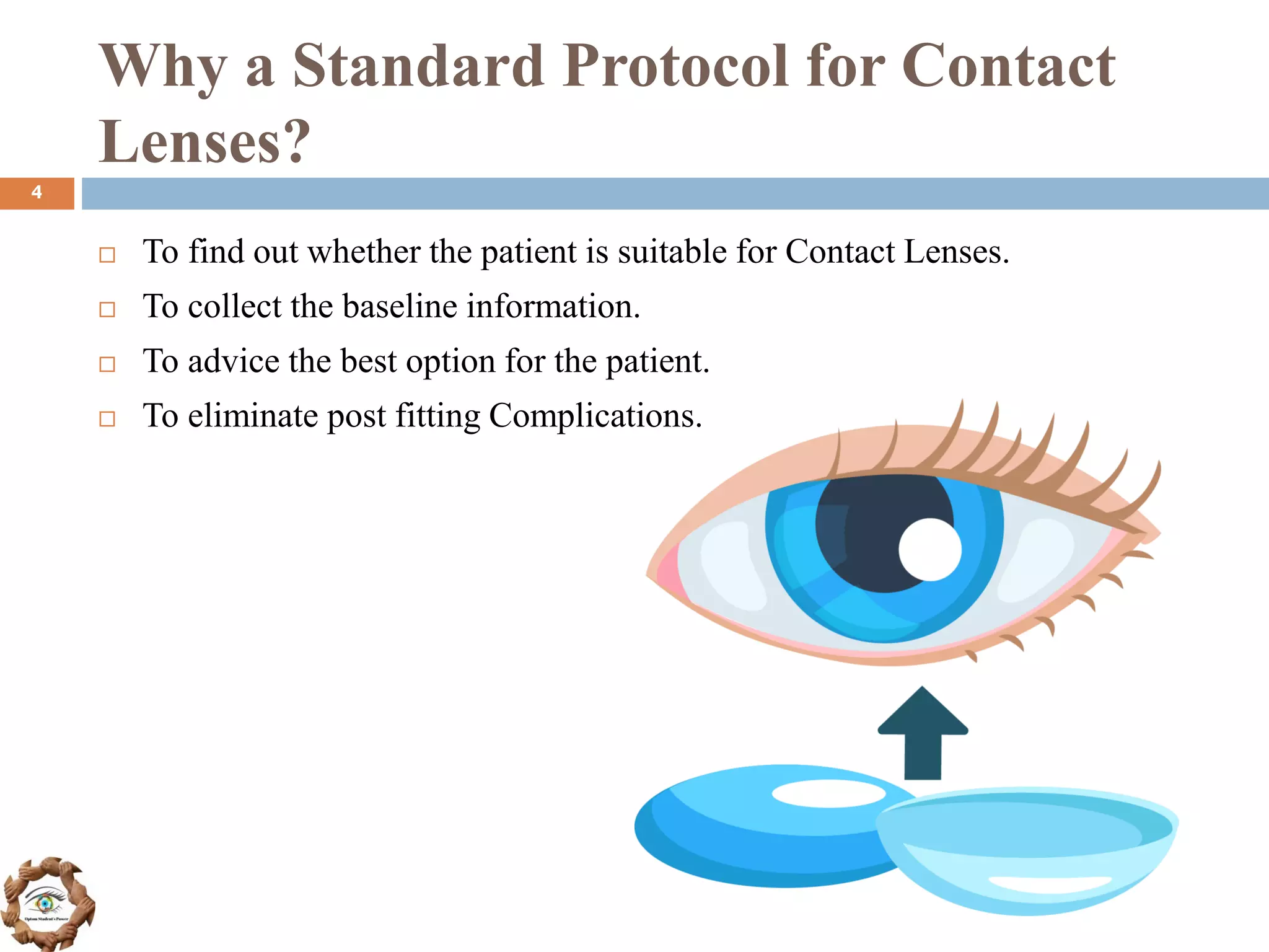
Lenses?
To find out whether the patient is suitable for Contact Lenses.
To collect the baseline information.
To advice the best option for the patient.
To eliminate post fitting Complications.
4
5.
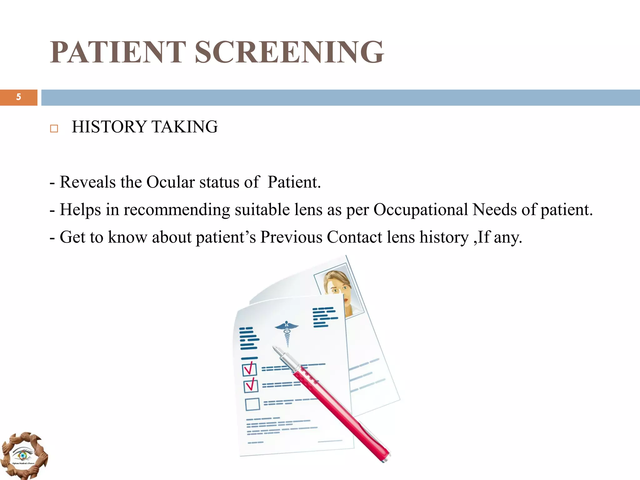
PATIENT SCREENING
HISTORYTAKING
- Reveals the Ocular status of Patient.
- Helps in recommending suitable lens as per Occupational Needs of patient.
- Get to know about patient’s Previous Contact lens history ,If any.
5
6.
PATIENT SCREENING
IdentifyWhy the Patient needs Contact Lenses?
- Cosmetic Purpose
- Therapeutic Purpose
- Occupational Requirements
- Pathological Condition
- Myopia Control
It will help in choosing the Best Contact Lenses for Patient.
6
7.
ANTERIOR SEGMENT
EXAMINATION
SlitLamp Biomicroscopy is used for examining the Anterior Segment of
Eye.
Biomicroscopy is necessary as it helps in diagnosing any anomalies
present that may cause Complications in Contact Lens wear.
7
8.
ANTERIOR SEGMENT
EXAMINATION
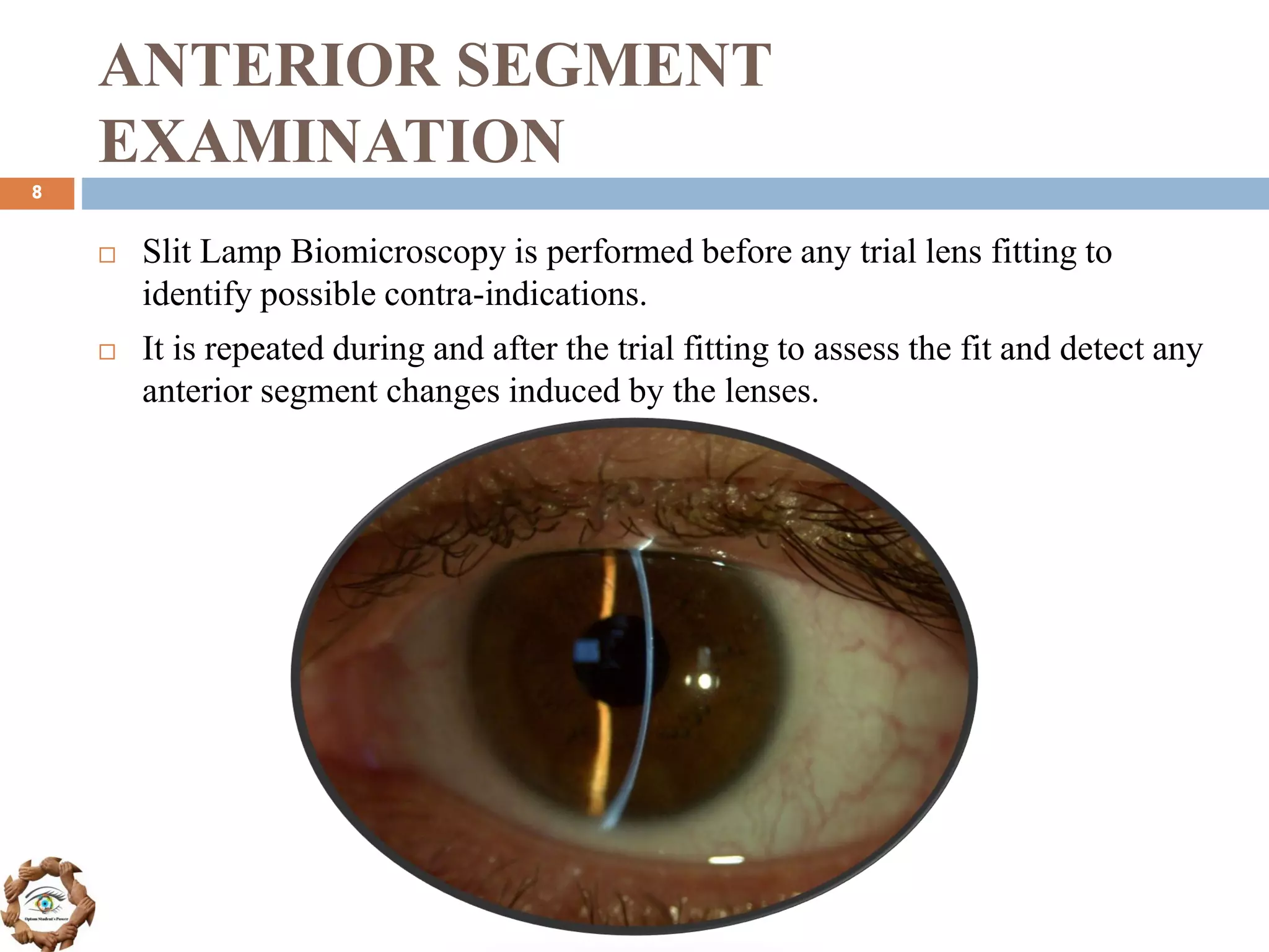
SlitLamp Biomicroscopy is performed before any trial lens fitting to
identify possible contra-indications.
It is repeated during and after the trial fitting to assess the fit and detect any
anterior segment changes induced by the lenses.
8
9.
ANTERIOR SEGMENT
EXAMINATION
A SlitLamp Examination is performed to assess the condition of:
Eyelids
Conjunctiva
Tears
Cornea
Anterior chamber
Iris and lens
9
10.
MEASUREMENTS OF OCULAR
DIMENSIONS
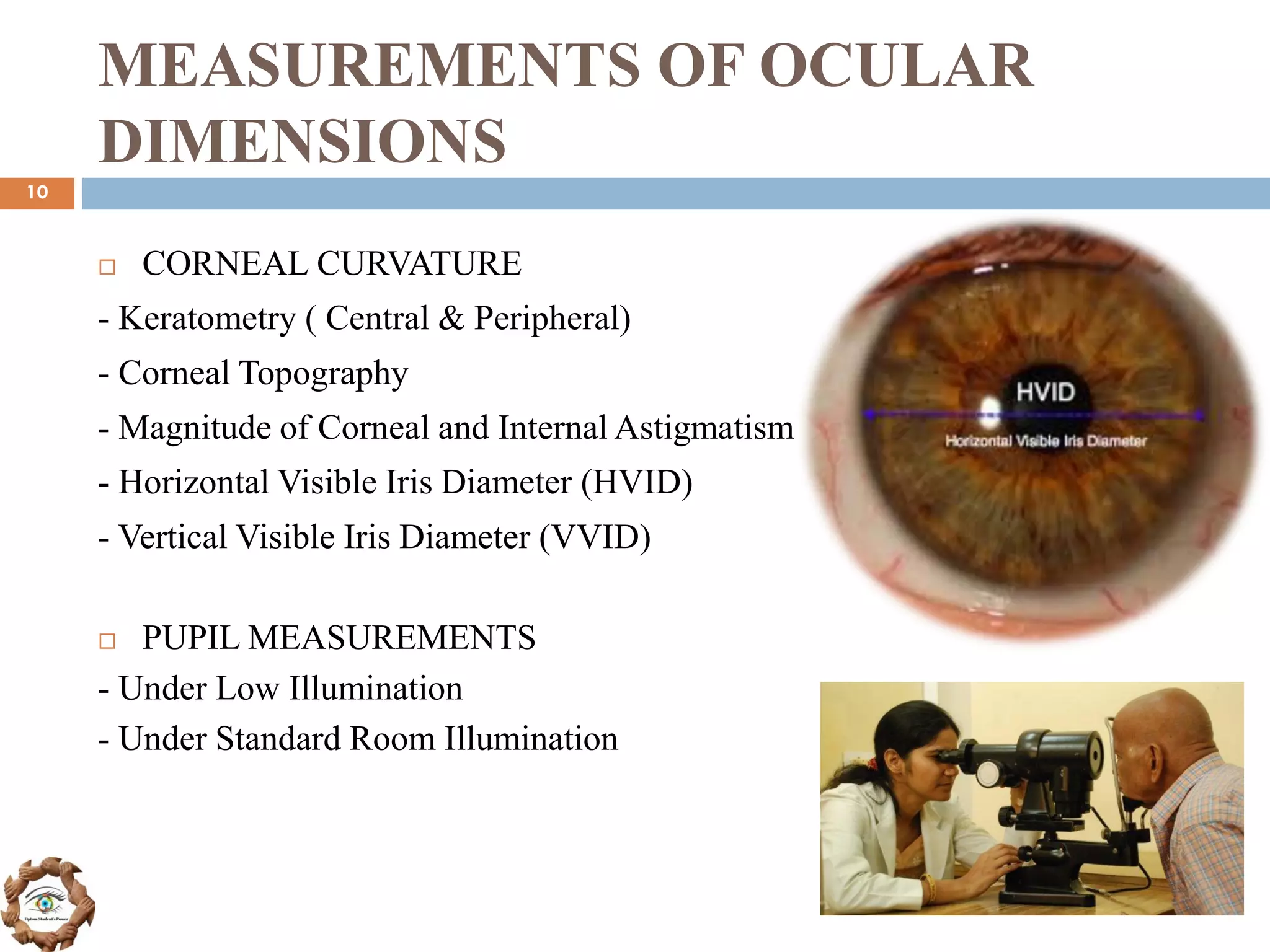
CORNEAL CURVATURE
- Keratometry ( Central & Peripheral)
- Corneal Topography
- Magnitude of Corneal and Internal Astigmatism
- Horizontal Visible Iris Diameter (HVID)
- Vertical Visible Iris Diameter (VVID)
PUPIL MEASUREMENTS
- Under Low Illumination
- Under Standard Room Illumination
10
11.
MEASUREMENTS OF OCULAR
DIMENSIONS
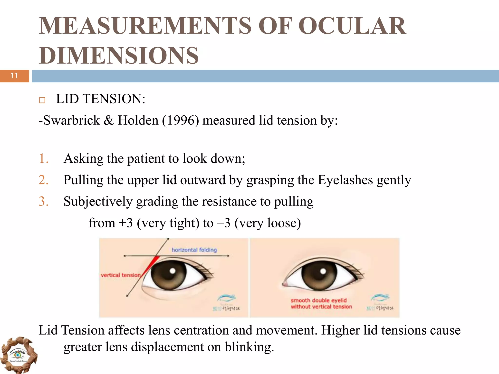
LID TENSION:
-Swarbrick & Holden (1996) measured lid tension by:
1. Asking the patient to look down;
2. Pulling the upper lid outward by grasping the Eyelashes gently
3. Subjectively grading the resistance to pulling
from +3 (very tight) to –3 (very loose)
Lid Tension affects lens centration and movement. Higher lid tensions cause
greater lens displacement on blinking.
11
12.
MEASUREMENTS OF OCULAR
DIMENSIONS
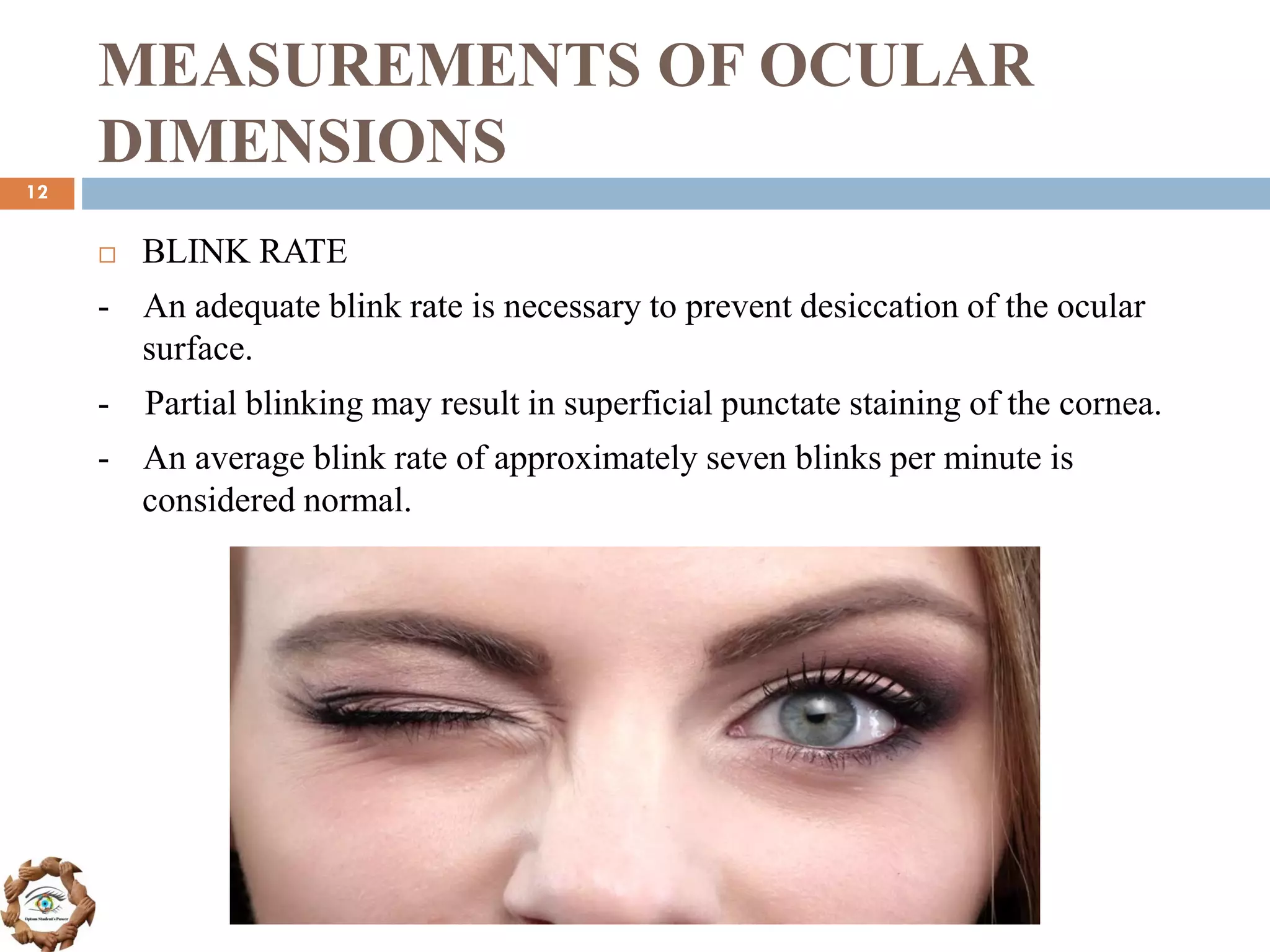
BLINK RATE
- An adequate blink rate is necessary to prevent desiccation of the ocular
surface.
- Partial blinking may result in superficial punctate staining of the cornea.
- An average blink rate of approximately seven blinks per minute is
considered normal.
12
13.
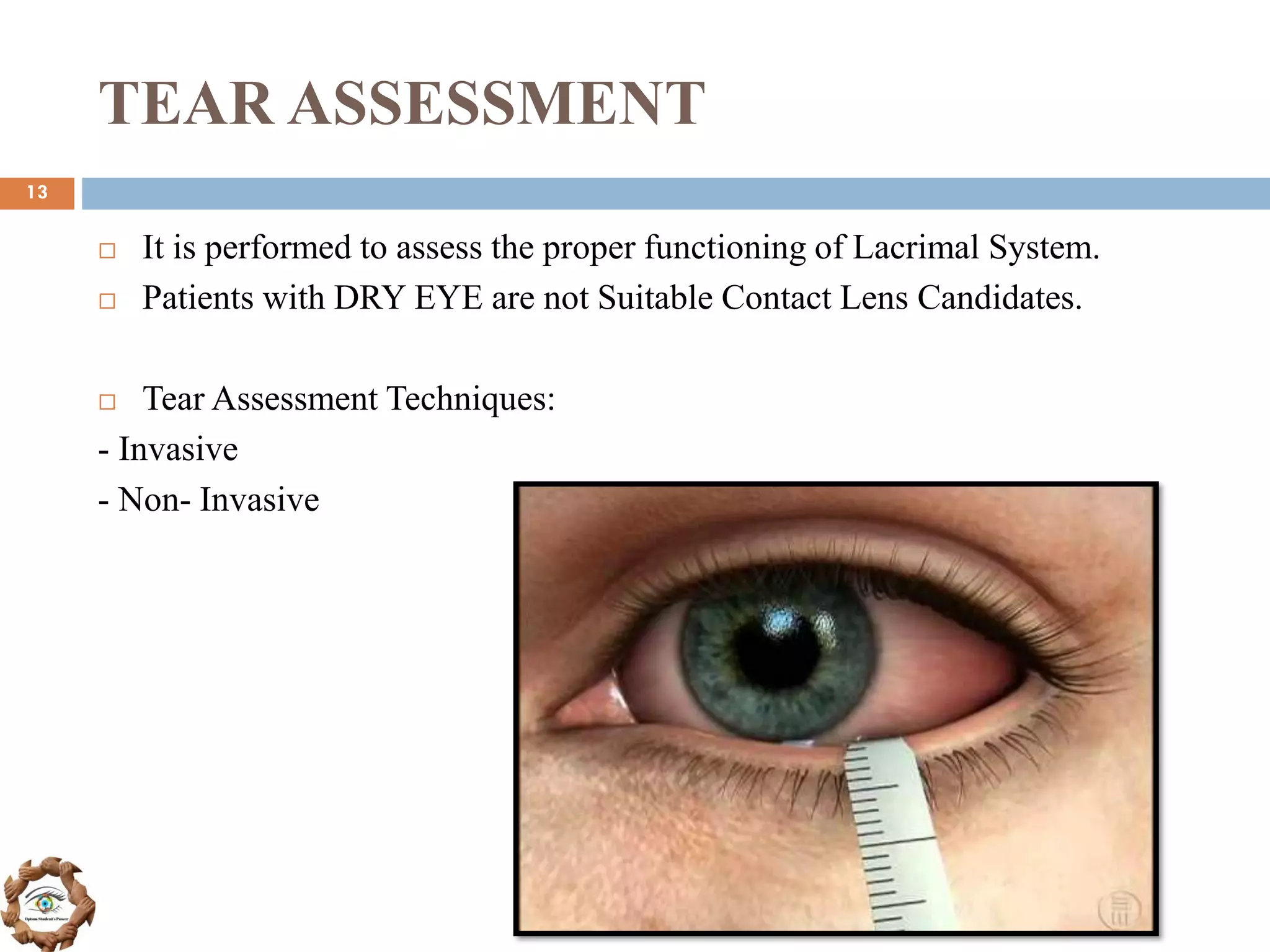
TEAR ASSESSMENT
Itis performed to assess the proper functioning of Lacrimal System.
Patients with DRY EYE are not Suitable Contact Lens Candidates.
Tear Assessment Techniques:
- Invasive
- Non- Invasive
13
14.
TEAR ASSESSMENT
Invasive:
-Break-Up-Time (BUT)
- Schirmer test
- Phenol-red thread test
- Rose bengal staining
Non invasive:
- Non-invasive Tear Break-Up-Time (NIBUT)
- Tear prism height
- Lipid layer evaluation
14
SPECTACLE REFRACTION
Baselinerefraction:
It includes both subjective and objective findings (autorefractor,
retinoscopy).
Vertex distance :
The spectacle plane is approximately 12 - 15 mm from the corneal apex. A
myope requires less minus power at the cornea than at the spectacle plane,
whereas the hyperope requires more plus power than spectacles.
Accommodation and Convergence:
Myopes have to converge and accommodate more in wearing contact
lenses than in spectacles.
Conversely, hyperopes have to accommodate and converge less when
wearing contact lenses.
16
17.
Let’s Summarise!
PATIENTSCREENING - Find out whether the person is suitable for
Contact Lenses or not.
ANTERIOR SEGMENT EXAMINATION – Using a Slit Lamp, Check for
any anomalies in Ocular Structures of Patient, to avoid Complications.
MEASUREMENTS OF OCULAR DIMENSIONS – Record and assess
the Corneal Curvature, Lid Tension, Pupil Measurements and Blink rate.
TEAR ASSESSMENT – It is important to perform as it helps in knowing
the status of lacrimal system, and identifying the Suitable CL candidate.
SPECTACLE REFRACTION – Baseline Refraction including vertex
distance etc. is required for getting adequate Vision through Contact
Lenses.
17
18.
REFERENCES
▪ Clinical Manualof Contact Lenses by Edward S. Bennett, Henry
▪ The IACLE Module 4
▪ The IACLE Module 3
▪ The Contact Lens Manual by Andrew Gasson, Judith Morris.
18


![Understanding Parkinson’s Disease: Causes, Symptoms, and Treatment [2025]](https://cdn.slidesharecdn.com/ss_thumbnails/understandingparkinson-251208102525-80ba3223-thumbnail.jpg?width=640&height=640&fit=bounds)


