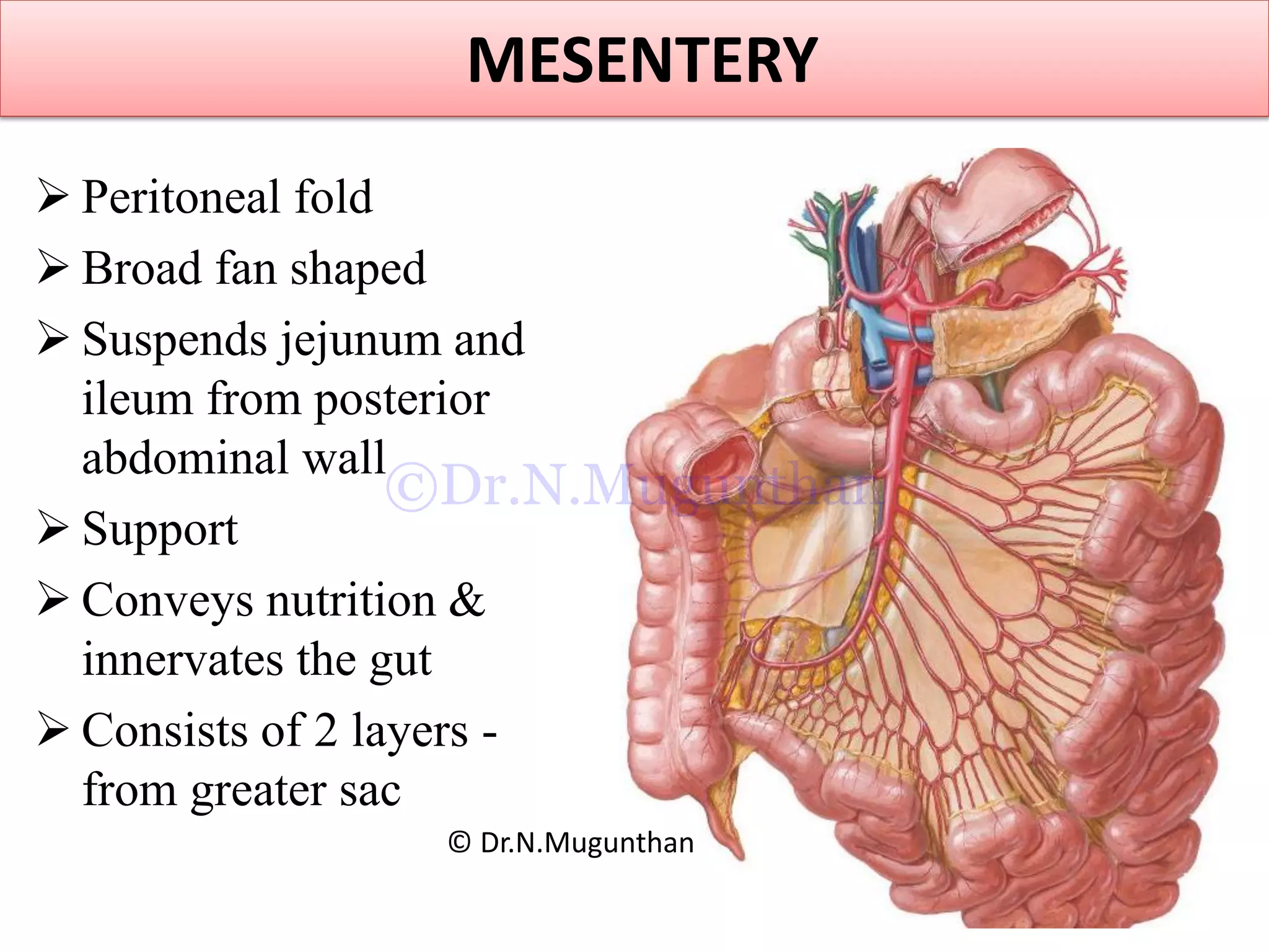
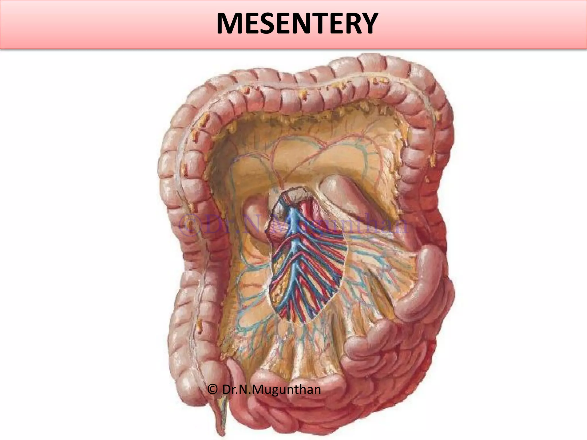
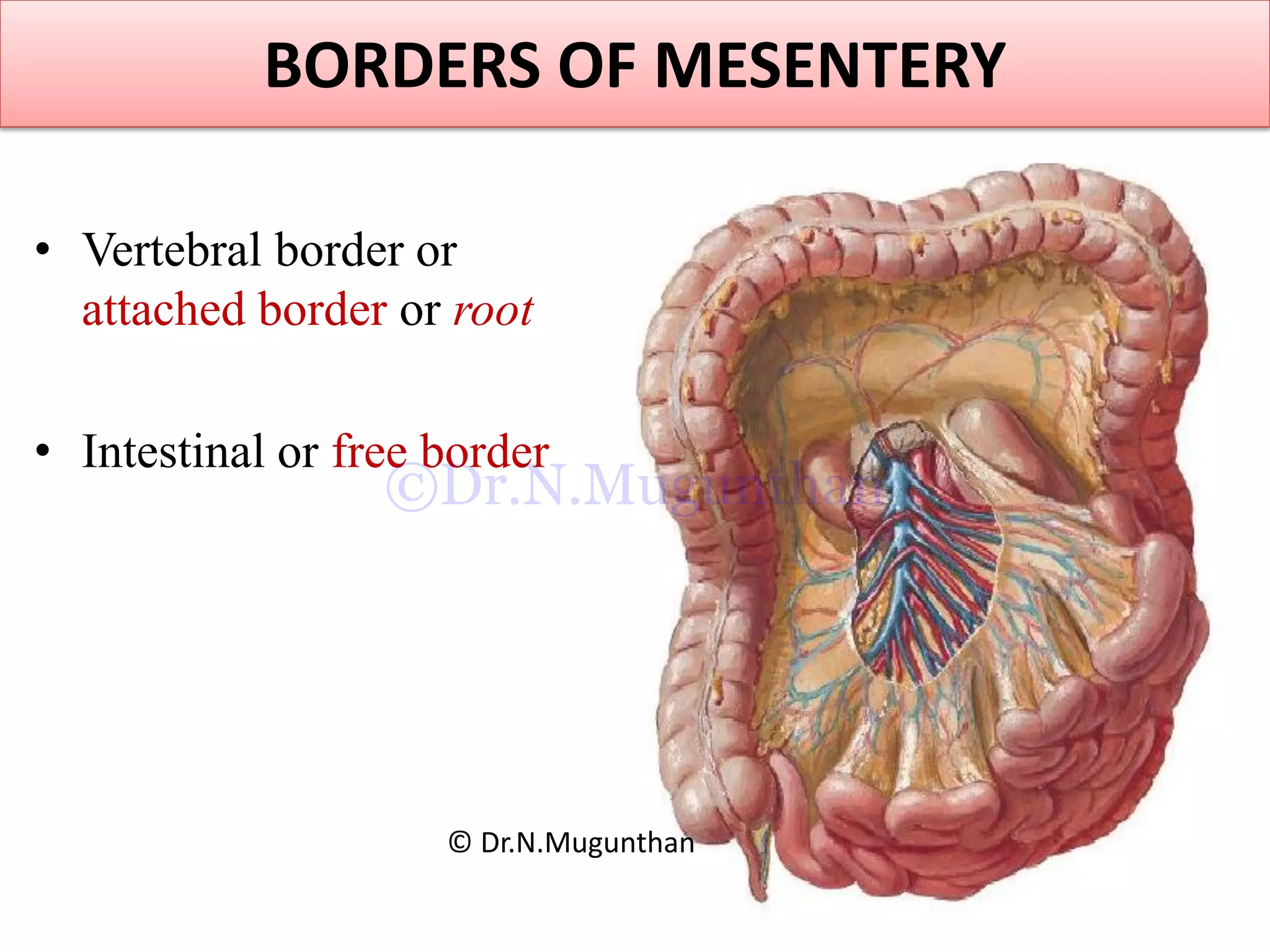
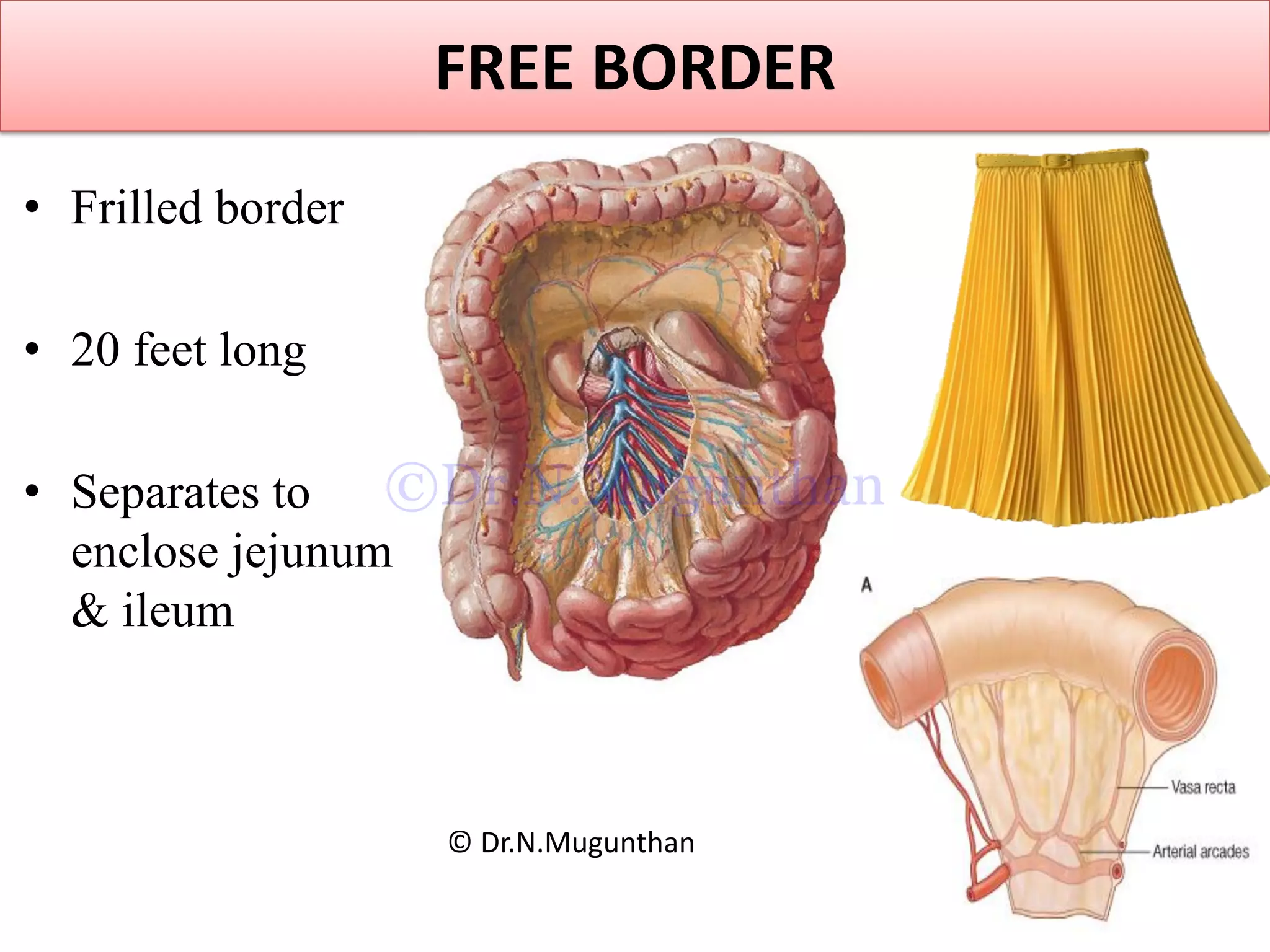
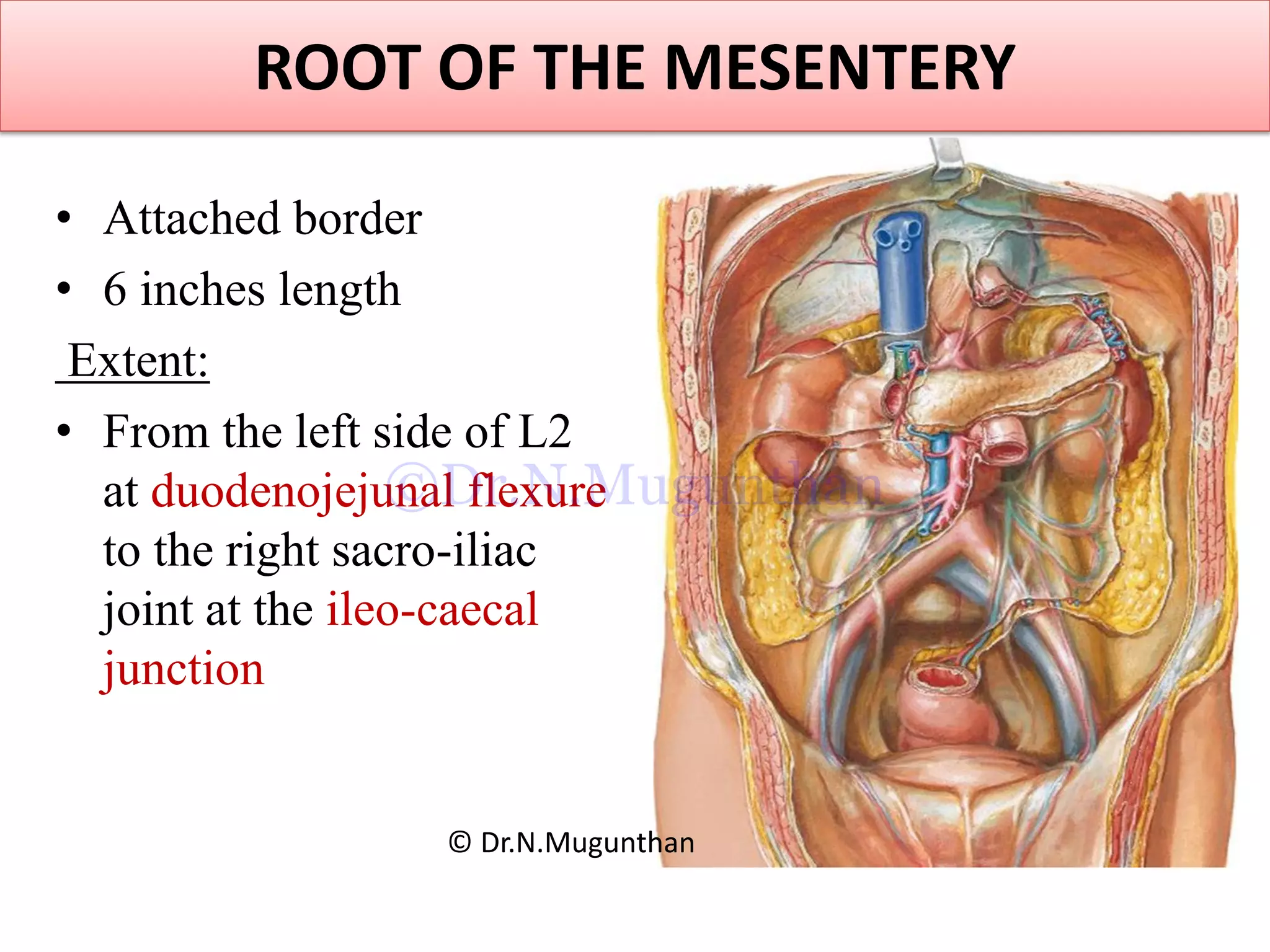
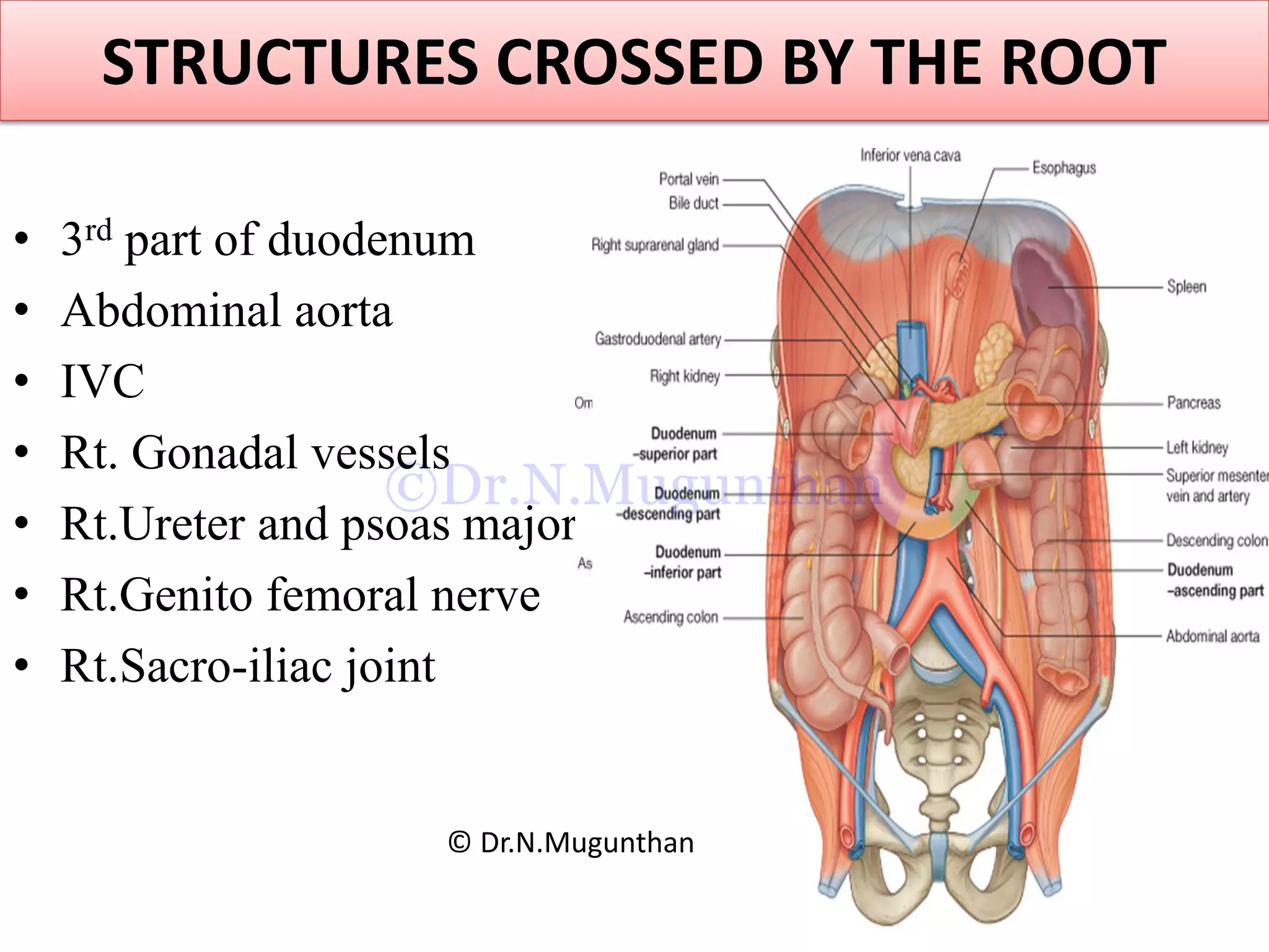
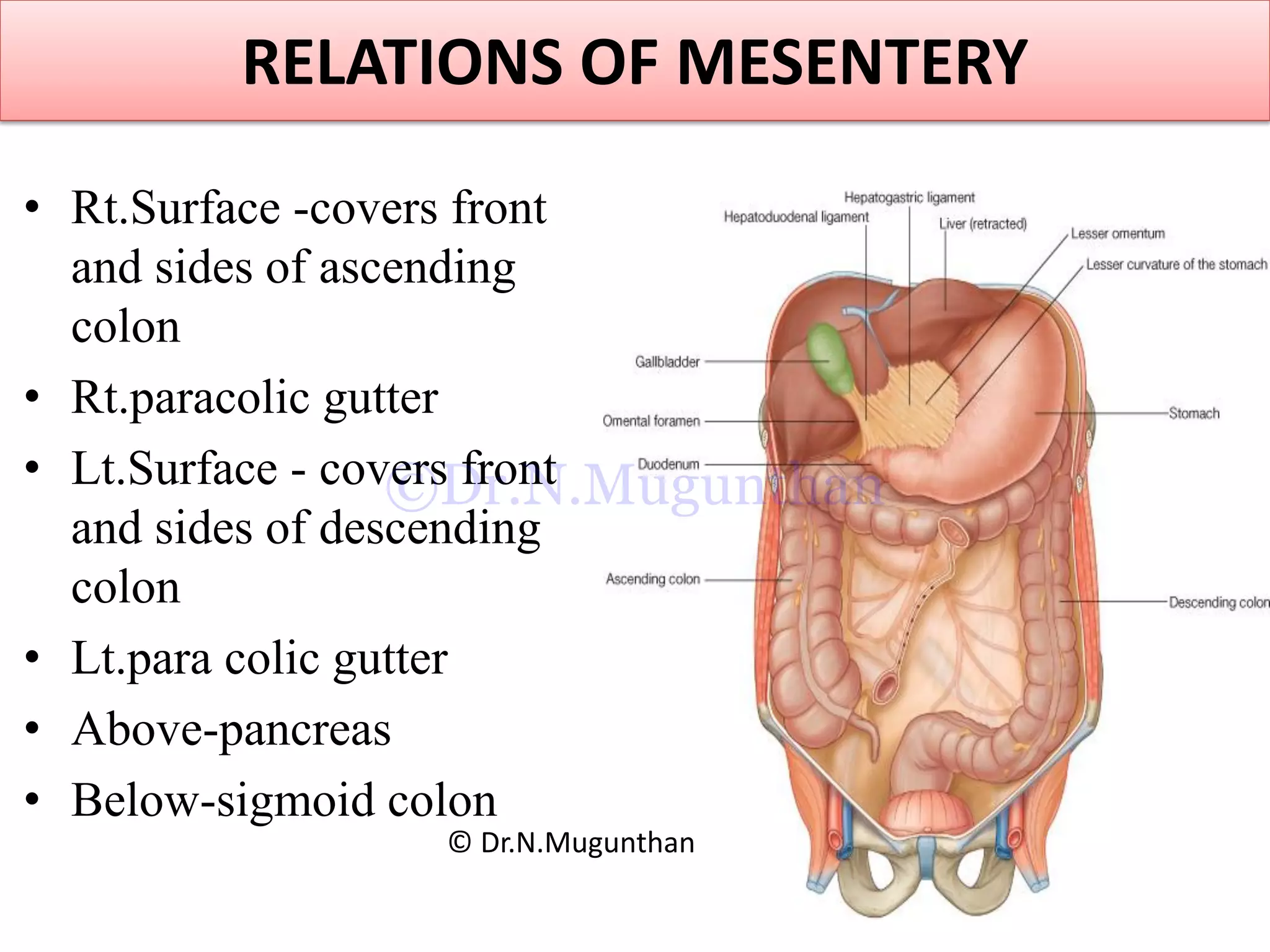
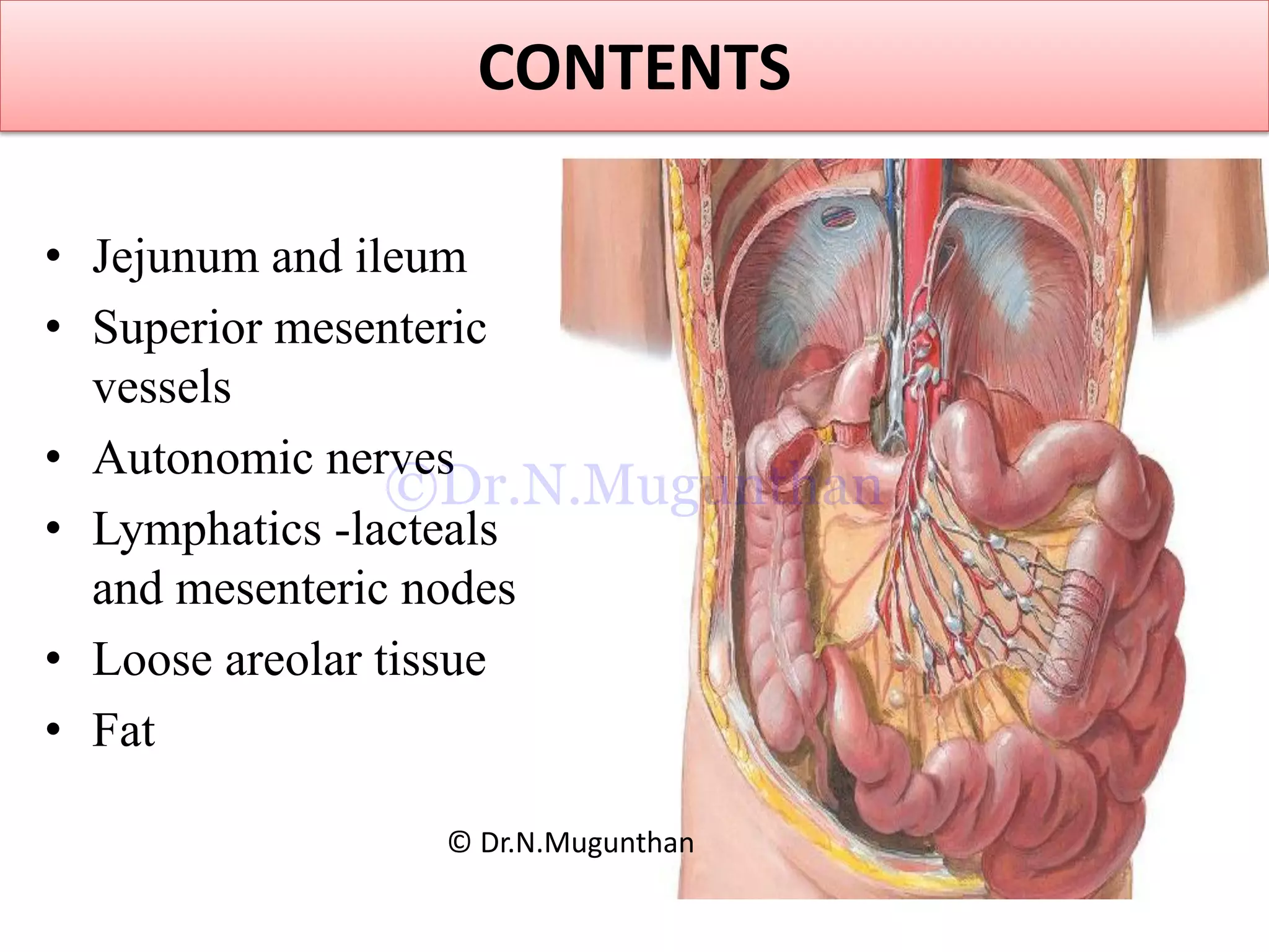
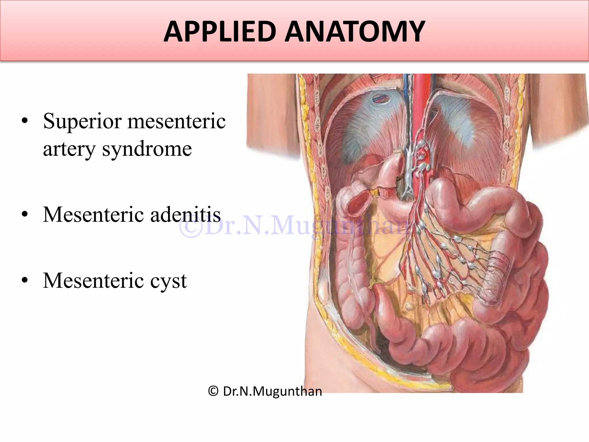
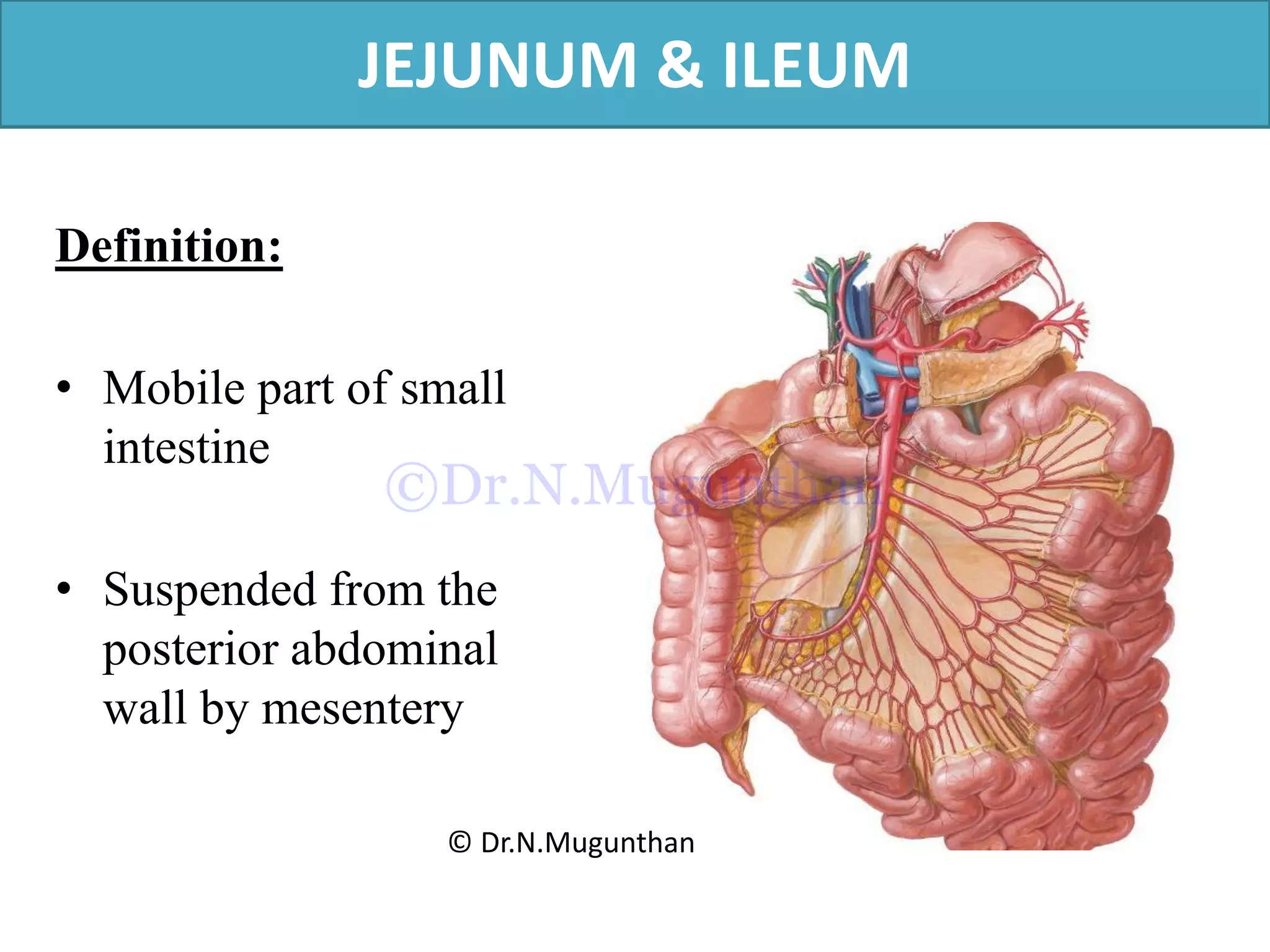
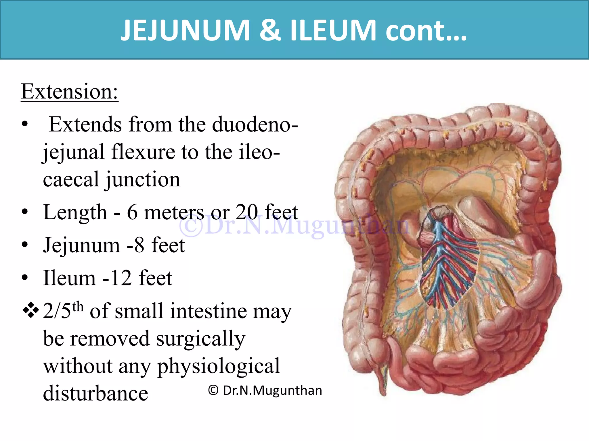
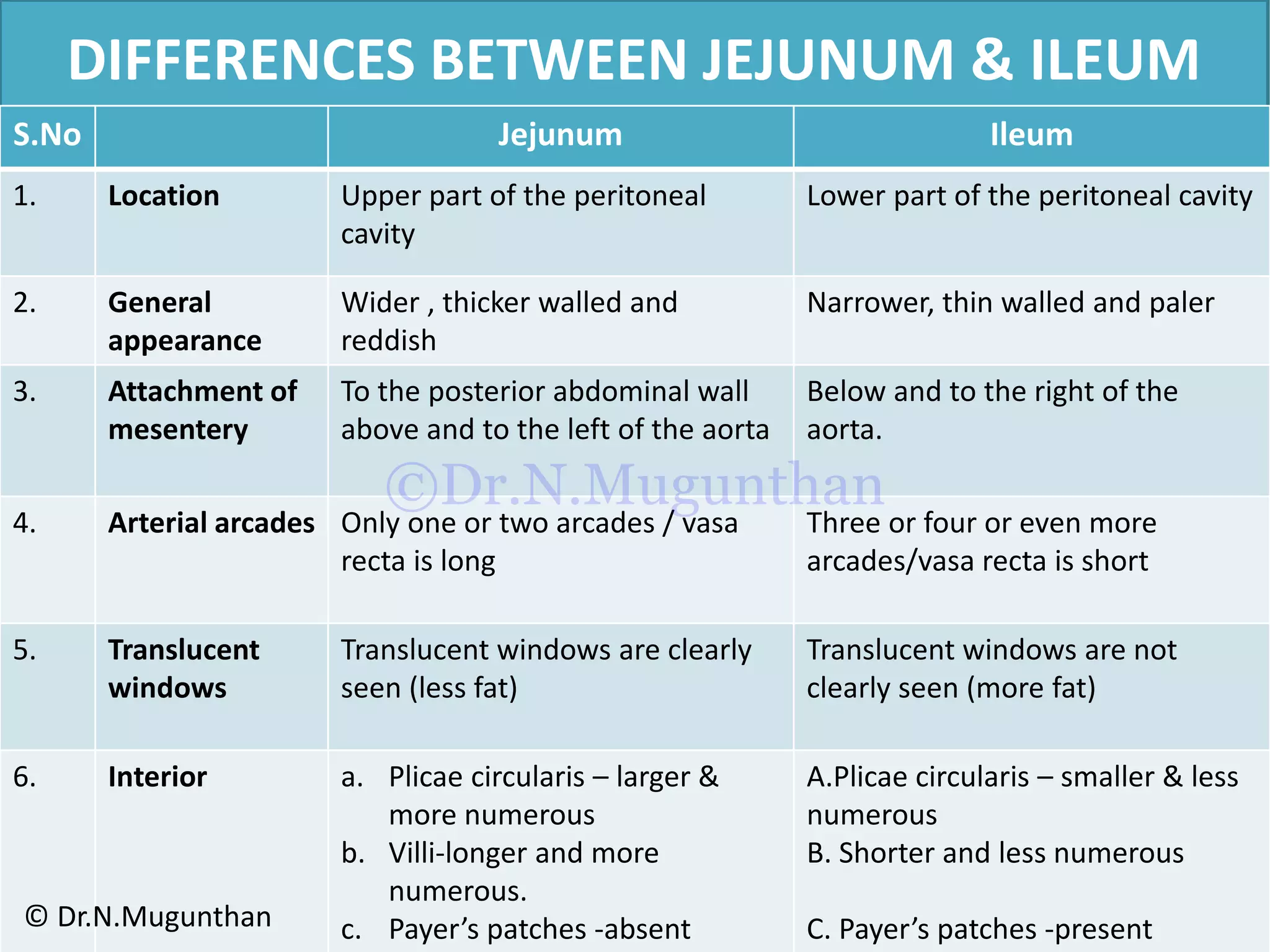
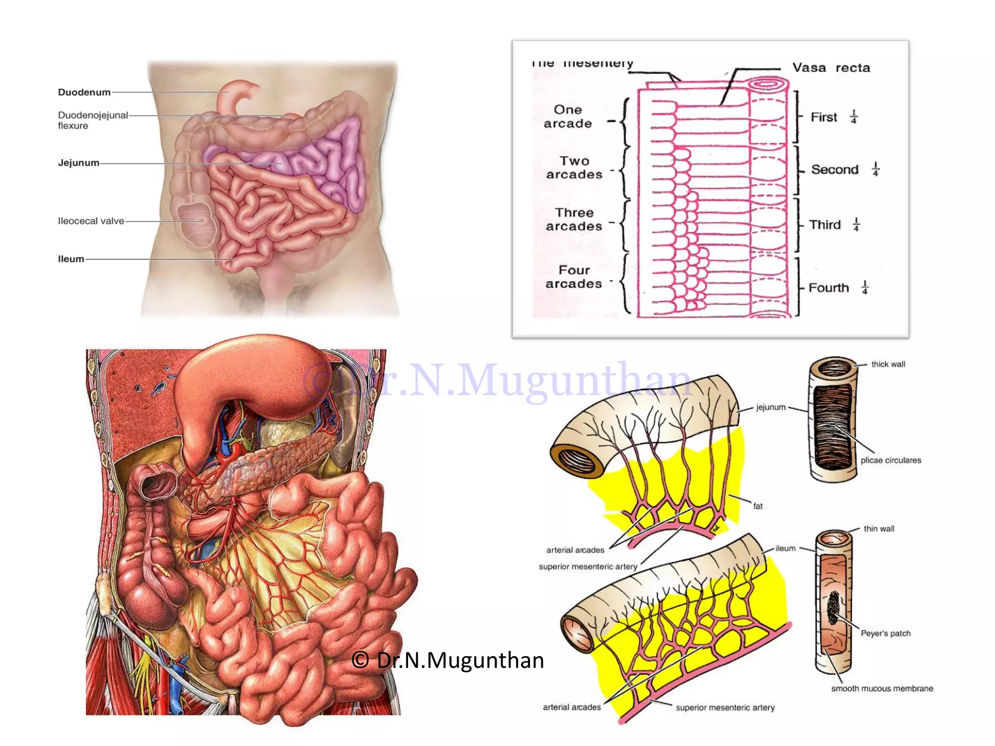
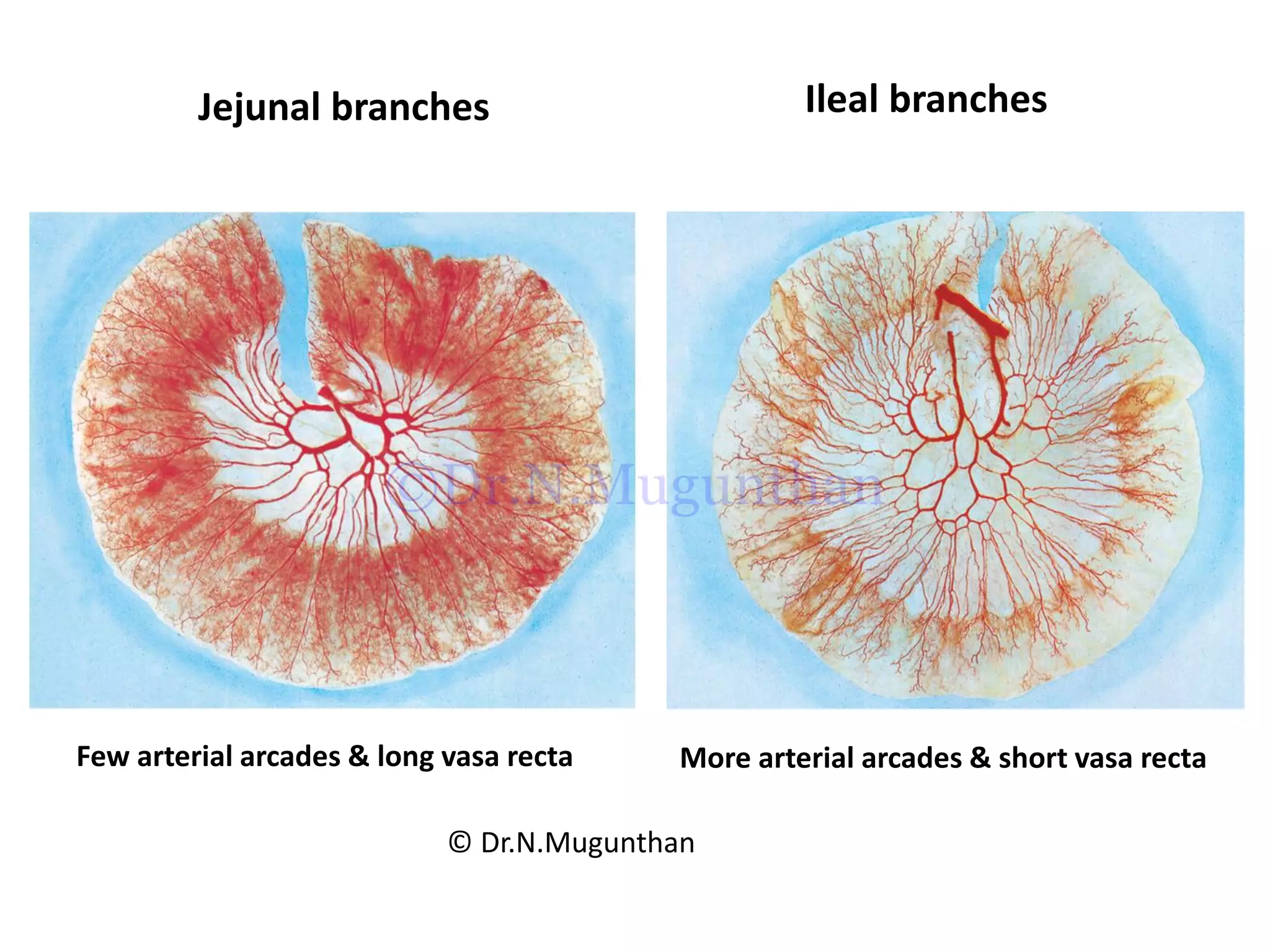
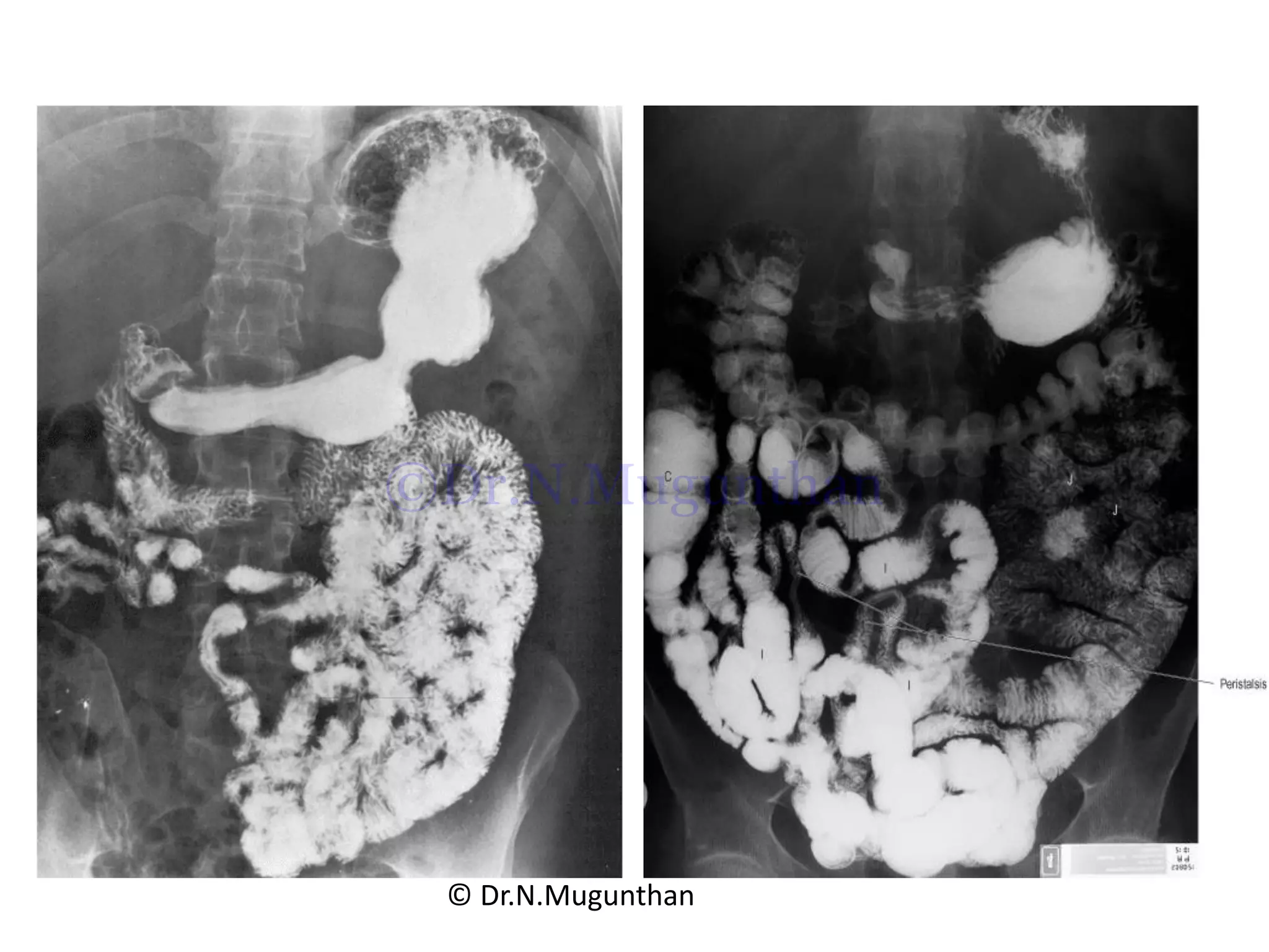
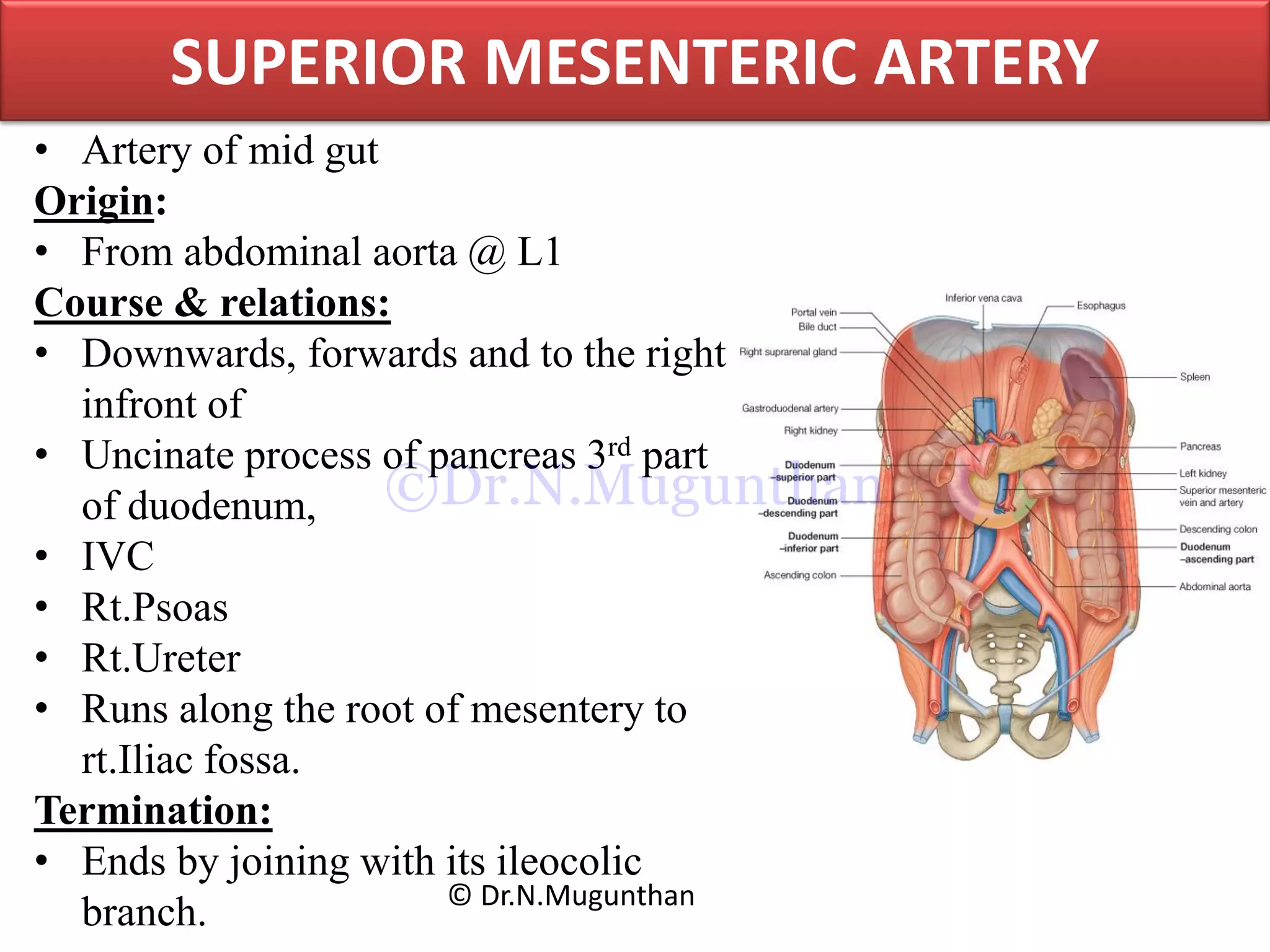
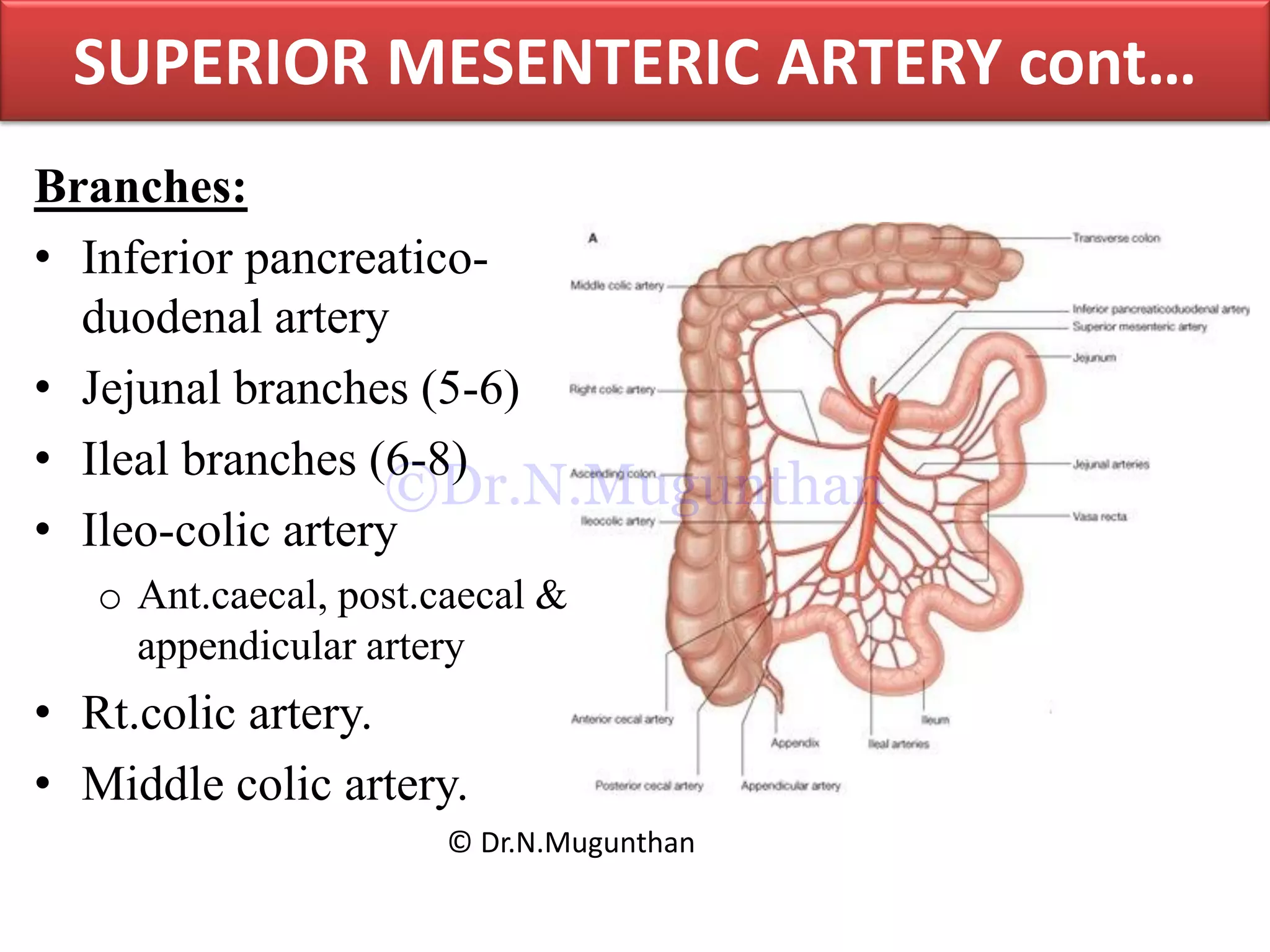
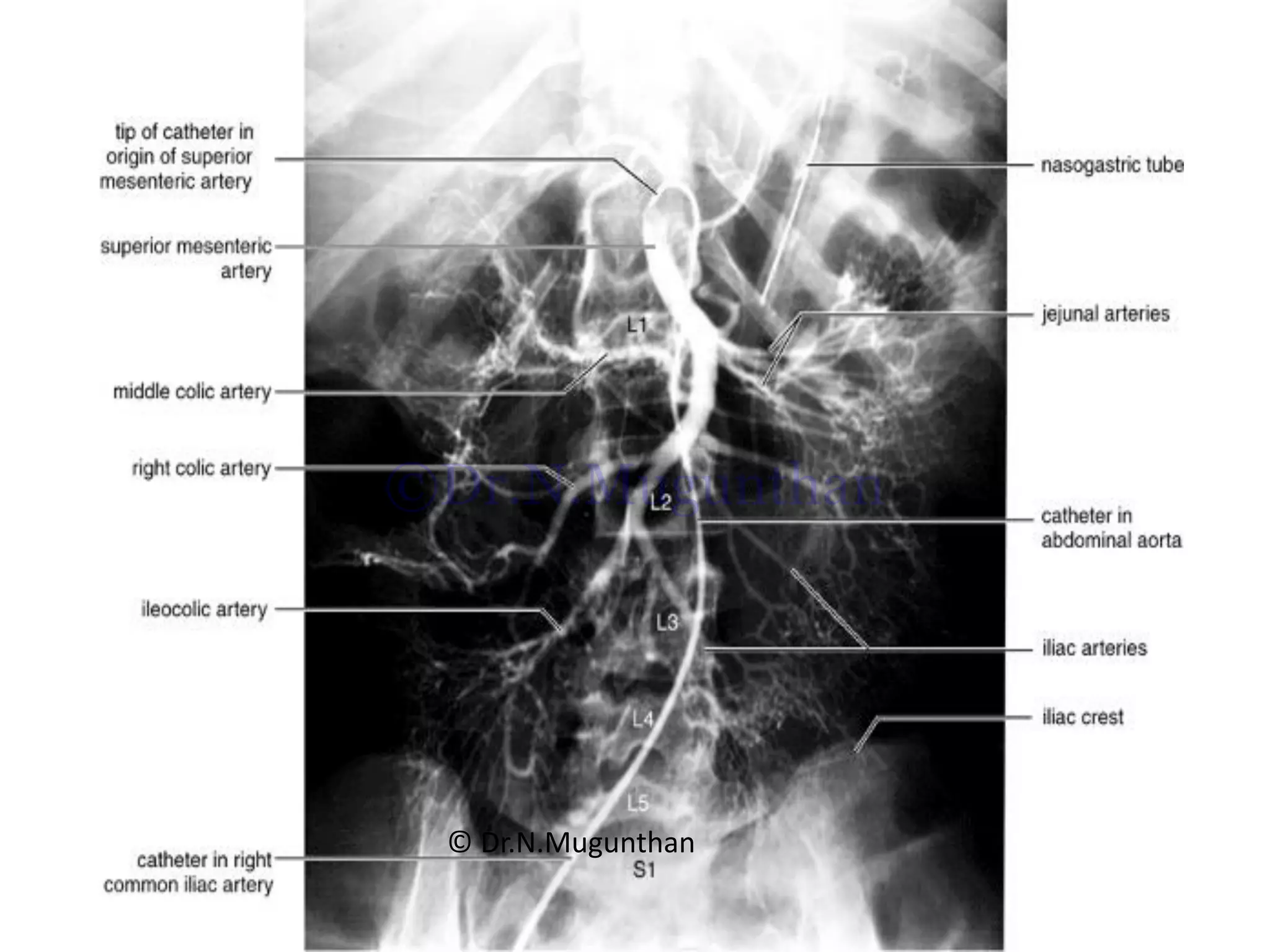
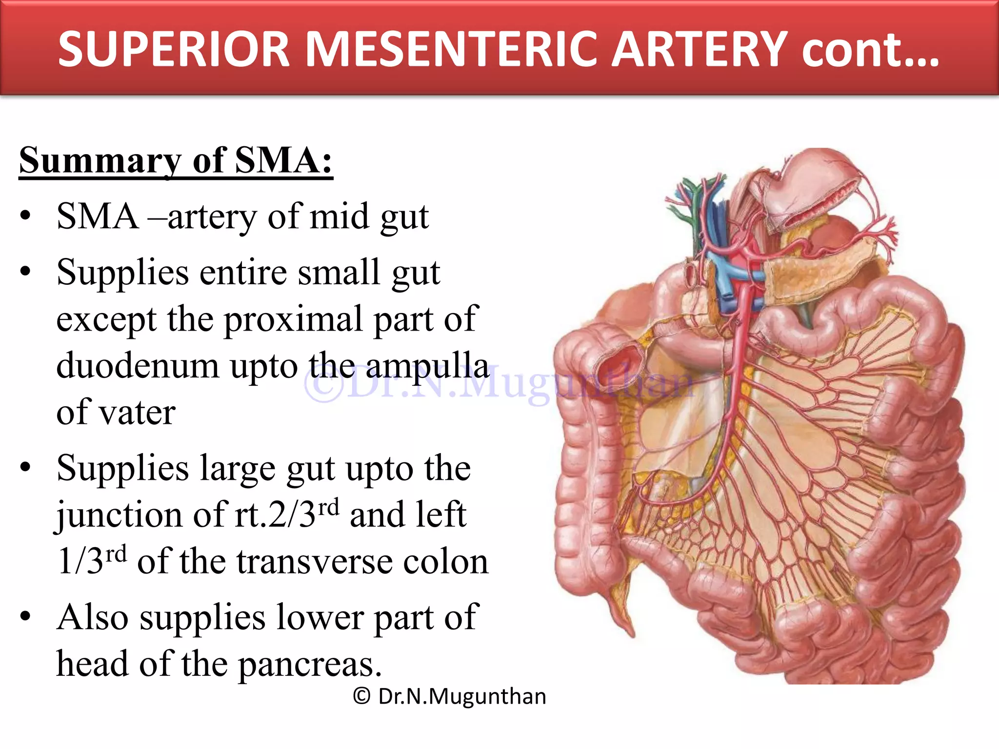
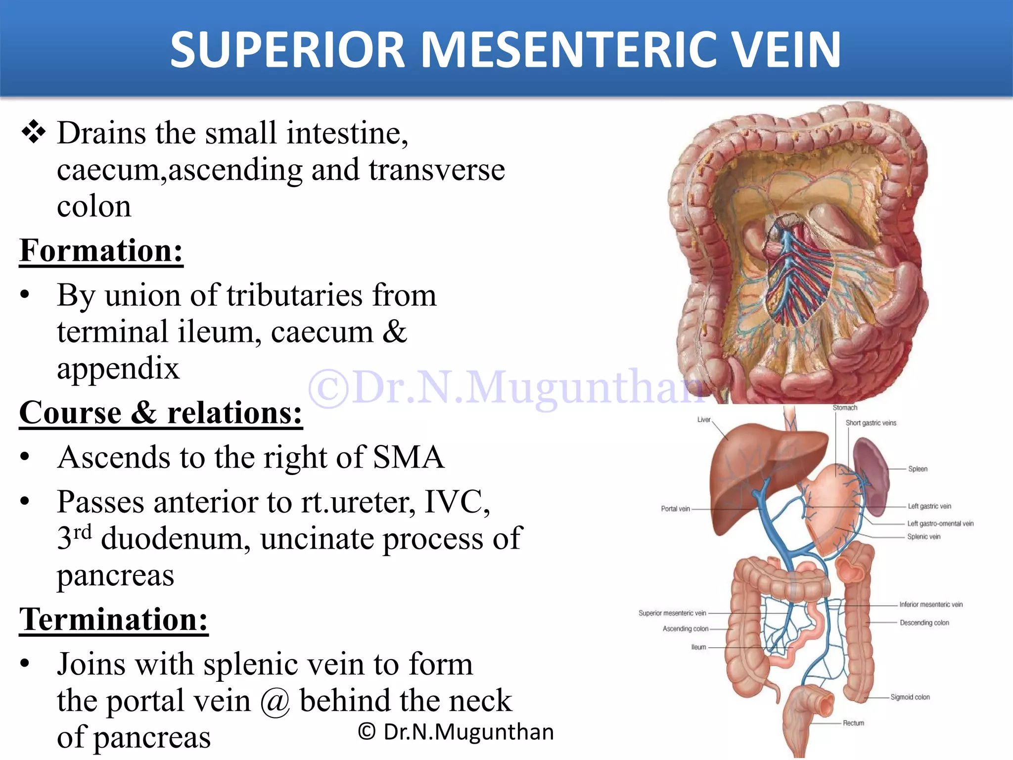
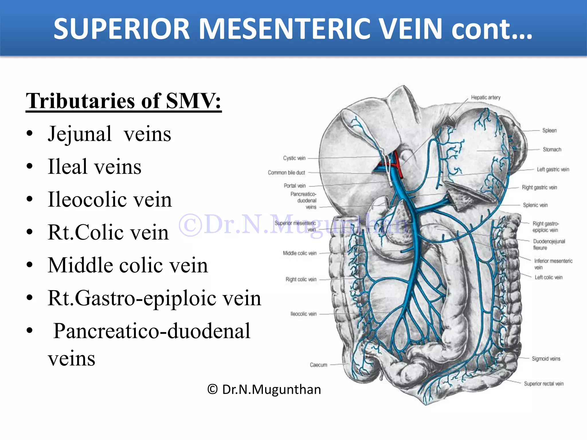
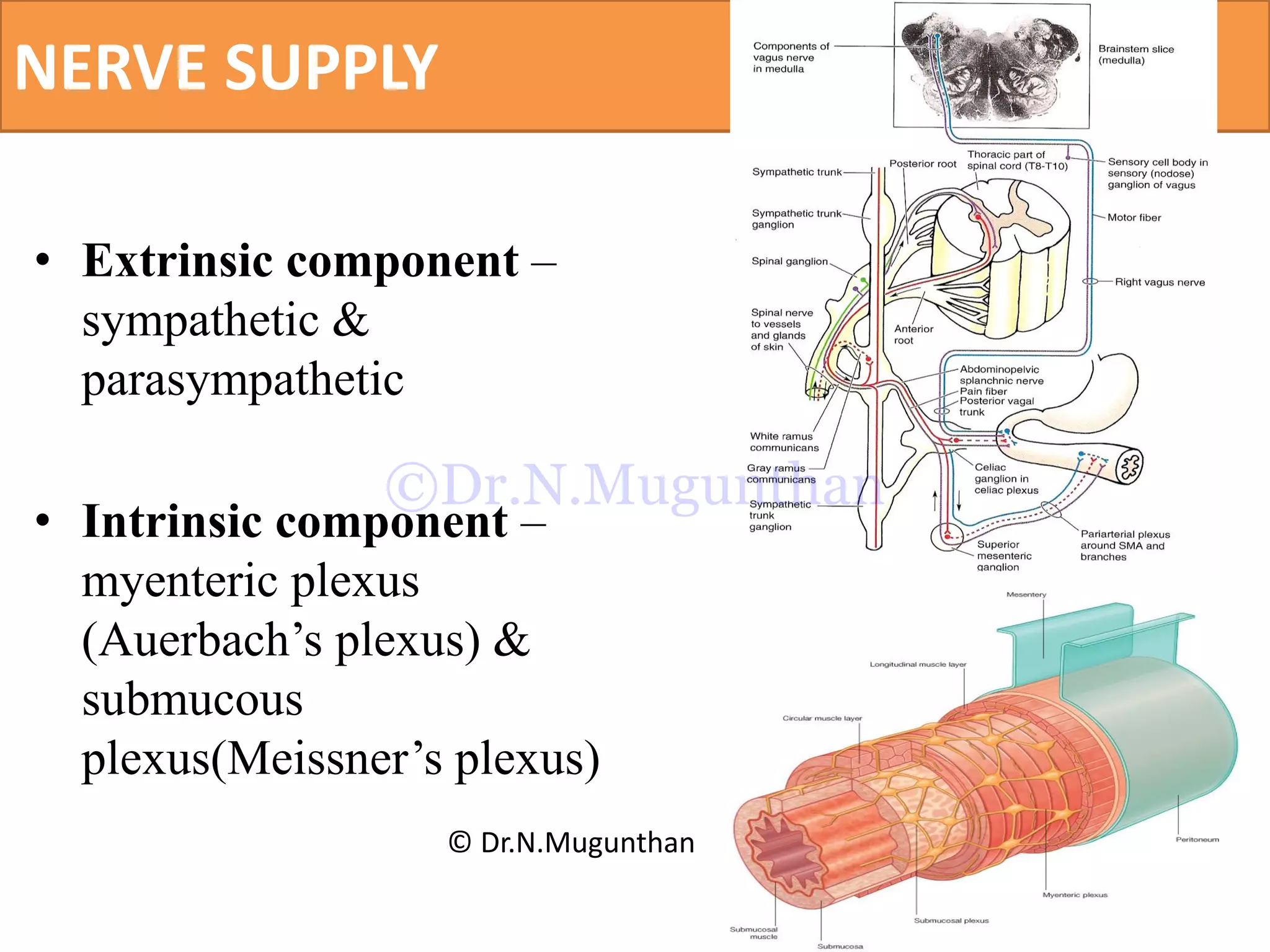
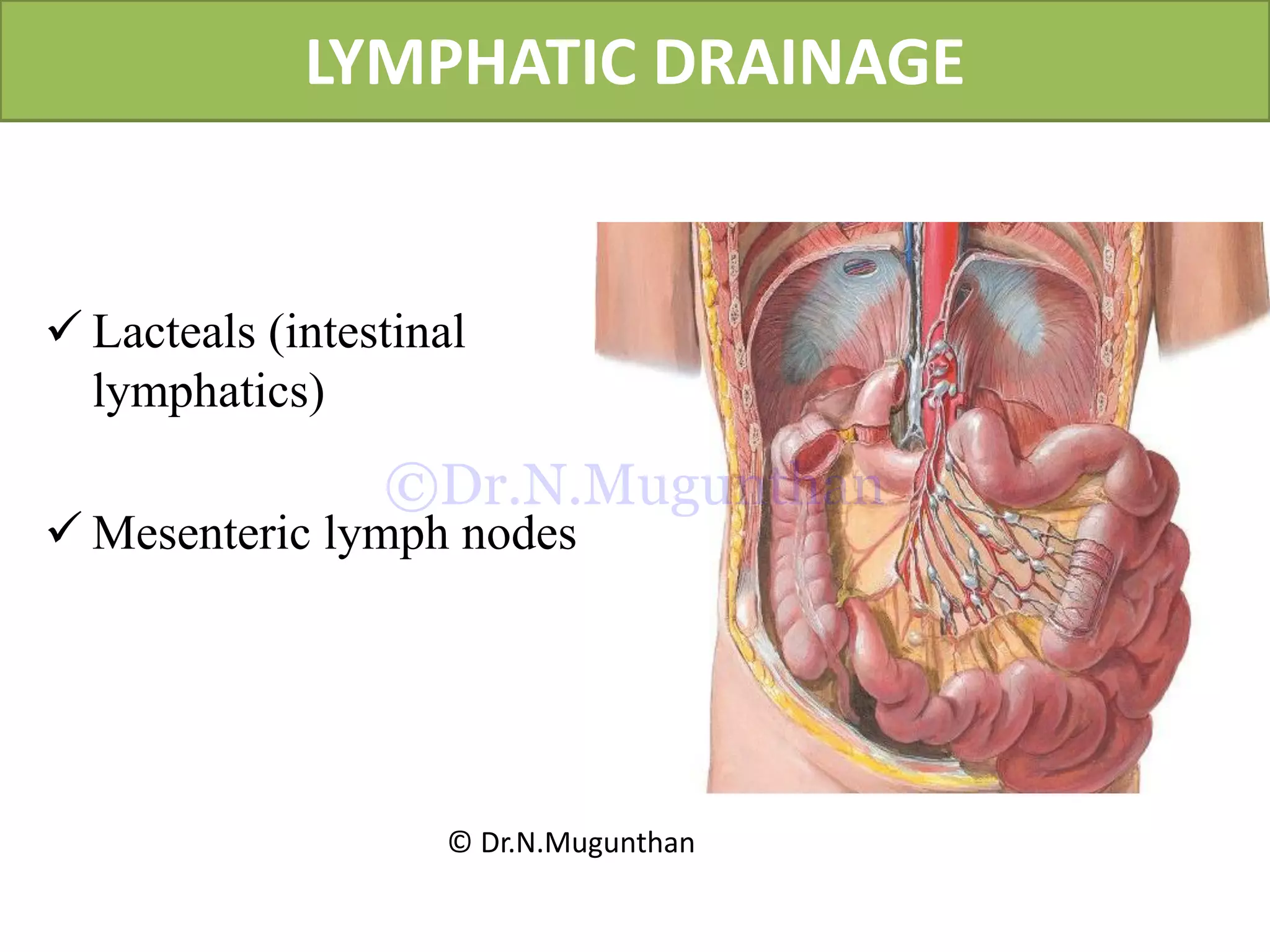
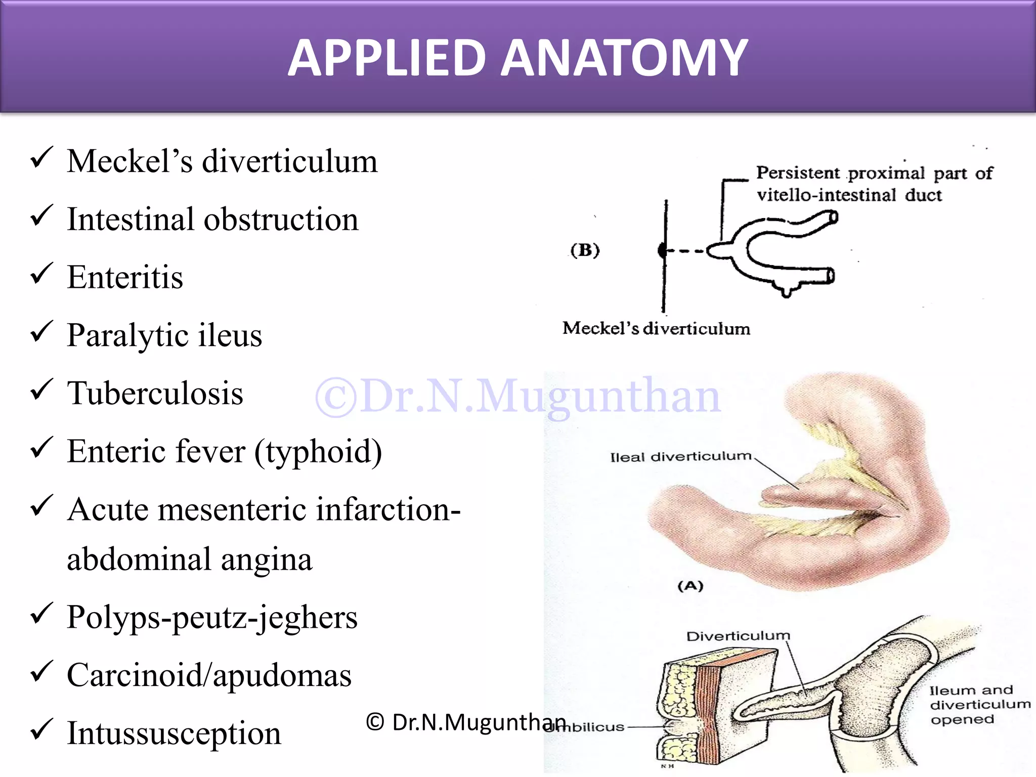
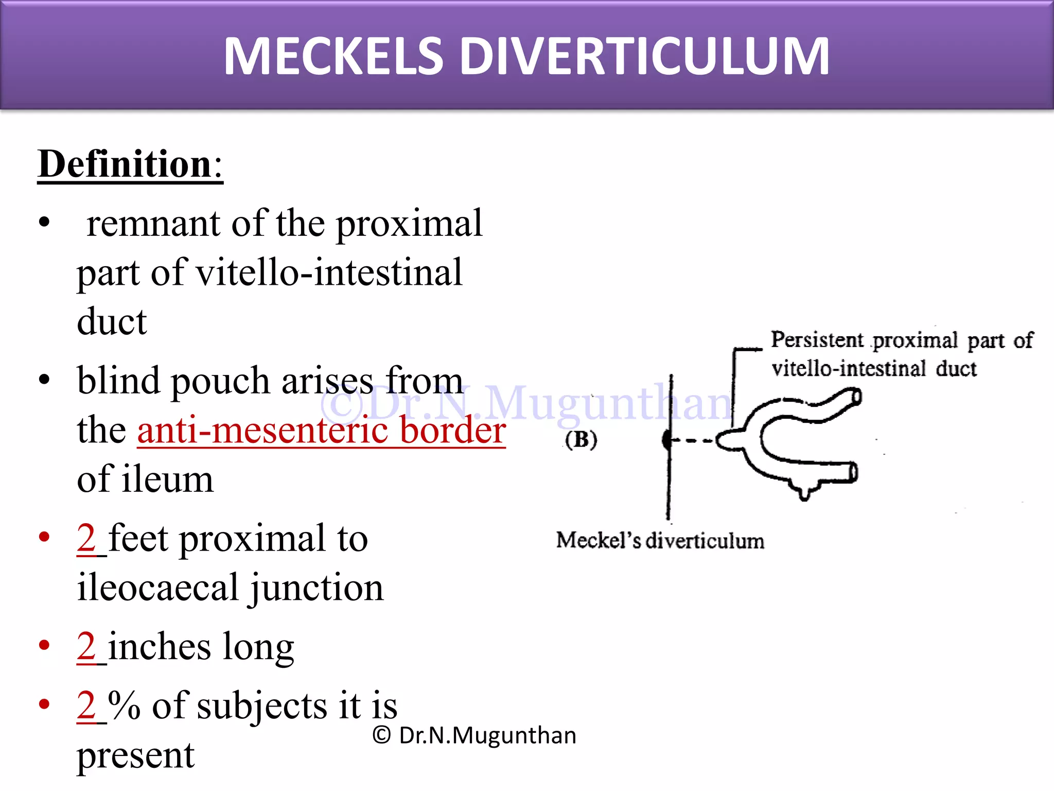
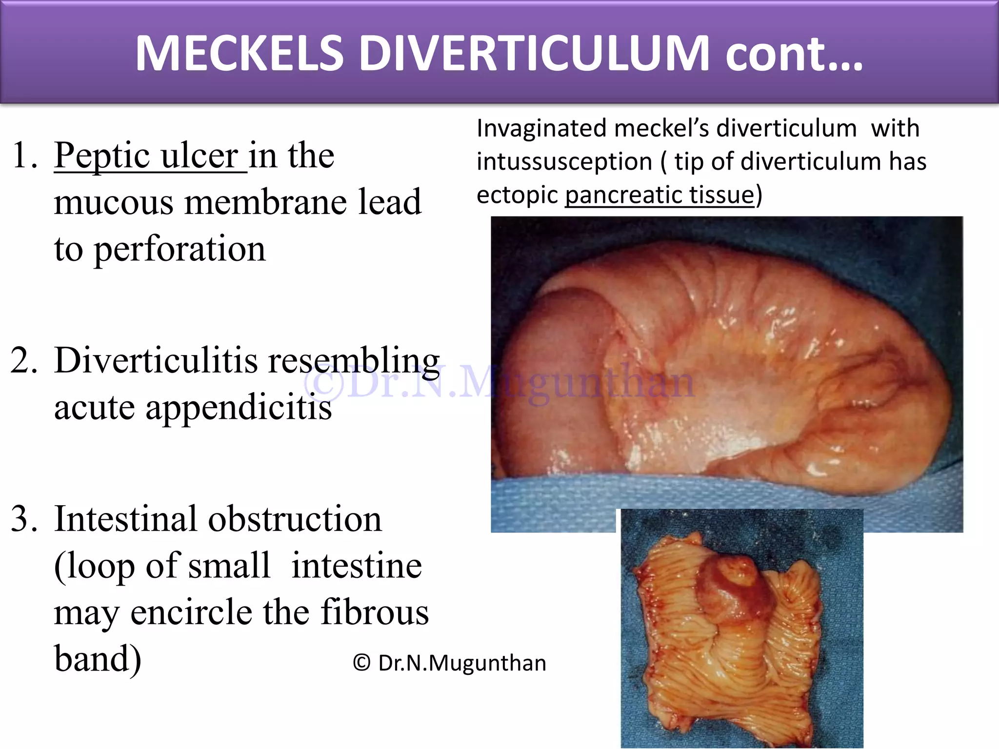
Dr. N.Mugunthan. MS, DNB, MNAMS, PhD.,
Professor
MGMC & RI
OBJECTIVES
Mesentery
Jejunum & ileum
Superior mesenteric artery
Applied anatomy
© Dr.N.Mugunthan
©Dr.N.Mugunthan
MESENTERY
 Peritoneal fold
 Broad fan shaped
 Suspends jejunum and
ileum from posterior
abdominal wall
 Support
 Conveys nutrition &
innervates the gut
 Consists of 2 layers -
from greater sac
© Dr.N.Mugunthan
©Dr.N.Mugunthan
MESENTERY
© Dr.N.Mugunthan
©Dr.N.Mugunthan
BORDERS OF MESENTERY
• Vertebral border or
attached border or root
• Intestinal or free border
© Dr.N.Mugunthan
©Dr.N.Mugunthan
FREE BORDER
• Frilled border
• 20 feet long
• Separates to
enclose jejunum
& ileum
© Dr.N.Mugunthan
©Dr.N.Mugunthan
ROOT OF THE MESENTERY
• Attached border
• 6 inches length
Extent:
• From the left side of L2
at duodenojejunal flexure
to the right sacro-iliac
joint at the ileo-caecal
junction
© Dr.N.Mugunthan
©Dr.N.Mugunthan
STRUCTURES CROSSED BY THE ROOT
• 3rd part of duodenum
• Abdominal aorta
• IVC
• Rt. Gonadal vessels
• Rt.Ureter and psoas major
• Rt.Genito femoral nerve
• Rt.Sacro-iliac joint
© Dr.N.Mugunthan
©Dr.N.Mugunthan
RELATIONS OF MESENTERY
• Rt.Surface -covers front
and sides of ascending
colon
• Rt.paracolic gutter
• Lt.Surface - covers front
and sides of descending
colon
• Lt.para colic gutter
• Above-pancreas
• Below-sigmoid colon
© Dr.N.Mugunthan
©Dr.N.Mugunthan
CONTENTS
• Jejunum and ileum
• Superior mesenteric
vessels
• Autonomic nerves
• Lymphatics -lacteals
and mesenteric nodes
• Loose areolar tissue
• Fat
© Dr.N.Mugunthan
©Dr.N.Mugunthan
APPLIED ANATOMY
• Superior mesenteric
artery syndrome
• Mesenteric adenitis
• Mesenteric cyst
© Dr.N.Mugunthan
©Dr.N.Mugunthan
JEJUNUM & ILEUM
Definition:
• Mobile part of small
intestine
• Suspended from the
posterior abdominal
wall by mesentery
© Dr.N.Mugunthan
©Dr.N.Mugunthan
Extension:
• Extends from the duodeno-
jejunal flexure to the ileo-
caecal junction
• Length - 6 meters or 20 feet
• Jejunum -8 feet
• Ileum -12 feet
2/5th of small intestine may
be removed surgically
without any physiological
disturbance
JEJUNUM & ILEUM cont…
© Dr.N.Mugunthan
©Dr.N.Mugunthan
DIFFERENCES BETWEEN JEJUNUM & ILEUM
S.No Jejunum Ileum
1. Location Upper part of the peritoneal
cavity
Lower part of the peritoneal cavity
2. General
appearance
Wider , thicker walled and
reddish
Narrower, thin walled and paler
3. Attachment of
mesentery
To the posterior abdominal wall
above and to the left of the aorta
Below and to the right of the
aorta.
4. Arterial arcades Only one or two arcades / vasa
recta is long
Three or four or even more
arcades/vasa recta is short
5. Translucent
windows
Translucent windows are clearly
seen (less fat)
Translucent windows are not
clearly seen (more fat)
6. Interior a. Plicae circularis – larger &
more numerous
b. Villi-longer and more
numerous.
c. Payer’s patches -absent
A.Plicae circularis – smaller & less
numerous
B. Shorter and less numerous
C. Payer’s patches -present© Dr.N.Mugunthan
©Dr.N.Mugunthan
© Dr.N.Mugunthan
©Dr.N.Mugunthan
Jejunal branches Ileal branches
Few arterial arcades & long vasa recta More arterial arcades & short vasa recta
© Dr.N.Mugunthan
©Dr.N.Mugunthan
STRUCTURE OF JEJUNUM & ILEUM
4 coats (from without
inwards)
1. Serous coat
2. Muscular coat
3. Submucous coat
4. Mucous coat
a. muscularis mucosa
b. lamina propria
c. epithelium © Dr.N.Mugunthan
©Dr.N.Mugunthan
© Dr.N.Mugunthan
©Dr.N.Mugunthan
SUPERIOR MESENTERIC ARTERY
• Artery of mid gut
Origin:
• From abdominal aorta @ L1
Course & relations:
• Downwards, forwards and to the right
infront of
• Uncinate process of pancreas 3rd part
of duodenum,
• IVC
• Rt.Psoas
• Rt.Ureter
• Runs along the root of mesentery to
rt.Iliac fossa.
Termination:
• Ends by joining with its ileocolic
branch. © Dr.N.Mugunthan
©Dr.N.Mugunthan
Branches:
• Inferior pancreatico-
duodenal artery
• Jejunal branches (5-6)
• Ileal branches (6-8)
• Ileo-colic artery
o Ant.caecal, post.caecal &
appendicular artery
• Rt.colic artery.
• Middle colic artery.
SUPERIOR MESENTERIC ARTERY cont…
© Dr.N.Mugunthan
©Dr.N.Mugunthan
© Dr.N.Mugunthan
©Dr.N.Mugunthan
Summary of SMA:
• SMA –artery of mid gut
• Supplies entire small gut
except the proximal part of
duodenum upto the ampulla
of vater
• Supplies large gut upto the
junction of rt.2/3rd and left
1/3rd of the transverse colon
• Also supplies lower part of
head of the pancreas.
SUPERIOR MESENTERIC ARTERY cont…
© Dr.N.Mugunthan
©Dr.N.Mugunthan
SUPERIOR MESENTERIC VEIN
 Drains the small intestine,
caecum,ascending and transverse
colon
Formation:
• By union of tributaries from
terminal ileum, caecum &
appendix
Course & relations:
• Ascends to the right of SMA
• Passes anterior to rt.ureter, IVC,
3rd duodenum, uncinate process of
pancreas
Termination:
• Joins with splenic vein to form
the portal vein @ behind the neck
of pancreas © Dr.N.Mugunthan
©Dr.N.Mugunthan
Tributaries of SMV:
• Jejunal veins
• Ileal veins
• Ileocolic vein
• Rt.Colic vein
• Middle colic vein
• Rt.Gastro-epiploic vein
• Pancreatico-duodenal
veins
SUPERIOR MESENTERIC VEIN cont…
© Dr.N.Mugunthan
©Dr.N.Mugunthan
NERVE SUPPLY
• Extrinsic component –
sympathetic &
parasympathetic
• Intrinsic component –
myenteric plexus
(Auerbach’s plexus) &
submucous
plexus(Meissner’s plexus)
© Dr.N.Mugunthan
©Dr.N.Mugunthan
LYMPHATIC DRAINAGE
 Lacteals (intestinal
lymphatics)
 Mesenteric lymph nodes
© Dr.N.Mugunthan
©Dr.N.Mugunthan
APPLIED ANATOMY
 Meckel’s diverticulum
 Intestinal obstruction
 Enteritis
 Paralytic ileus
 Tuberculosis
 Enteric fever (typhoid)
 Acute mesenteric infarction-
abdominal angina
 Polyps-peutz-jeghers
 Carcinoid/apudomas
 Intussusception © Dr.N.Mugunthan
©Dr.N.Mugunthan
© Dr.N.Mugunthan
©Dr.N.Mugunthan
MECKELS DIVERTICULUM
Definition:
• remnant of the proximal
part of vitello-intestinal
duct
• blind pouch arises from
the anti-mesenteric border
of ileum
• 2 feet proximal to
ileocaecal junction
• 2 inches long
• 2 % of subjects it is
present
© Dr.N.Mugunthan
©Dr.N.Mugunthan
MECKELS DIVERTICULUM cont…
1. Peptic ulcer in the
mucous membrane lead
to perforation
2. Diverticulitis resembling
acute appendicitis
3. Intestinal obstruction
(loop of small intestine
may encircle the fibrous
band)
Invaginated meckel’s diverticulum with
intussusception ( tip of diverticulum has
ectopic pancreatic tissue)
© Dr.N.Mugunthan
©Dr.N.Mugunthan
SUMMARY
Mesentery
Root of mesentery & structures crossed by it
Superior mesenteric artery
Branches
Applied anatomy
Meckel’s diverticulum
© Dr.N.Mugunthan
©Dr.N.Mugunthan
Thank u 4 Listening

Mesentery, jejunum, ileum & superior mesenteric artery -PDF Lecture Notes -Dr.N.Mugunthan

  • 1.
    Dr. N.Mugunthan. MS,DNB, MNAMS, PhD., Professor MGMC & RI
  • 2.
    OBJECTIVES Mesentery Jejunum & ileum Superiormesenteric artery Applied anatomy © Dr.N.Mugunthan ©Dr.N.Mugunthan
  • 3.
    MESENTERY  Peritoneal fold Broad fan shaped  Suspends jejunum and ileum from posterior abdominal wall  Support  Conveys nutrition & innervates the gut  Consists of 2 layers - from greater sac © Dr.N.Mugunthan ©Dr.N.Mugunthan
  • 4.
  • 5.
    BORDERS OF MESENTERY •Vertebral border or attached border or root • Intestinal or free border © Dr.N.Mugunthan ©Dr.N.Mugunthan
  • 6.
    FREE BORDER • Frilledborder • 20 feet long • Separates to enclose jejunum & ileum © Dr.N.Mugunthan ©Dr.N.Mugunthan
  • 7.
    ROOT OF THEMESENTERY • Attached border • 6 inches length Extent: • From the left side of L2 at duodenojejunal flexure to the right sacro-iliac joint at the ileo-caecal junction © Dr.N.Mugunthan ©Dr.N.Mugunthan
  • 8.
    STRUCTURES CROSSED BYTHE ROOT • 3rd part of duodenum • Abdominal aorta • IVC • Rt. Gonadal vessels • Rt.Ureter and psoas major • Rt.Genito femoral nerve • Rt.Sacro-iliac joint © Dr.N.Mugunthan ©Dr.N.Mugunthan
  • 9.
    RELATIONS OF MESENTERY •Rt.Surface -covers front and sides of ascending colon • Rt.paracolic gutter • Lt.Surface - covers front and sides of descending colon • Lt.para colic gutter • Above-pancreas • Below-sigmoid colon © Dr.N.Mugunthan ©Dr.N.Mugunthan
  • 10.
    CONTENTS • Jejunum andileum • Superior mesenteric vessels • Autonomic nerves • Lymphatics -lacteals and mesenteric nodes • Loose areolar tissue • Fat © Dr.N.Mugunthan ©Dr.N.Mugunthan
  • 11.
    APPLIED ANATOMY • Superiormesenteric artery syndrome • Mesenteric adenitis • Mesenteric cyst © Dr.N.Mugunthan ©Dr.N.Mugunthan
  • 12.
    JEJUNUM & ILEUM Definition: •Mobile part of small intestine • Suspended from the posterior abdominal wall by mesentery © Dr.N.Mugunthan ©Dr.N.Mugunthan
  • 13.
    Extension: • Extends fromthe duodeno- jejunal flexure to the ileo- caecal junction • Length - 6 meters or 20 feet • Jejunum -8 feet • Ileum -12 feet 2/5th of small intestine may be removed surgically without any physiological disturbance JEJUNUM & ILEUM cont… © Dr.N.Mugunthan ©Dr.N.Mugunthan
  • 14.
    DIFFERENCES BETWEEN JEJUNUM& ILEUM S.No Jejunum Ileum 1. Location Upper part of the peritoneal cavity Lower part of the peritoneal cavity 2. General appearance Wider , thicker walled and reddish Narrower, thin walled and paler 3. Attachment of mesentery To the posterior abdominal wall above and to the left of the aorta Below and to the right of the aorta. 4. Arterial arcades Only one or two arcades / vasa recta is long Three or four or even more arcades/vasa recta is short 5. Translucent windows Translucent windows are clearly seen (less fat) Translucent windows are not clearly seen (more fat) 6. Interior a. Plicae circularis – larger & more numerous b. Villi-longer and more numerous. c. Payer’s patches -absent A.Plicae circularis – smaller & less numerous B. Shorter and less numerous C. Payer’s patches -present© Dr.N.Mugunthan ©Dr.N.Mugunthan
  • 15.
  • 16.
    Jejunal branches Ilealbranches Few arterial arcades & long vasa recta More arterial arcades & short vasa recta © Dr.N.Mugunthan ©Dr.N.Mugunthan
  • 17.
    STRUCTURE OF JEJUNUM& ILEUM 4 coats (from without inwards) 1. Serous coat 2. Muscular coat 3. Submucous coat 4. Mucous coat a. muscularis mucosa b. lamina propria c. epithelium © Dr.N.Mugunthan ©Dr.N.Mugunthan
  • 18.
  • 19.
    SUPERIOR MESENTERIC ARTERY •Artery of mid gut Origin: • From abdominal aorta @ L1 Course & relations: • Downwards, forwards and to the right infront of • Uncinate process of pancreas 3rd part of duodenum, • IVC • Rt.Psoas • Rt.Ureter • Runs along the root of mesentery to rt.Iliac fossa. Termination: • Ends by joining with its ileocolic branch. © Dr.N.Mugunthan ©Dr.N.Mugunthan
  • 20.
    Branches: • Inferior pancreatico- duodenalartery • Jejunal branches (5-6) • Ileal branches (6-8) • Ileo-colic artery o Ant.caecal, post.caecal & appendicular artery • Rt.colic artery. • Middle colic artery. SUPERIOR MESENTERIC ARTERY cont… © Dr.N.Mugunthan ©Dr.N.Mugunthan
  • 21.
  • 22.
    Summary of SMA: •SMA –artery of mid gut • Supplies entire small gut except the proximal part of duodenum upto the ampulla of vater • Supplies large gut upto the junction of rt.2/3rd and left 1/3rd of the transverse colon • Also supplies lower part of head of the pancreas. SUPERIOR MESENTERIC ARTERY cont… © Dr.N.Mugunthan ©Dr.N.Mugunthan
  • 23.
    SUPERIOR MESENTERIC VEIN Drains the small intestine, caecum,ascending and transverse colon Formation: • By union of tributaries from terminal ileum, caecum & appendix Course & relations: • Ascends to the right of SMA • Passes anterior to rt.ureter, IVC, 3rd duodenum, uncinate process of pancreas Termination: • Joins with splenic vein to form the portal vein @ behind the neck of pancreas © Dr.N.Mugunthan ©Dr.N.Mugunthan
  • 24.
    Tributaries of SMV: •Jejunal veins • Ileal veins • Ileocolic vein • Rt.Colic vein • Middle colic vein • Rt.Gastro-epiploic vein • Pancreatico-duodenal veins SUPERIOR MESENTERIC VEIN cont… © Dr.N.Mugunthan ©Dr.N.Mugunthan
  • 25.
    NERVE SUPPLY • Extrinsiccomponent – sympathetic & parasympathetic • Intrinsic component – myenteric plexus (Auerbach’s plexus) & submucous plexus(Meissner’s plexus) © Dr.N.Mugunthan ©Dr.N.Mugunthan
  • 26.
    LYMPHATIC DRAINAGE  Lacteals(intestinal lymphatics)  Mesenteric lymph nodes © Dr.N.Mugunthan ©Dr.N.Mugunthan
  • 27.
    APPLIED ANATOMY  Meckel’sdiverticulum  Intestinal obstruction  Enteritis  Paralytic ileus  Tuberculosis  Enteric fever (typhoid)  Acute mesenteric infarction- abdominal angina  Polyps-peutz-jeghers  Carcinoid/apudomas  Intussusception © Dr.N.Mugunthan ©Dr.N.Mugunthan
  • 28.
  • 29.
    MECKELS DIVERTICULUM Definition: • remnantof the proximal part of vitello-intestinal duct • blind pouch arises from the anti-mesenteric border of ileum • 2 feet proximal to ileocaecal junction • 2 inches long • 2 % of subjects it is present © Dr.N.Mugunthan ©Dr.N.Mugunthan
  • 30.
    MECKELS DIVERTICULUM cont… 1.Peptic ulcer in the mucous membrane lead to perforation 2. Diverticulitis resembling acute appendicitis 3. Intestinal obstruction (loop of small intestine may encircle the fibrous band) Invaginated meckel’s diverticulum with intussusception ( tip of diverticulum has ectopic pancreatic tissue) © Dr.N.Mugunthan ©Dr.N.Mugunthan
  • 31.
    SUMMARY Mesentery Root of mesentery& structures crossed by it Superior mesenteric artery Branches Applied anatomy Meckel’s diverticulum © Dr.N.Mugunthan ©Dr.N.Mugunthan
  • 32.
    Thank u 4Listening