
Recommended
More Related Content
Similar to Embryonic Development of Stomach, Liver, Pancreas and Gallbladder
Similar to Embryonic Development of Stomach, Liver, Pancreas and Gallbladder (20)
Recently uploaded
Recently uploaded (20)
Embryonic Development of Stomach, Liver, Pancreas and Gallbladder
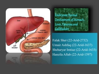
- 1. EmbryonicNormal Development of Stomach, Liver, Pancreas and Gallbladder Falak Sher (22-Arid-2732) Umair Ashfaq (22-Arid-1637) Sheharyar Imtiaz (22-Arid-1633) Hanzila Aftab (22-Arid-1597)
- 2. EMBRYONIC NORMAL DEVELOPMENT OF STOMACH Falak Sher (22-ARID-2732)
- 3. Common Steps of Stomach Development In early embryological development, stomach can be seen as a fusiform dilation of caudal part of the foregut . It is attached to the dorsal abdominal wall by the dorsal abdominal mesogastrium , and to the ventral wall by the ventral mesogastrium
- 4. Cont... Dorsal region of the stomach grows at faster rate than ventral region. Due to this, it changes morphologically, resulting in the formation of a greater dorsal curvature and lesser ventral curvature .At cranial aspect of greater curvature, the growth give rise to the fundus of a simple Stomach.
- 5. Rotations of Stomach Stomach undergoes two rotations during its development. 1-Longitudinal Rotation 2- Antero-Posterior Rotation
- 6. Longitudinal Rotation(1st Rotation) Stomach rotates clockwise at the angle of 90⁰ in such a way that left side moves to the ventral position and right side moves to the dorsal position. At this stage of development, Stomach is C-Shaped, flattened dorso-ventrally with greater curvature and lesser curvature.
- 7. Antero-Posterior Rotation (2nd Rotation) Stomach moves through an angle of 45⁰ in such a way that cardiac region moves slightly downward and left side while pyloric region moves upward and right side. Hence, stomach gets its final position and size in this way
- 8. Bovine Stomach Development The bovine stomach undergoes a complex process of embryonic development, resulting in the formation of several distinct regions that play different roles in the digestion of food. The development of the bovine stomach can be broadly divided into several stages, including gastrulation, the formation of the primitive gut tube, and the differentiation of the stomach regions.
- 9. Overview of Embryonic Development of Bovine Stomach Rumen: The rumen is the largest compartment of the bovine stomach. It develops from the dorsal and left side of the foregut. The rumen plays a crucial role in the fermentation of ingested plant
- 10. Reticulum: The reticulum is located caudal to the rumen and forms adjacent to it. It develops from the ventral part of the foregut. The reticulum functions in the regurgitation and remastication of food, aiding in the breakdown of plant material.
- 11. Omasum: The omasum develops from the caudal part of the foregut and lies to the right of the rumen. It functions primarily in the absorption of water, electrolytes, and some fermentation products.
- 12. Abomasum: The abomasum, often referred to as the "true stomach," is the most caudal region of the bovine stomach. It develops from the ventral part of the foregut. The abomasum secretes gastric juices and enzymes, allowing for the breakdown of proteins and further digestion.
- 13. Throughout embryonic development, the different stomach regions continue to grow and differentiate. After birth, the bovine stomach undergoes further changes in structure and function to accommodate the transition from a milk-based diet to a diet consisting primarily of plant material.
- 19. Embryology of Liver Organogenesis the development of the organs take place from the third to the eighth week during Embryogenesis. The derivatives or portions of the body that develop from the Endoderm germ layer include parts of the digestive tract, the respiratory tract, the urinary tract and the several internal organs.
- 20. Development of Liver The liver develops as a hollow ventral diverticulum from the caudal region of the foregut. The diverticulum divides into two parts: Cranial (hepatic) Caudal (cystic)
- 21. Cont... The gallbladder, bile duct and liver begin to develop 4th week of embryogenesis as a ventral bud from the most caudal aspect of foregut. This bud is called Hepatic Diverticulum and it grows between the layers of Ventral mesentry.
- 22. Components of Liver Formation 1 • Primitive Endoderm • Foregut-Midgut junction 2 • Septum Transversum • Hepatic Diverticulum • Cystic Primordium • Gallbladder 3 • Common bile duct • Hepatic Ducts • Liver/ Gallbladder
- 23. Cont... The Hepatic bud penetrates the Septum Transversum. The Mesenchyme of Septum Transversum induces this Endoderm to proliferate to branch and to form the Glandular Epithelium of the liver. The portion of Hepatic Diverticulum continues to function as the Drainage duct of the liver and the branch from this duct produces the Gallbladder.
- 24. Cont... The Hepatic Endodermal cells undergo a morphological transition from columnar to Pseudostratified resulting in thickening into the early Liver bud. Hepatic Stellate cells are derived from Mesenchyme.
- 25. Embryological Development of various organs
- 26. Cont... After migration of Hepatoblasts into the Septum transversum mesenchyme, the hepatic architecture begins to be established, with Liver sinusoids and bile canaliculi appearing. The liver bud separates into the lobes.
- 27. Cont... The left Umbilical vein becomes the Ductus venosus and the right Vitelline vein becomes the Portal vein. The bipotential hepatoblasts begin differentiating into Biliary epithelial cells and hepatocytes.
- 28. . The cells from cords (Line of cells) which interact with Vitelline and Umbilical veins in Septum Transversum to form the Hepatic Sinusoids. Hepatopoietic cells, Kuffer cells and Connective tissue from Septum Transversum.
- 29. Functions of Liver From 2nd month of Gestation, embryonic liver starts forming blood. Bile production starts at 12th week and give dark colour to the meconium. The liver’s initial embryonic function is cardiovascular as it is a Vascular connection between Placental vessels and Heart.
- 30. EMBRYONIC NORMAL DEVELOPMENT OF PANCREAS Sheharyar Imtiaz (22-Arid-1633)
- 31. Pancreas The pancreas is J-shaped, lobulated, soft, retroperitoneal organ. Its size is about 12-15cm. It is located transversely, a bit obliquely on the posterior abdominal wall behind the stomach. Its weight in adults is about 70-110 g.
- 33. Cont... Its development depends upon the pancreas specific genes which then decides the development of pancreas in different time periods. Its development period varies from species to species. Like in Human , it is developed just prior 26 days of gestation. Like in different ruminants it is formed in the third trimester.
- 34. The pancreas originates from two separate endoderm diverticula, each of which elongates, branches, and then forms acini in typical glandular fashion. one diverticulum arises ventrally as a bud of the hepatic diverticulum, it forms the pancreatic duct and right lobe of the pancreas the other diverticulum arises dorsally from the duodenum (minor duodenal papilla) and forms the accessory pancreatic duct and the left lobe of the pancreas
- 36. o As the right and left lobes cross one another during development, they fuse to from the body of the pancreas; also, the duct systems anastomose to form a common drainage system. o The endocrine (islet) cells of the pancreas also develop from the endoderm of the diverticula.
- 39. Two outpouchings of the Endodermal linings of the Duodenum Ventral Bud >> Lower part of the head >> Uncinated Process Dorsal Bud >> Upper part of the head >> Neck >> Body & Tail Summary : Development of Pancreas Week 7 to20- Pancreatic hormones secretion increases, small amount maternal insulin. Week 10- Glucagon (alpha) differentiate first, Somatostatin (delta), insulin (beta) cells differentiate, insulin secretion begins. Week 15- Glucagon detectable in fetal Plasma.
- 40. EMBRYONIC NORMAL DEVELOPMENT OF GALLBLADDER Hanzila Aftab (22-ARID-1597)
- 41. Definition of the Gallbladder In vertebrates, the gallbladder, also known as the cholecyst, is a small hollow organ where bile is stored and concentrated before it is released into the small intestine. In humans, the pear- shaped gallbladder lies beneath the liver, although the structure and position of the gallbladder can vary significantly among animal species
- 42. Cont... It receives and stores bile, produced by the liver, via the common hepatic duct, and releases it via the common bile duct into the duodenum, where the bile helps in the digestion of fats.
- 43. Structure of the Gallbladder The gallbladder is a hollow organ that sits in a shallow depression below the right lobe of the liver, which is grey-blue in life. In adults, the gallbladder measures approximately 7 to 10 centimeters (2.8 to 3.9 inches) in length and 4 centimetres (1.6 in) in diameter when fully distended. The gallbladder has a capacity of about 50 millilitres (1.8 imperial fluid ounces)
- 44. Cont... The gallbladder is shaped like a pear, with its tip opening into the cystic duct. The gallbladder is divided into three sections: the fundus, body, and neck. The fundus is the rounded base, angled so that it faces the abdominal wall. The body lies in a depression in the surface of the lower liver. The neck tapers and is continuous with the cystic duct, part of the biliary tree.
- 47. Embryonic Development of Gallbladder The gallbladder develops from an endodermal outpouching of the embryonic gut tube. Early in development, the human embryo has three germ layers and abuts an embryonic yolk sac. During the second week of embryogenesis, as the embryo grows, it begins to surround and envelop portions of this sac.
- 48. Cont... The enveloped portions form the basis for the adult gastrointestinal tract. Sections of this foregut begin to differentiate into the organs of the gastrointestinal tract, such as the esophagus, stomach, and intestines.
- 49. Cont... During the fourth week of embryological development, the stomach rotates. The stomach, originally lying in the midline of the embryo, rotates so that its body is on the left. This rotation also affects the part of the gastrointestinal tube immediately below the stomach, which will go on to become the duodenum.
- 50. By the end of the fourth week, the developing duodenum begins to spout a small outpouching on its right side, the hepatic diverticulum, which will go on to become the biliary tree. Just below this is a second outpouching, known as the cystic diverticulum, that will eventually develop into the gallbladder
- 52. Gallbladder Removal A cholecystectomy is a procedure in which the gallbladder is removed. It may be removed because of recurrent gallstones and is considered an elective procedure. A cholecystectomy may be an open procedure, or a laparoscopic one.
- 53. In the surgery, the gallbladder is removed from the neck to the fundus, and so bile will drain directly from the liver into the biliary tree. About 30 percent of patients may experience some degree of indigestion following the procedure, although severe complications are much rarer. About 10 percent of surgeries lead to a chronic condition of postcholecystectomy syndrome.
- 54. Other Animals Most vertebrates have gallbladders, but the form and arrangement of the bile ducts may vary considerably. In many species, for example, there are several separate ducts running to the intestine, rather than the single common bile duct found in humans. Several species of Mammals (including Horses, Deer, Rats and Laminoids), several species of birds lampreys and all invertebrates do not have a gallbladder.
- 55. Cont... The bile from several species of bears is used in traditional Chinese medicine; bile bears are kept alive in captivity while their bile is extracted, in an industry characterized by animal cruelty.