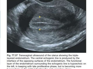
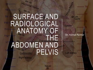
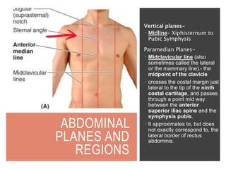
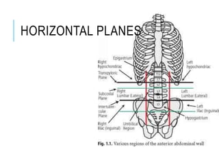
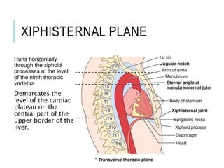
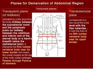
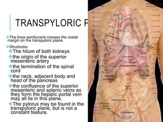
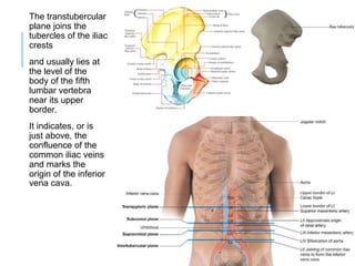
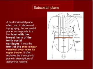
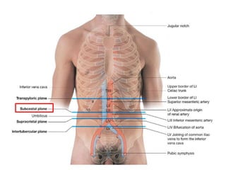
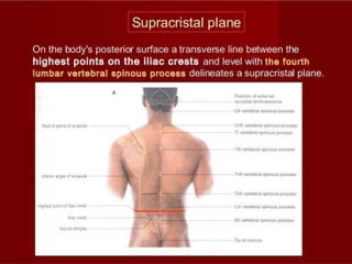
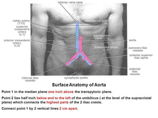
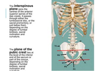
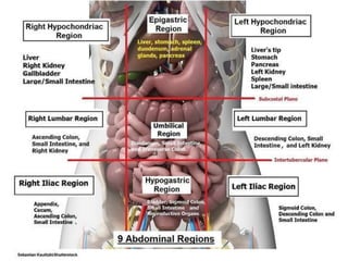
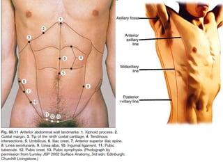
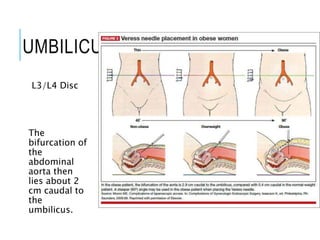
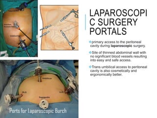
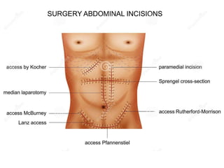
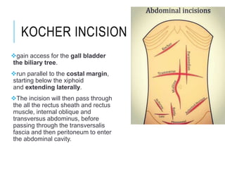
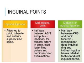
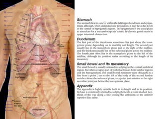
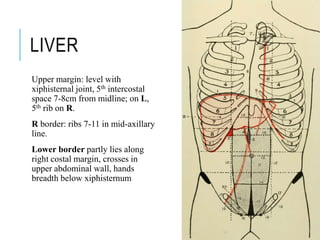
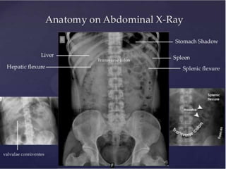
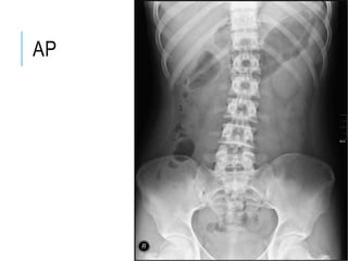
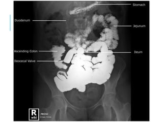
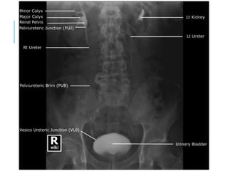
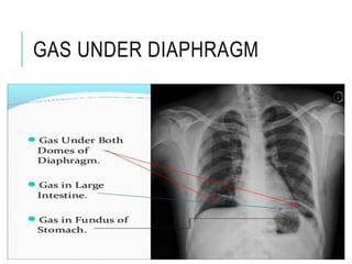
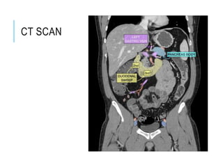
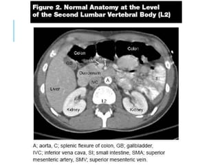
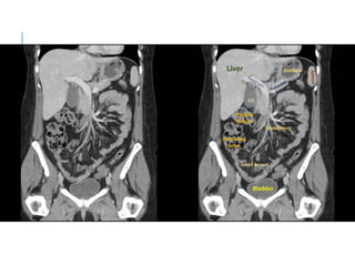
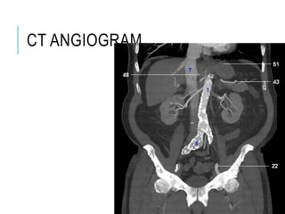
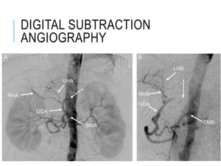
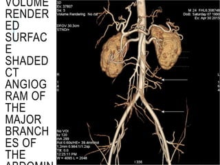
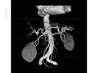
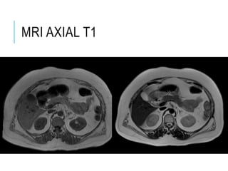
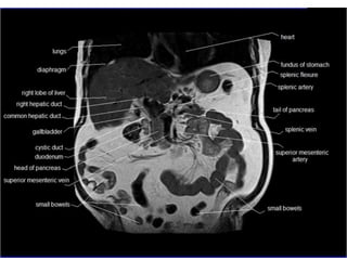
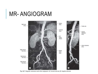
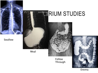
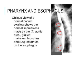
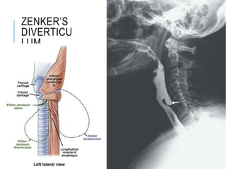
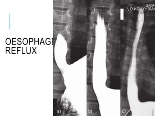
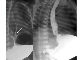
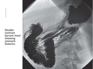
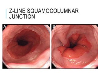
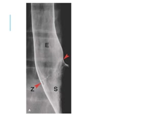
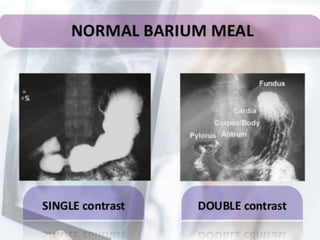
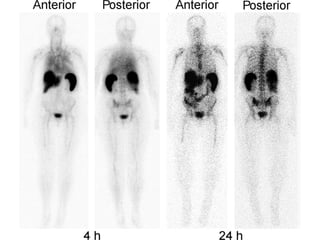
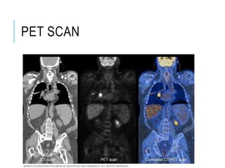
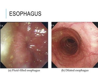
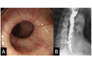
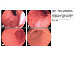
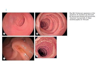
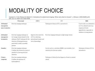
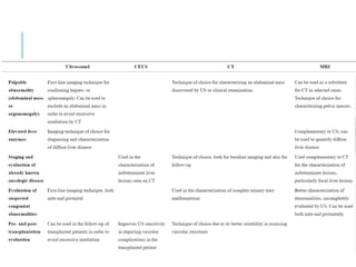
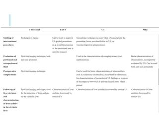
SURFACE AND
RADIOLOGICAL
ANATOMY OF
THE
ABDOMEN AND
PELVIS
Dr. Komal Parmar
ABDOMINAL
PLANES AND
REGIONS
Vertical planes-
 Midline- Xiphisternum to
Pubic Symphysis
Paramedian Planes-
 Midclavicular line (also
sometimes called the lateral
or the mammary line).- the
midpoint of the clavicle
 crosses the costal margin just
lateral to the tip of the ninth
costal cartilage, and passes
through a point mid way
between the anterior
superior iliac spine and the
symphysis pubis.
 It approximates to, but does
not exactly correspond to, the
lateral border of rectus
abdominis.
HORIZONTAL PLANES
XIPHISTERNAL PLANE
Runs horizontally
through the xiphoid
processes at the level
of the ninth thoracic
vertebra
Demarcates the
level of the cardiac
plateau on the
central part of the
upper border of the
liver.
TRANSPYLORIC PLANE
The linea semilunaris crosses the costal
margin on the transpyloric plane.
Structures-
The hilum of both kidneys
the origin of the superior
mesenteric artery
the termination of the spinal
cord
the neck, adjacent body and
head of the pancreas
the confluence of the superior
mesenteric and splenic veins as
they form the hepatic portal vein
may all lie in this plane.
The pylorus may be found in the
transpyloric plane, but is not a
constant feature.
The transtubercular
plane joins the
tubercles of the iliac
crests
and usually lies at
the level of the
body of the fifth
lumbar vertebra
near its upper
border.
It indicates, or is
just above, the
confluence of the
common iliac veins
and marks the
origin of the inferior
vena cava.
The interspinous
plane joins the
centres of the anterior
superior spines of the
iliac crests. It passes
through either the
lumbosacral disc, or the
sacral promontory, or
just below them,
depending on the
degree of lumbar
lordosis, sacral
inclination and
curvature.
The plane of the
pubic crest lies at
the level of the inferior
end of the sacrum or
part of the coccyx,
depending on the
degree of lumbar
lordosis, sacral
inclination and
ABDOMINAL REGIONS
SOFT TISSUE
LANDMARKS
UMBILICUS
L3/L4 Disc
The
bifurcation of
the
abdominal
aorta then
lies about 2
cm caudal to
the
umbilicus.
LAPAROSCOPI
C SURGERY
PORTALS
primary access to the peritoneal
cavity during laparoscopic surgery.
Site of thinnest abdominal wall with
no significant blood vessels resulting
into easy and safe access.
Trans umbilical access to peritoneal
cavity is also cosmetically and
ergonomically better.
RECTUS ABDOMINIS
Rectus Abdominis
Linea Alba
Linea Semilunaris
Median vs
Paramedian
Incisions-
Incidence of early
complications was
more in paramedian
incision but midline
incision is associated
with more chances of
development of
incisional hernia.
KOCHER INCISION
gain access for the gall bladder
the biliary tree.
run parallel to the costal margin,
starting below the xiphoid
and extending laterally.
The incision will then pass through
the all the rectus sheath and rectus
muscle, internal oblique and
transversus abdominus, before
passing through the transversalis
fascia and then peritoneum to enter
the abdominal cavity.
CHEVRON AND
MERCEDEZ BENZ
INCISIONS
MC BURNEY’S= GRIDION
INCISION
LANZ
The position of the
incision is based
upon the location of
the McBurney
point, which is a
point one third of the
distance from the
anterior superior iliac
spine (ASIS) to the
umbilicus.
Place the incision
(1.5-5.0 cm in length,
depending on the
patient's age)
between the first
third and the second
third of the distance
from the ASIS to the
umbilicus,
PFANNELSTEIL
INCISION
A Pfannenstiel
incision or pubic
incision is a type
of abdominal
surgical incision
that allows access
to the abdomen.
It is used for
gynecologic and
orthopedics
surgeries, and it is
the most common
method for
performing
Caesarian sections
INGUINAL POINTS
INTRA-
ABDOMINAL
VISCERA
LIVER
Upper margin: level with
xiphisternal joint, 5th intercostal
space 7-8cm from midline; on L,
5th rib on R.
R border: ribs 7-11 in mid-axillary
line.
Lower border partly lies along
right costal margin, crosses in
upper abdominal wall, hands
breadth below xiphisternum
PANCREAS
The surface projection of
the head of the pancreas
lies within the duodenal
curve.
The neck lies in the
transpyloric plane, behind
the pylorus in the midline.
The body passes obliquely
up and to the left for
approximately 10 cm, its
left part lying a little above
the transpyloric plane.
The tail lies a little above
and to the left of the
intersection of the
transpyloric and left lateral
planes.
KIDNEY
Anterior: The right kidney lies 1.25 cm lower than
the left.
On the anterior surface, the centre of the hilum lies in
the transpyloric plane 5 cm from the midline and
slightly medial to the tip of the ninth costal cartilage.
From the hilum, the outline of the anterior surface can
be drawn 11 cm long and 4.5 cm broad, the upper pole
being 2.5 cm, and the lower 7.5 cm, from the midline
Posterior- the posterior surface, the centre of the
hilum lies opposite the lower border of the spinous
process of the first lumbar vertebra and 5 cm from the
midline.
The outline of the posterior surface can be traced
similarly to the anterior surface.
The lower pole is 2.5 cm above the summit of the iliac
crest.
The kidneys are 2.5 cm lower in the standing than in
the supine
ABDOMINAL AORTA
Supracristal Plane
Interspinous plane
RADIOLOGY OF
ABDOMEN
SALIVARY GLANDS
CT, MRI
SIALOGRA
PHY
injection of a small
amount of contrast
medium into the
salivary duct of a
single gland,
followed by
routine X-
ray projections
ABDOMINAL X-
RAY
AP
PA
GAS UNDER DIAPHRAGM
CT SCAN
CT ANGIOGRAM
DIGITAL SUBTRACTION
ANGIOGRAPHY
VOLUME
RENDER
ED
SURFAC
E
SHADED
CT
ANGIOG
RAM OF
THE
MAJOR
BRANCH
ES OF
THE
MRI AXIAL T1
MR- ANGIOGRAM
BARIUM STUDIES
Swallow
Meal
Follow
Through
Enema
PHARYNX AND ESOPHAGUS
ZENKER’S
DIVERTICU
LUM
OESOPHAGEAL
REFLUX
Double
contrast
barium meal
showing
stomach
features.
Z-LINE SQUAMOCOLUMNAR
JUNCTION
Z- Line
ENTEROCLYSIS: SMALL
BOWEL ENEMA
BARIUM
FOLLOW
THROUGH
BARIUM ENEMA
BARIUM
ENEMA
PERITONEUM
PERITONEAL CAVITY
PERITONITIS
INTESTINA
L
OBSTRUCT
ION
Air Fluid Levels
PET SCAN
ENDOSCOPY
ESOPHAGUS
LIVER
ERCP (ENDOSCOPIC
RETROGRADE CHOLANGIO-
PANCREATOGRAPHY)
KIDNEY AND URETERS
MODALITY OF CHOICE
Caraiani C, Yi D, Petresc B, Dietrich C. Indications for abdominal imaging: When and what to choose?. J Ultrason. 2020;20(80):e43-
e54. doi:10.15557/JoU.2020.0008
Abdomen  surface marking and radiology

Abdomen surface marking and radiology