During the fourth to eighth weeks of human development:
- All major external and internal structures are established. The main organ systems start to develop and the embryo takes on a distinctly human appearance.
- Rapid differentiation of tissues and organs occurs. Exposure to teratogens during this period can cause major birth defects.
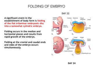
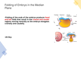
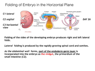
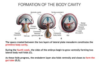
- Folding of the embryo occurs in the median and horizontal planes, establishing the basic body form with a head, trunk and tail region.