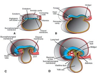
DEVELOPMENT OF DIGESTIVE
SYSTEM
DR SHEHU H.
Vocabulary
• Competency: The ability of a cell or group of cells to
respond to a particular signal
• Endoderm: layer of post-gastrulation embryo that gives rise
to the epithelium of the gut tube and gut-derived organs
• Mesoderm/Mesenchyme: layer of post-gastrulation
embryo that gives rise to the surrounding tissue of the gut
tube
• Cell-cell communication: Passage of signals between two
groups of cells (usually endoderm and mesoderm); these
signals are crucial in the specification of cell fate and
proliferation/differentiation decisions
Gastrulation:
• Epiblast cells migrate
through the primitive
streak.
• Definitive (embryonic)
endoderm cells
displace the
hypoblast.
• Mesoderm spreads
between endoderm
and ectoderm.
Specific regions of the epiblast
migrate through the streak at
different levels and assume
different positions within
the embryo:
Cranial to caudal:
Notochord (n)
Paraxial mesoderm (pm)
Intermediate mesoderm
(im)
*Lateral plate mesoderm
(lpm)
Extraembryonic mesoderm
(eem
Outline
• Introduction
• Foregut
• Midgut
• Hindgut
• Clinical correlates/applied anatomy
Introduction
The alimentary or digestive system develops
between Wk4-8. The system is made up of the
digestive tube (epithelium), with an upper opening
(stomodeum), a lower exit (proctodeum), muscles
& connective tissues as well the digestive glands.
The epithelium & glands of primitive gut of
digestive tube are derived from endoderm. The
stomodeum & proctodeum are part of the tube
derived from ectoderm. The muscles, connective
tissue & other layers of the wall of tract are derived
from splanchnic mesoderm.
With the establishment of the head and tail
folds, part of the cavity of the definitive yolk sac
is enclosed within the embryo to form the
primitive gut.
The primitive gut is in free communication with
the rest of the yolk sac. The part of the gut
cranial to this communication is the foregut; the
part caudal to the communication is the
hindgut, while the intervening
part is the midgut.
The communication between foregut and
midgut is called anterior intestinal portal which
is represented in the adult by the termination of
bile duct in the second part of duodenum.
The communication between the midgut and
hindgut is called posterior intestinal portal
which corresponds in the adult to the junction
of right two-thirds with the left one-third of
transverse colon
Parts of the primitive gut
The foregut is in the head fold of the embryo. Cranially,
the foregut is separated from the stomodeum by the
buccopharyngeal membrane. The hindgut is in the tail
fold of the embryo. Caudally, the hindgut is separated
from the proctodeum by the cloacal membrane. At a later
stage of development, the buccopharyngeal and cloacal
membranes disappear, and the foregut and hindgut are
in communication with stomodeum and proctodeum
Respectively. Thus, the gut communicates with the
exterior. The midgut during early embryonic
period communicates with the extraembryonic part of
yolk sac via vitellointestinal duct. The vitellointestinal
duct disappears by 5th week of development.
The digestive tube is itself is divided into 3 parts:
1. Foregut (upper part)
2. Midgut (middle part)
3. Hindgut (lower part)
Foregut: results from the incorporation i.e.
integration of the yolk sac due to head folding
• Midgut: results from the incorporate of the
yolk sac due to lateral folding (translaminal
lateral)
• Hindgut: results from the incorporation of the
yolk sac due to tail folding.
The developing endoderm (yellow) is initially open to the yolk
sac (the cardiac region is initially most anterior)…
Cranio-caudal folding at both ends of the embryo and lateral
folding at the sides of the embryo bring the endoderm inside
and form the gut tube
Folding creates the anterior and posterior intestinal
portals (foregut and hindgut, respectively)
Juxtaposition of ectoderm and endoderm at:
Oropharyngeal (buccopharyngeal) membrane -
future mouth. Cloacal membrane - future anus
Foregut
The foregut extends from the buccopharyngeal membrane to
the duodenum where the liver bud arises (the anterior
intestinal portal)
• The arterial supply of foregut is the coeliac axis
• The derivatives of the foregut are:
1. Pharynx (primordial)
2. Esophagus
3. Stomach
4. Duodenum, proximal to the opening of the bile duct (i.e. 1st
part +1st ½ of 2nd part up to major duodenal papilla)
5. Pancreas
6. Liver & biliary system/ apparatus (hepatic ducts, gallbladder
& bile duct).
Development of Esophagus
• It develops immediately caudal to primordial pharynx as a short tube
which elongates rapidly because of growth & descent of the heart &
lungs reaching its final relative length by the week 7.
• The epithelium (derived from endoderm) of the tube proliferates &
temporarily obliterates (partly or completely) its lumen. It is followed
by apoptosis of the cells & recanalization by the end of embryonic
period (Wk8). Thus by Wk9, the lumen is open again. This occlusion &
recanalization process occurs THROUGHOUT the tube (esophagus to
anus) & errors in this process can occur in anywhere along the tube
resulting in stenosis (narrowing of the lumen or even outright
occlusion) in that region.
• The striated muscles in the upper 3rd part develop from
mesenchyme in the caudal pharyngeal arches while the smooth
muscle in the lower part is derived from splanchnic mesoderm
Applied embryology: Congenital abnormality:
a) Esophagus atresia: 1:3000/1:4500. 1/3rd
premature birth. Usually associated with trachea-
esophageal fistula. Causes (i) Deviation of the
tracheoeshageal septum in a posterior direction, (ii)
Failure of recanalization of the esophagus by Wk8
resulting in polyhydraminos.
b) Esophageal stenosis: Distal 1/3rd of esophagus.
Causes (a) Failure to recanalize (b) Failure of blood
vessels to develop which leads to atrophy of
affected segments.
c) Short esophagus: due to failure of the esophagus
to elongate resulting in congenital hiatal hernia
Development of stomach
• The stomach appears initially, as a fusiform dilation
caudal to esophagus by Wk4 having 2 borders (ventral &
dorsal).
• It later enlarges ventrodorsally with the dorsal border
grow faster than the ventral border. The dorsal border
becomes the greater curvature while the ventral the
lesser curvature
• At the same time, it rotates 90 degrees to the right on
its vertical axis in the midline, manoeuvring in a counter-
clockwise ‘’pronating direction’’ resulting in lesser
curvature facing right & greater curvature facing left, the
left surface facing anterior while right surface posteriorly.
This explains why the left vagus nerve innervates the ant.
Surface of the stomach and right vagus nerve post.
Surface.
Initially the two ends (cranial and caudal) of the
stomach lie in the midline. But during rotation:
the cranial end (cardiac orifice) moves to the left
and slightly downward; the caudal end (pylorus)
moves to the right and upward. After rotation,
stomach assumes its final position with its long
axis running from above left to below right; the
pylorus to shift to right and the cardiac orifice to
shift to the left.
Changes in the position and shape of stomach
Congenital abnormality of the stomach
– These are very rare.
– Congenital hypertrophic pyloric stenosis:
Male- 1:150; Female- 1:170. It is hereditary &
common in homozygotic twins.
Development of Duodenum
• Begins in Wk4, its is derived from 2 sources:
(i) Caudal part of foregut & (ii) Cranial part of
midgut, both parts grow very rapidly & form a C-
shaped loop that projects ventrally. The Junction of
the 2 parts is at the apex of the embryonic loop.
• With the development & rotation of the stomach,
the duodenal loop rotates 90 degrees counter
clockwise to the right such that it is convex to the
right & comes to lie largely retroperitoneally.
• Proliferation of the epithelial cells occurs so the
lumen gets obliterated, recanalization occurs by the
end of 8th week
Congenital abnormalities
• Duodenal duplication
• Duodenal stenosis: results from: Non recanalization or
annular pancreas; usually occurs at the 3rd or 4th part of
the duodenal
• Duodenal diverticula: The vitelline duct persistence
• Duodenal atresia: This usually occurs after opening of
the bile duct at the 2nd & 3rd part. Is associated with
(“CAP-D”):
– CVS abnormalities,
– Annular pancreas,
– Prematurity &
– Down syndrome.
Annular pancreas surrounding the duodenum
Midgut
• By Wk5 the midgut is a simple loop suspended from the
dorsal abdominal wall by an elongated dorsal mesentery
• It communicates ventrally with the yolk sac through the
vitelline duct
• The artery of the midgut is the superior mesenteric
artery.
Derivatives of the midgut:
1. Pancreas (lower part of head & uncinate process)
2. Duodenum (distal to the opening of bile duct)
3. Jejunum
4. Ileum
5. Caecum & appendix
6. Ascending colon &
7. Transverse colon (proximal 2/3rd )
Sequential process of development of midgut
(‘Mnenonic: Elijah Hastenly Rotated Ronkes Forms’)
a) Elongation of midgut loop
b) Herniation of midgut loop
c) Rotation of midgut loop
d) Return of midgut loop
e) Fixation of intestines
• Elongation: The midgut elongates, forming a
ventral, U-shaped loop of gut that projects into the
remains of the extraembryonic coelom in the
proximal part of the umbilical cord.
• Herniation & growth: By Wk6, due to no space
in abdomen, the midgut loop projects into the
primitive umbilical cord forming a
physiological umbilical hernia. The apex of
midgut loop communicates with the yolk sac
by a narrow stalk till Wk10, it has (cranial &
caudal limb). The cranial limb grow rapidly to
form duodenum, jejunum & ileum; caudal
limb grows very little developing a swelling
the caecal diverticulum that marks the future
caecum.
• Rotation of midgut loop: It is a 90 degrees counter
clock wise rotation around the axis of the superior
mesenteric artery. This brings the cranial limb (small
intestine) of the midgut loop to the right and the
caudal limb (large intestine) to the left. During rotation,
the cranial limb also elongates and forms intestinal
loops (eg primordia of jejunum and ileum), elongation
of the small intestinal loops continues and the jejunum
and ileum form a number of coiled loops. The large
intestine elongates but does not participate in the
coiling phenomenum.
• Return of midgut loop: The return into the abdomen
occur by Wk12 resultng in a reduction of the umbilical
hernia & a further counter clock wise rotation of 180O.
Possible cause of the return are: (i) increase in the size
of the abdominal cavity & (ii) decrease in the sizes of
the liver & kidney.
The small intestine returns first passing posteriorly
to superior mesenteric artery to occupy the central
& left part of abdomen. This is followed by the large
intestine. The caecum, (widest part) returns last to
be located caudal to the right lobe of the liver &
then descend to lie in the right iliac fossa.
• Fixation of intestines:
The duodenum with the pancreas falls to the right
& becomes plastered to the posterior abdominal
wall, its adjacent layers of peritoneum fuse &
mesenteries disappear, thus becomes
retroperitoneal structures this event is due to the
rotations of stomach & duodenum.
The small intestine (jejunum & ileum) retains
only its fan shaped dorsal mesentery & acquires
a new line of attachment that passes from
duodenojejunal junction obliquely downwards
across to the ileocaecal junction.
The large intestine (ascending & transverse
anterior 2/3rd), during rotation & return of
midgut, the ascending as well as the descending
are retroperitoneal structures, their mesenteries
fuse with the posterior abdominal wall,
ascending colon plastered to the right,
descending colon to the left. The transverse
colon is intraperitoneal retaining its mesentery.
Congenital abnormalities:
• The congenital anomalies involving the midgut are very
common & usually resulting from: (a) incomplete rotation &
(b) incomplete fixation
[Mnemonics: “SUROMI.VGC”
– Stenosis, Atresia & Fistula
– Umbilical hernia
– Congenital umbilical hernia (CUH)- covered by skin
– Rotations: Non rotation, Reversed rotation & Mixed rotation
– Omphalocoel – similar to CUH but covered by amnion
- meckel’s diverticulum
– Intestinal duplication
– Vitelline duct defects: e.g cyst, sinus, fistula, diverticulum,
stenosis atresia
– Gastrochisis
– Caecum: Sub-hepatic Caecum, Mobile Caecum
Errors of rotation. (A) Nonrotation. Coils of small intestine lie
in the right half of the abdomen, and colon in the left half; (B)
Reversed rotation. The duodenum lies anterior to the superior
mesenteric artery, and the colon crosses behind it.
Pancreas
• Pancreas appears as 2 buds, dorsal & ventral, from the caudal part of
foregut region that is developing into the duodenum. The buds grow
within the dorsal & ventral mesenteries respectively
• Dorsal bud is larger, appears first & lies cranial to the smaller ventral
bud, rotation of stomach & duodenum carry the ventral bud dorsally
along with the bile duct. Ventral bud comes to lie posterior to the
dorsal bud & later fuses with it & their ducts anastomose.
• The ducts of ventral bud & distal part of duct of dorsal bud form the
main pancreatic duct that opens on the major duodenal papilla. The
proximal part of duct of the dorsal bud often persists as the accessory
pancreatic duct that opens separately on the minor duodenal papilla
• Dorsal bud forms most of pancreas. Ventral bud gives rise to the
uncinate process & part of the head of pancreas
• Finally pancreas comes to lie retroperitoneally plastered horizontal
along the posterior abdominal wall.
• Note that rotation of the duodenum brings
the ventral and dorsal pancreas together
• Aberrations in this process may result in an
annular pancreas, which can constrict the
duodenum.
• Also, since the dorsal and ventral pancreas
arise by different mechanisms, it’s possible
that one or the other may be absent in the
adult.
Congenital abnormalities of pancreas
• Heterotropic pancreas: when pancreatic tissue is found in
either the stomach, duodenum or in Meckel’s diverticulum
• Annular pancreas: a bifid ventral bud may cause this
condition. It is rare (males more). Leads to duodenal
obstruction
• Meckel’s diverticulum: Is as a result of the persistence of the
diverticulum in the vitelline duct. They are found on the anti-
mesenteric border of the ileum
– It occurs approximately in 2% population
– It is 2 inches in length
– It is 2 feet long from the ileocaecal junction
– It has 2 aberrant tissues (1. Gastric tissue. 2. Pancreatic
tissue
– It therefore mimics 2 clinical conditions: Gastric tissue
(Gastric/Peptic ulcer) & Pancreatic tissue (Pancreatitis).
Development of Liver
• Liver appears in Wk4 as a ventral bud called hepatic
diverticulum/ bud, from the caudal part of foregut. The bud
grows into septum transversum (which is forming the
ventral mesentery in this region).
• Hepatic bud divides into 2 parts: larger cranial part (pars
hepatica) is primordium of liver & smaller caudal part (pars
cystica) gives rise to gall bladder & cystic duct.
• Liver structural origins: (a) Hepatic cords (hepatocytes) &
epithelial lining of the intrahepatic biliary system are
derived from proliferation of endodermal cells of hepatic
buds (b) Hepatic sinusoids are derived from vitelline veins
they anastomose with hepatic cords (c) Kupffer cells &
connective tissue are derived from mesenchyme of septum
transversum
Liver grows rapidly by Wk5-10 & fills a large part of
the abdominal cavity, By Wk8-9 the liver has grown
to about 10% of total body weight (TBW).
• Hematopoeisis begins by Wk6 giving a dark color
to liver & accounts much for the large size of the
liver at Wk8 (neonate liver is 4% of TBW; compared
to adult liver which is 2.5-3.4% of TBW)
• Hepatic cells begin to form bile by Wk12-13.
• Liver divides into equal halves: Initially right & left
lobes are of the same size, later right grows larger.
Left forms caudate & quadrate lobes. In the
definitive liver, caudate & quadrate lobes are
classified as the right lobe but embryological they
are part of left lobe.
Biliary apparatus
• The rounded portion of the pars cystic forms the
gallbladder while the stalk forms the cystic duct
• The tract connecting hepatic & cystic ducts to the
duodenum is the bile duct, which initially
attaches to the ventral aspect of duodenum but
as duodenum rotates, the entrance of the bile
duct is carried around to the dorsal aspect of the
duodenum.
• The ducts become occluded initially, but are later
recanalized
• By Wk13-14, bile pigments enter the duodenum
giving its content a dark green color called
meconeum.
• Congenital abnormalities of the liver are rare & may
include:
– (i) variations in: lobulation, hepatic duct, bile duct,
cystic duct (are common),
– (ii) extra-hepatic biliary atresia
Highlights:
• Wk4 (buds appear)
• Wk5-10 (rapid growth filling abdomen)
• Wk6 (hematopoeisis starts)
• Wk8-9 (10% TBW)
• Wk12-13 (bile)
• Wk13-14 (bile enter duodenum)
• Neonate 4% TBW
• Adult 2.5-3.4% TBW
Hindgut
The hindgut follows the midgut, in the embryo &
extends from the posterior intestinal portal to the cloacal membrane
It is supplied by the inferior mesenteric artery (L3)
Derivatives:
1. Transverse colon (distal 1/3rd)
2. Descending colon
3. Pelvic/sigmoid colon
4. Rectum
5. Anal canal (upper 2/3rd of above the pectinate
(dentate) line
6. Part of the urogenital system (i.e. epithelium of the
bladder & urethra)
Highlight: The anterior intestinal portal is the foregut to
midgut transition while the posterior intestinal portal is
the midgut to hindgut transition.
Cloaca
• Cloaca is an endoderm-lined cavity that is in direct contact
with the surface ectoderm (proctodeum or anal pit) with no
intervening mesoderm.
• The terminal portion of the hindgut enters into the posterior
region of the cloaca -(primitive anorectal canal).
• Cloaca, receives the:
– Allantois ventrally (primitive urogenital sinus) &
– Mesonephric ducts laterally
• Septation of the cloaca
During development, a ridge of mesenchyme, the Urorectal
septum forms a partition between allantois & hindgut. This
septum is a complex of 2 septal systems: (i) Tourneux fold
(central) that migrates downwards to meet & (ii) Rathke folds
(lateral) which arise from the lateral walls.
• By end of Wk7, urorectal septum fuses with
cloaca membrane separating Cloaca into the:
Primitive urogenital sinus (anterior part) &
Anorectal canal (posterior part). The point
where urorectal septum intersects the cloacal
membrane becomes the perineal body.
• Cloaca membrane is divided into (i) a larger
anterior urogenital membrane & (ii) a smaller
posterior anal membrane.
• Development of the hind gut
Anal canal
• By Wk9, proliferations of mesenchyme around the edges of
anal membrane & produces raised prominences called anal
folds forming a shallow depression called anal pit or
proctodeum lined by ectoderm.
• Soon after, the anal membrane, at the bottom of anal pit
ruptures to establish the anal canal, its upper 2/3rd is derived
from hindgut endoderm; lower 1/3rd from the anal pit
ectoderm.
• The junction between the hindgut endoderm & anal pit
ectoderm is indicated by the pectinate line, at the level of the
anal valves. At this line, the epithelium changes from
columnar to stratified squamous epithelium.
• As a result of the different embryologic origins of the upper
& lower parts of the anal canal, the blood vessels, lymphatic
drainages & nerve supply differs in the various parts of the
canal.
• Hilton's white line is slightly below the pectinate
line, a landmark for border between internal &
external anal sphincter muscles
Fixation of the hindgut
• During the rotation of the midgut, the descending
colon is displaced to the left & is fixed in a
retroperitoneal position.
• The sigmoid colon remains intraperitoneal in
nature, but its mesentery is shortened during
fixation of the descending colon.
• The dorsal mesenteries of rectum & anal canal
disappear, as they become retroperitoneal organs.
Clinical correlates of hindgut
• Anal membrane may fail to
breakdown leading to an
imperforate anus
• Hirschsprung disease occurs
when parasympathetic
ganglia are absent in the
walls of the colon. It results
from mutations in RET gene,
a tyrosine kinase receptors
involved in neural crest cell
migration
THANKYOU

DEVELOPMENT OF DIGESTIVE SYSTEM.pptx

  • 1.
  • 2.
    Vocabulary • Competency: Theability of a cell or group of cells to respond to a particular signal • Endoderm: layer of post-gastrulation embryo that gives rise to the epithelium of the gut tube and gut-derived organs • Mesoderm/Mesenchyme: layer of post-gastrulation embryo that gives rise to the surrounding tissue of the gut tube • Cell-cell communication: Passage of signals between two groups of cells (usually endoderm and mesoderm); these signals are crucial in the specification of cell fate and proliferation/differentiation decisions
  • 3.
    Gastrulation: • Epiblast cellsmigrate through the primitive streak. • Definitive (embryonic) endoderm cells displace the hypoblast. • Mesoderm spreads between endoderm and ectoderm.
  • 4.
    Specific regions ofthe epiblast migrate through the streak at different levels and assume different positions within the embryo: Cranial to caudal: Notochord (n) Paraxial mesoderm (pm) Intermediate mesoderm (im) *Lateral plate mesoderm (lpm) Extraembryonic mesoderm (eem
  • 5.
    Outline • Introduction • Foregut •Midgut • Hindgut • Clinical correlates/applied anatomy
  • 6.
    Introduction The alimentary ordigestive system develops between Wk4-8. The system is made up of the digestive tube (epithelium), with an upper opening (stomodeum), a lower exit (proctodeum), muscles & connective tissues as well the digestive glands. The epithelium & glands of primitive gut of digestive tube are derived from endoderm. The stomodeum & proctodeum are part of the tube derived from ectoderm. The muscles, connective tissue & other layers of the wall of tract are derived from splanchnic mesoderm.
  • 7.
    With the establishmentof the head and tail folds, part of the cavity of the definitive yolk sac is enclosed within the embryo to form the primitive gut. The primitive gut is in free communication with the rest of the yolk sac. The part of the gut cranial to this communication is the foregut; the part caudal to the communication is the hindgut, while the intervening part is the midgut.
  • 8.
    The communication betweenforegut and midgut is called anterior intestinal portal which is represented in the adult by the termination of bile duct in the second part of duodenum. The communication between the midgut and hindgut is called posterior intestinal portal which corresponds in the adult to the junction of right two-thirds with the left one-third of transverse colon
  • 9.
    Parts of theprimitive gut
  • 10.
    The foregut isin the head fold of the embryo. Cranially, the foregut is separated from the stomodeum by the buccopharyngeal membrane. The hindgut is in the tail fold of the embryo. Caudally, the hindgut is separated from the proctodeum by the cloacal membrane. At a later stage of development, the buccopharyngeal and cloacal membranes disappear, and the foregut and hindgut are in communication with stomodeum and proctodeum Respectively. Thus, the gut communicates with the exterior. The midgut during early embryonic period communicates with the extraembryonic part of yolk sac via vitellointestinal duct. The vitellointestinal duct disappears by 5th week of development.
  • 11.
    The digestive tubeis itself is divided into 3 parts: 1. Foregut (upper part) 2. Midgut (middle part) 3. Hindgut (lower part) Foregut: results from the incorporation i.e. integration of the yolk sac due to head folding • Midgut: results from the incorporate of the yolk sac due to lateral folding (translaminal lateral) • Hindgut: results from the incorporation of the yolk sac due to tail folding.
  • 12.
    The developing endoderm(yellow) is initially open to the yolk sac (the cardiac region is initially most anterior)… Cranio-caudal folding at both ends of the embryo and lateral folding at the sides of the embryo bring the endoderm inside and form the gut tube
  • 13.
    Folding creates theanterior and posterior intestinal portals (foregut and hindgut, respectively) Juxtaposition of ectoderm and endoderm at: Oropharyngeal (buccopharyngeal) membrane - future mouth. Cloacal membrane - future anus
  • 14.
    Foregut The foregut extendsfrom the buccopharyngeal membrane to the duodenum where the liver bud arises (the anterior intestinal portal) • The arterial supply of foregut is the coeliac axis • The derivatives of the foregut are: 1. Pharynx (primordial) 2. Esophagus 3. Stomach 4. Duodenum, proximal to the opening of the bile duct (i.e. 1st part +1st ½ of 2nd part up to major duodenal papilla) 5. Pancreas 6. Liver & biliary system/ apparatus (hepatic ducts, gallbladder & bile duct).
  • 16.
    Development of Esophagus •It develops immediately caudal to primordial pharynx as a short tube which elongates rapidly because of growth & descent of the heart & lungs reaching its final relative length by the week 7. • The epithelium (derived from endoderm) of the tube proliferates & temporarily obliterates (partly or completely) its lumen. It is followed by apoptosis of the cells & recanalization by the end of embryonic period (Wk8). Thus by Wk9, the lumen is open again. This occlusion & recanalization process occurs THROUGHOUT the tube (esophagus to anus) & errors in this process can occur in anywhere along the tube resulting in stenosis (narrowing of the lumen or even outright occlusion) in that region. • The striated muscles in the upper 3rd part develop from mesenchyme in the caudal pharyngeal arches while the smooth muscle in the lower part is derived from splanchnic mesoderm
  • 18.
    Applied embryology: Congenitalabnormality: a) Esophagus atresia: 1:3000/1:4500. 1/3rd premature birth. Usually associated with trachea- esophageal fistula. Causes (i) Deviation of the tracheoeshageal septum in a posterior direction, (ii) Failure of recanalization of the esophagus by Wk8 resulting in polyhydraminos. b) Esophageal stenosis: Distal 1/3rd of esophagus. Causes (a) Failure to recanalize (b) Failure of blood vessels to develop which leads to atrophy of affected segments. c) Short esophagus: due to failure of the esophagus to elongate resulting in congenital hiatal hernia
  • 20.
    Development of stomach •The stomach appears initially, as a fusiform dilation caudal to esophagus by Wk4 having 2 borders (ventral & dorsal). • It later enlarges ventrodorsally with the dorsal border grow faster than the ventral border. The dorsal border becomes the greater curvature while the ventral the lesser curvature • At the same time, it rotates 90 degrees to the right on its vertical axis in the midline, manoeuvring in a counter- clockwise ‘’pronating direction’’ resulting in lesser curvature facing right & greater curvature facing left, the left surface facing anterior while right surface posteriorly. This explains why the left vagus nerve innervates the ant. Surface of the stomach and right vagus nerve post. Surface.
  • 21.
    Initially the twoends (cranial and caudal) of the stomach lie in the midline. But during rotation: the cranial end (cardiac orifice) moves to the left and slightly downward; the caudal end (pylorus) moves to the right and upward. After rotation, stomach assumes its final position with its long axis running from above left to below right; the pylorus to shift to right and the cardiac orifice to shift to the left.
  • 22.
    Changes in theposition and shape of stomach
  • 23.
    Congenital abnormality ofthe stomach – These are very rare. – Congenital hypertrophic pyloric stenosis: Male- 1:150; Female- 1:170. It is hereditary & common in homozygotic twins.
  • 24.
    Development of Duodenum •Begins in Wk4, its is derived from 2 sources: (i) Caudal part of foregut & (ii) Cranial part of midgut, both parts grow very rapidly & form a C- shaped loop that projects ventrally. The Junction of the 2 parts is at the apex of the embryonic loop. • With the development & rotation of the stomach, the duodenal loop rotates 90 degrees counter clockwise to the right such that it is convex to the right & comes to lie largely retroperitoneally. • Proliferation of the epithelial cells occurs so the lumen gets obliterated, recanalization occurs by the end of 8th week
  • 25.
    Congenital abnormalities • Duodenalduplication • Duodenal stenosis: results from: Non recanalization or annular pancreas; usually occurs at the 3rd or 4th part of the duodenal • Duodenal diverticula: The vitelline duct persistence • Duodenal atresia: This usually occurs after opening of the bile duct at the 2nd & 3rd part. Is associated with (“CAP-D”): – CVS abnormalities, – Annular pancreas, – Prematurity & – Down syndrome.
  • 26.
  • 27.
    Midgut • By Wk5the midgut is a simple loop suspended from the dorsal abdominal wall by an elongated dorsal mesentery • It communicates ventrally with the yolk sac through the vitelline duct • The artery of the midgut is the superior mesenteric artery. Derivatives of the midgut: 1. Pancreas (lower part of head & uncinate process) 2. Duodenum (distal to the opening of bile duct) 3. Jejunum 4. Ileum 5. Caecum & appendix 6. Ascending colon & 7. Transverse colon (proximal 2/3rd )
  • 28.
    Sequential process ofdevelopment of midgut (‘Mnenonic: Elijah Hastenly Rotated Ronkes Forms’) a) Elongation of midgut loop b) Herniation of midgut loop c) Rotation of midgut loop d) Return of midgut loop e) Fixation of intestines • Elongation: The midgut elongates, forming a ventral, U-shaped loop of gut that projects into the remains of the extraembryonic coelom in the proximal part of the umbilical cord.
  • 29.
    • Herniation &growth: By Wk6, due to no space in abdomen, the midgut loop projects into the primitive umbilical cord forming a physiological umbilical hernia. The apex of midgut loop communicates with the yolk sac by a narrow stalk till Wk10, it has (cranial & caudal limb). The cranial limb grow rapidly to form duodenum, jejunum & ileum; caudal limb grows very little developing a swelling the caecal diverticulum that marks the future caecum.
  • 30.
    • Rotation ofmidgut loop: It is a 90 degrees counter clock wise rotation around the axis of the superior mesenteric artery. This brings the cranial limb (small intestine) of the midgut loop to the right and the caudal limb (large intestine) to the left. During rotation, the cranial limb also elongates and forms intestinal loops (eg primordia of jejunum and ileum), elongation of the small intestinal loops continues and the jejunum and ileum form a number of coiled loops. The large intestine elongates but does not participate in the coiling phenomenum. • Return of midgut loop: The return into the abdomen occur by Wk12 resultng in a reduction of the umbilical hernia & a further counter clock wise rotation of 180O. Possible cause of the return are: (i) increase in the size of the abdominal cavity & (ii) decrease in the sizes of the liver & kidney.
  • 31.
    The small intestinereturns first passing posteriorly to superior mesenteric artery to occupy the central & left part of abdomen. This is followed by the large intestine. The caecum, (widest part) returns last to be located caudal to the right lobe of the liver & then descend to lie in the right iliac fossa. • Fixation of intestines: The duodenum with the pancreas falls to the right & becomes plastered to the posterior abdominal wall, its adjacent layers of peritoneum fuse & mesenteries disappear, thus becomes retroperitoneal structures this event is due to the rotations of stomach & duodenum.
  • 33.
    The small intestine(jejunum & ileum) retains only its fan shaped dorsal mesentery & acquires a new line of attachment that passes from duodenojejunal junction obliquely downwards across to the ileocaecal junction. The large intestine (ascending & transverse anterior 2/3rd), during rotation & return of midgut, the ascending as well as the descending are retroperitoneal structures, their mesenteries fuse with the posterior abdominal wall, ascending colon plastered to the right, descending colon to the left. The transverse colon is intraperitoneal retaining its mesentery.
  • 34.
    Congenital abnormalities: • Thecongenital anomalies involving the midgut are very common & usually resulting from: (a) incomplete rotation & (b) incomplete fixation [Mnemonics: “SUROMI.VGC” – Stenosis, Atresia & Fistula – Umbilical hernia – Congenital umbilical hernia (CUH)- covered by skin – Rotations: Non rotation, Reversed rotation & Mixed rotation – Omphalocoel – similar to CUH but covered by amnion - meckel’s diverticulum – Intestinal duplication – Vitelline duct defects: e.g cyst, sinus, fistula, diverticulum, stenosis atresia – Gastrochisis – Caecum: Sub-hepatic Caecum, Mobile Caecum
  • 36.
    Errors of rotation.(A) Nonrotation. Coils of small intestine lie in the right half of the abdomen, and colon in the left half; (B) Reversed rotation. The duodenum lies anterior to the superior mesenteric artery, and the colon crosses behind it.
  • 37.
    Pancreas • Pancreas appearsas 2 buds, dorsal & ventral, from the caudal part of foregut region that is developing into the duodenum. The buds grow within the dorsal & ventral mesenteries respectively • Dorsal bud is larger, appears first & lies cranial to the smaller ventral bud, rotation of stomach & duodenum carry the ventral bud dorsally along with the bile duct. Ventral bud comes to lie posterior to the dorsal bud & later fuses with it & their ducts anastomose. • The ducts of ventral bud & distal part of duct of dorsal bud form the main pancreatic duct that opens on the major duodenal papilla. The proximal part of duct of the dorsal bud often persists as the accessory pancreatic duct that opens separately on the minor duodenal papilla • Dorsal bud forms most of pancreas. Ventral bud gives rise to the uncinate process & part of the head of pancreas • Finally pancreas comes to lie retroperitoneally plastered horizontal along the posterior abdominal wall.
  • 39.
    • Note thatrotation of the duodenum brings the ventral and dorsal pancreas together • Aberrations in this process may result in an annular pancreas, which can constrict the duodenum. • Also, since the dorsal and ventral pancreas arise by different mechanisms, it’s possible that one or the other may be absent in the adult.
  • 40.
    Congenital abnormalities ofpancreas • Heterotropic pancreas: when pancreatic tissue is found in either the stomach, duodenum or in Meckel’s diverticulum • Annular pancreas: a bifid ventral bud may cause this condition. It is rare (males more). Leads to duodenal obstruction • Meckel’s diverticulum: Is as a result of the persistence of the diverticulum in the vitelline duct. They are found on the anti- mesenteric border of the ileum – It occurs approximately in 2% population – It is 2 inches in length – It is 2 feet long from the ileocaecal junction – It has 2 aberrant tissues (1. Gastric tissue. 2. Pancreatic tissue – It therefore mimics 2 clinical conditions: Gastric tissue (Gastric/Peptic ulcer) & Pancreatic tissue (Pancreatitis).
  • 41.
    Development of Liver •Liver appears in Wk4 as a ventral bud called hepatic diverticulum/ bud, from the caudal part of foregut. The bud grows into septum transversum (which is forming the ventral mesentery in this region). • Hepatic bud divides into 2 parts: larger cranial part (pars hepatica) is primordium of liver & smaller caudal part (pars cystica) gives rise to gall bladder & cystic duct. • Liver structural origins: (a) Hepatic cords (hepatocytes) & epithelial lining of the intrahepatic biliary system are derived from proliferation of endodermal cells of hepatic buds (b) Hepatic sinusoids are derived from vitelline veins they anastomose with hepatic cords (c) Kupffer cells & connective tissue are derived from mesenchyme of septum transversum
  • 42.
    Liver grows rapidlyby Wk5-10 & fills a large part of the abdominal cavity, By Wk8-9 the liver has grown to about 10% of total body weight (TBW). • Hematopoeisis begins by Wk6 giving a dark color to liver & accounts much for the large size of the liver at Wk8 (neonate liver is 4% of TBW; compared to adult liver which is 2.5-3.4% of TBW) • Hepatic cells begin to form bile by Wk12-13. • Liver divides into equal halves: Initially right & left lobes are of the same size, later right grows larger. Left forms caudate & quadrate lobes. In the definitive liver, caudate & quadrate lobes are classified as the right lobe but embryological they are part of left lobe.
  • 44.
    Biliary apparatus • Therounded portion of the pars cystic forms the gallbladder while the stalk forms the cystic duct • The tract connecting hepatic & cystic ducts to the duodenum is the bile duct, which initially attaches to the ventral aspect of duodenum but as duodenum rotates, the entrance of the bile duct is carried around to the dorsal aspect of the duodenum. • The ducts become occluded initially, but are later recanalized • By Wk13-14, bile pigments enter the duodenum giving its content a dark green color called meconeum.
  • 46.
    • Congenital abnormalitiesof the liver are rare & may include: – (i) variations in: lobulation, hepatic duct, bile duct, cystic duct (are common), – (ii) extra-hepatic biliary atresia Highlights: • Wk4 (buds appear) • Wk5-10 (rapid growth filling abdomen) • Wk6 (hematopoeisis starts) • Wk8-9 (10% TBW) • Wk12-13 (bile) • Wk13-14 (bile enter duodenum) • Neonate 4% TBW • Adult 2.5-3.4% TBW
  • 47.
    Hindgut The hindgut followsthe midgut, in the embryo & extends from the posterior intestinal portal to the cloacal membrane It is supplied by the inferior mesenteric artery (L3) Derivatives: 1. Transverse colon (distal 1/3rd) 2. Descending colon 3. Pelvic/sigmoid colon 4. Rectum 5. Anal canal (upper 2/3rd of above the pectinate (dentate) line 6. Part of the urogenital system (i.e. epithelium of the bladder & urethra) Highlight: The anterior intestinal portal is the foregut to midgut transition while the posterior intestinal portal is the midgut to hindgut transition.
  • 48.
    Cloaca • Cloaca isan endoderm-lined cavity that is in direct contact with the surface ectoderm (proctodeum or anal pit) with no intervening mesoderm. • The terminal portion of the hindgut enters into the posterior region of the cloaca -(primitive anorectal canal). • Cloaca, receives the: – Allantois ventrally (primitive urogenital sinus) & – Mesonephric ducts laterally • Septation of the cloaca During development, a ridge of mesenchyme, the Urorectal septum forms a partition between allantois & hindgut. This septum is a complex of 2 septal systems: (i) Tourneux fold (central) that migrates downwards to meet & (ii) Rathke folds (lateral) which arise from the lateral walls.
  • 49.
    • By endof Wk7, urorectal septum fuses with cloaca membrane separating Cloaca into the: Primitive urogenital sinus (anterior part) & Anorectal canal (posterior part). The point where urorectal septum intersects the cloacal membrane becomes the perineal body. • Cloaca membrane is divided into (i) a larger anterior urogenital membrane & (ii) a smaller posterior anal membrane.
  • 50.
    • Development ofthe hind gut
  • 51.
    Anal canal • ByWk9, proliferations of mesenchyme around the edges of anal membrane & produces raised prominences called anal folds forming a shallow depression called anal pit or proctodeum lined by ectoderm. • Soon after, the anal membrane, at the bottom of anal pit ruptures to establish the anal canal, its upper 2/3rd is derived from hindgut endoderm; lower 1/3rd from the anal pit ectoderm. • The junction between the hindgut endoderm & anal pit ectoderm is indicated by the pectinate line, at the level of the anal valves. At this line, the epithelium changes from columnar to stratified squamous epithelium. • As a result of the different embryologic origins of the upper & lower parts of the anal canal, the blood vessels, lymphatic drainages & nerve supply differs in the various parts of the canal.
  • 52.
    • Hilton's whiteline is slightly below the pectinate line, a landmark for border between internal & external anal sphincter muscles Fixation of the hindgut • During the rotation of the midgut, the descending colon is displaced to the left & is fixed in a retroperitoneal position. • The sigmoid colon remains intraperitoneal in nature, but its mesentery is shortened during fixation of the descending colon. • The dorsal mesenteries of rectum & anal canal disappear, as they become retroperitoneal organs.
  • 53.
    Clinical correlates ofhindgut • Anal membrane may fail to breakdown leading to an imperforate anus • Hirschsprung disease occurs when parasympathetic ganglia are absent in the walls of the colon. It results from mutations in RET gene, a tyrosine kinase receptors involved in neural crest cell migration
  • 55.