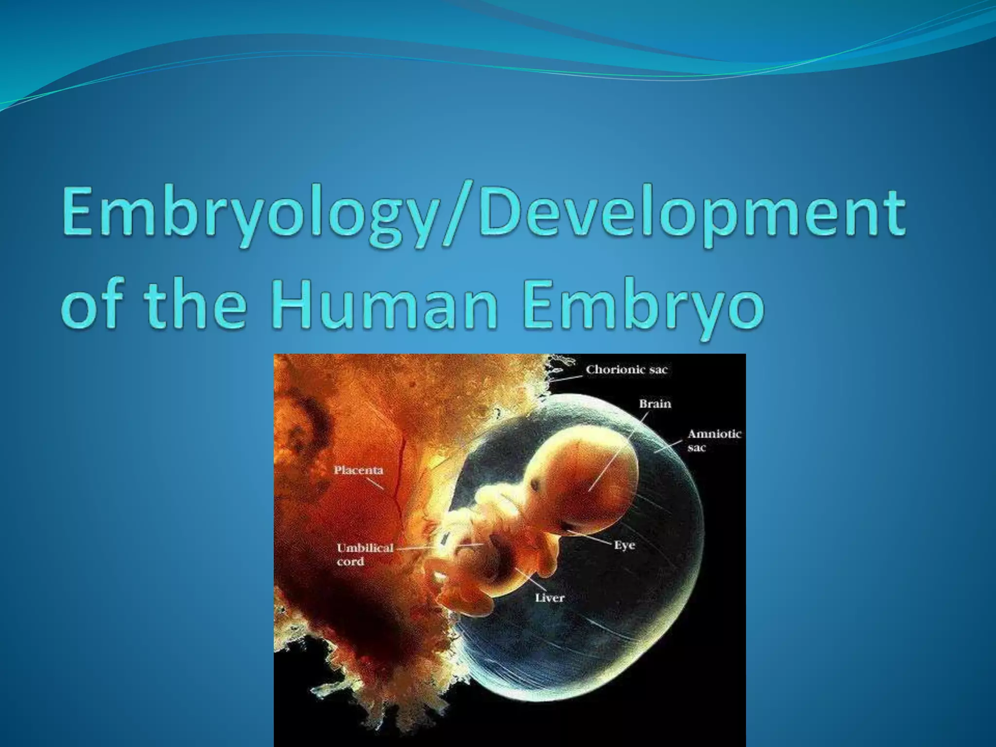
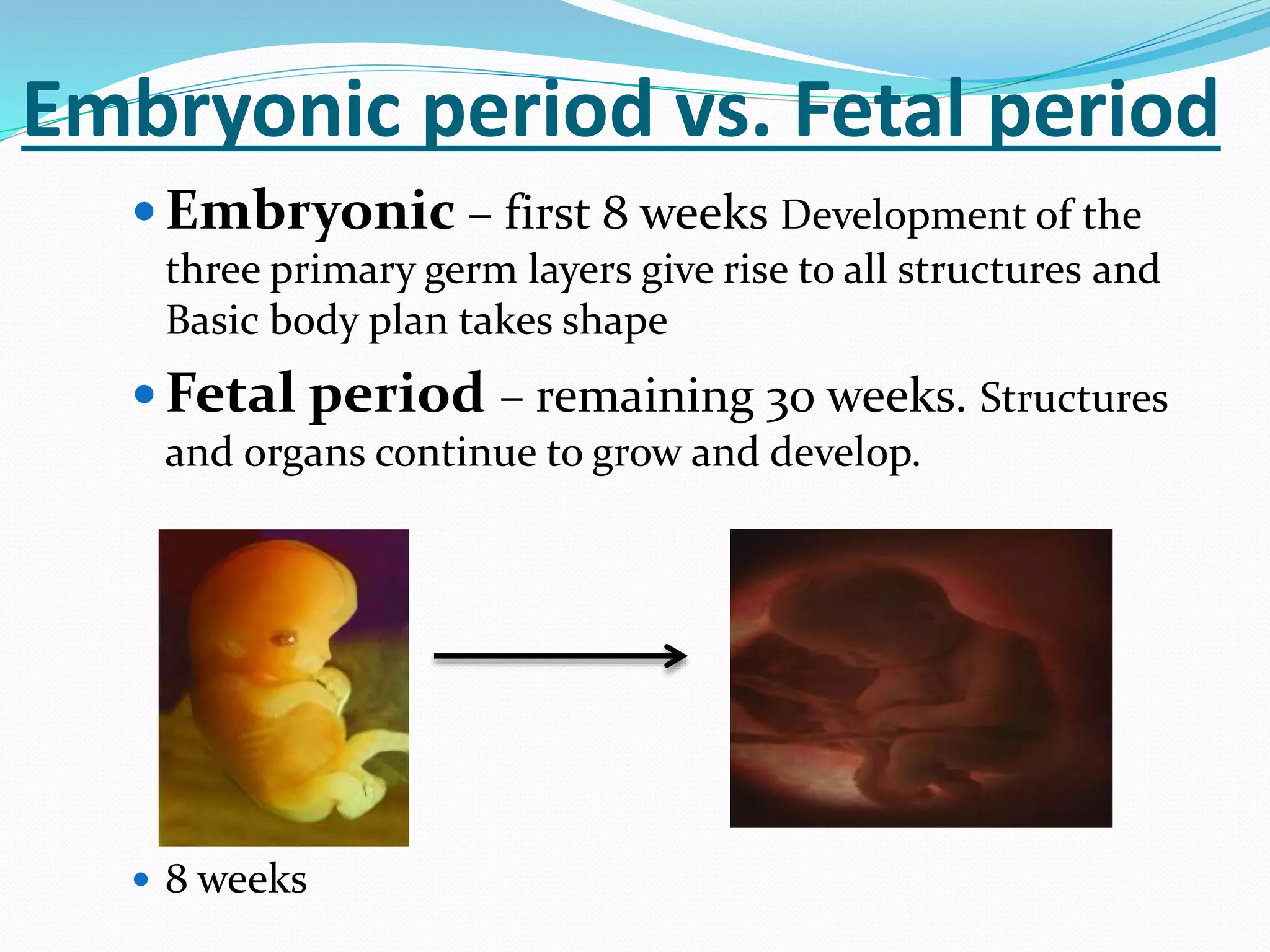
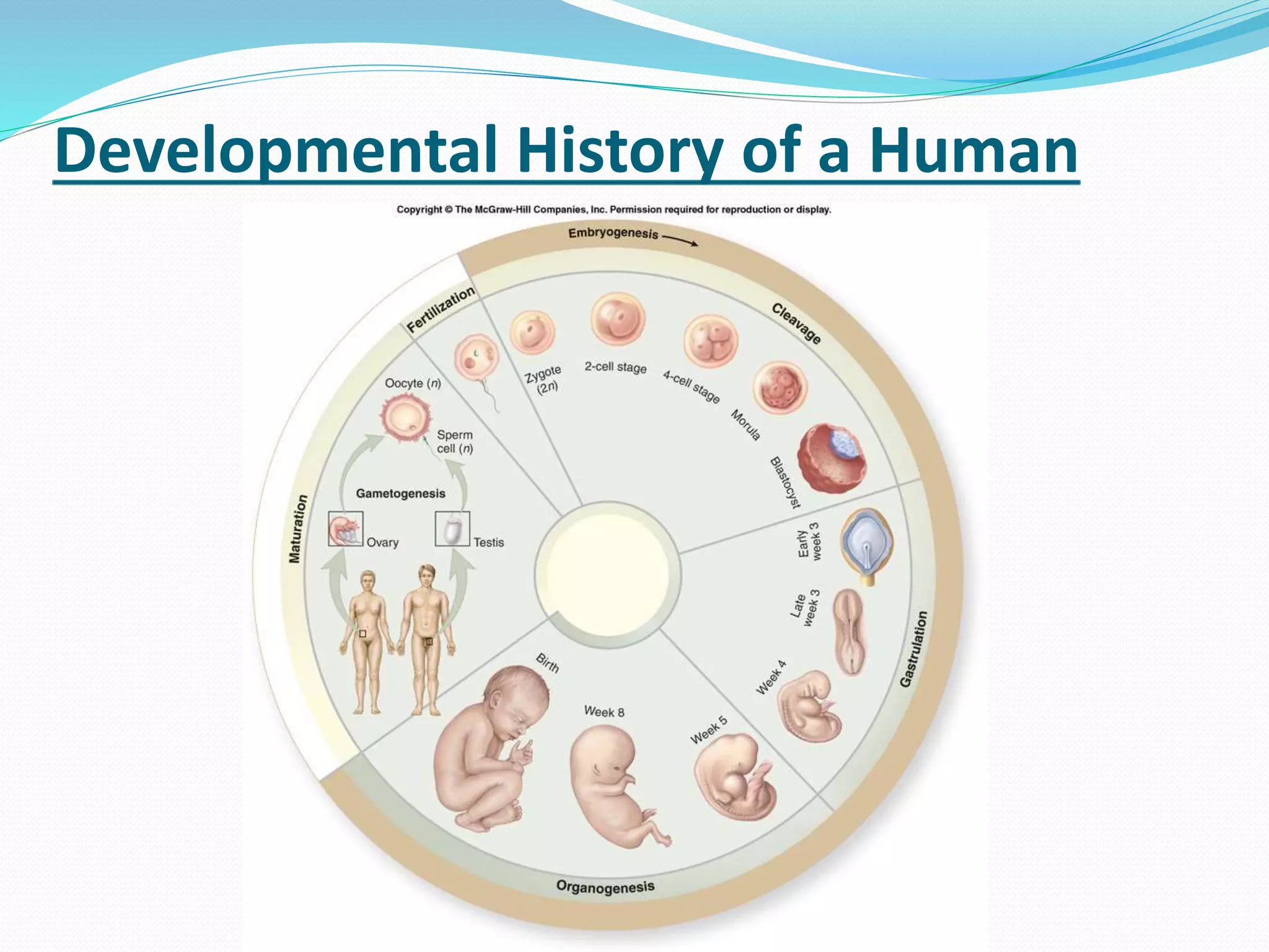
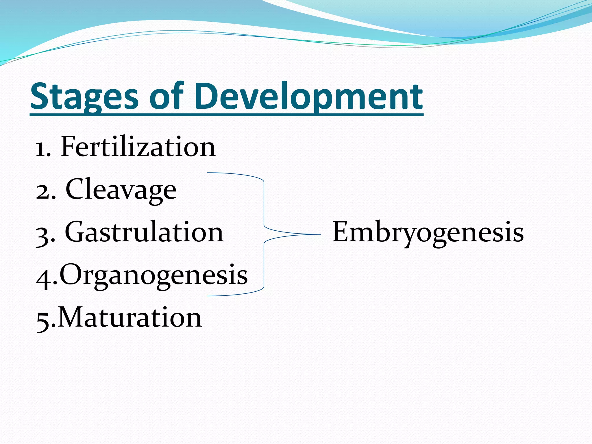
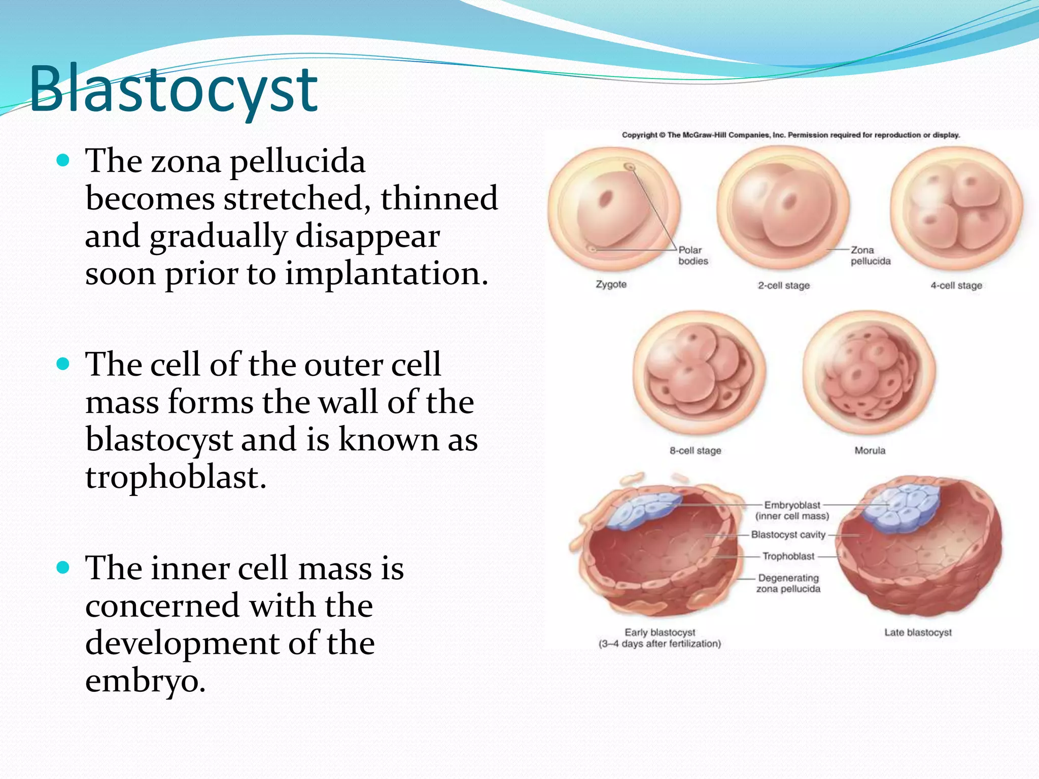
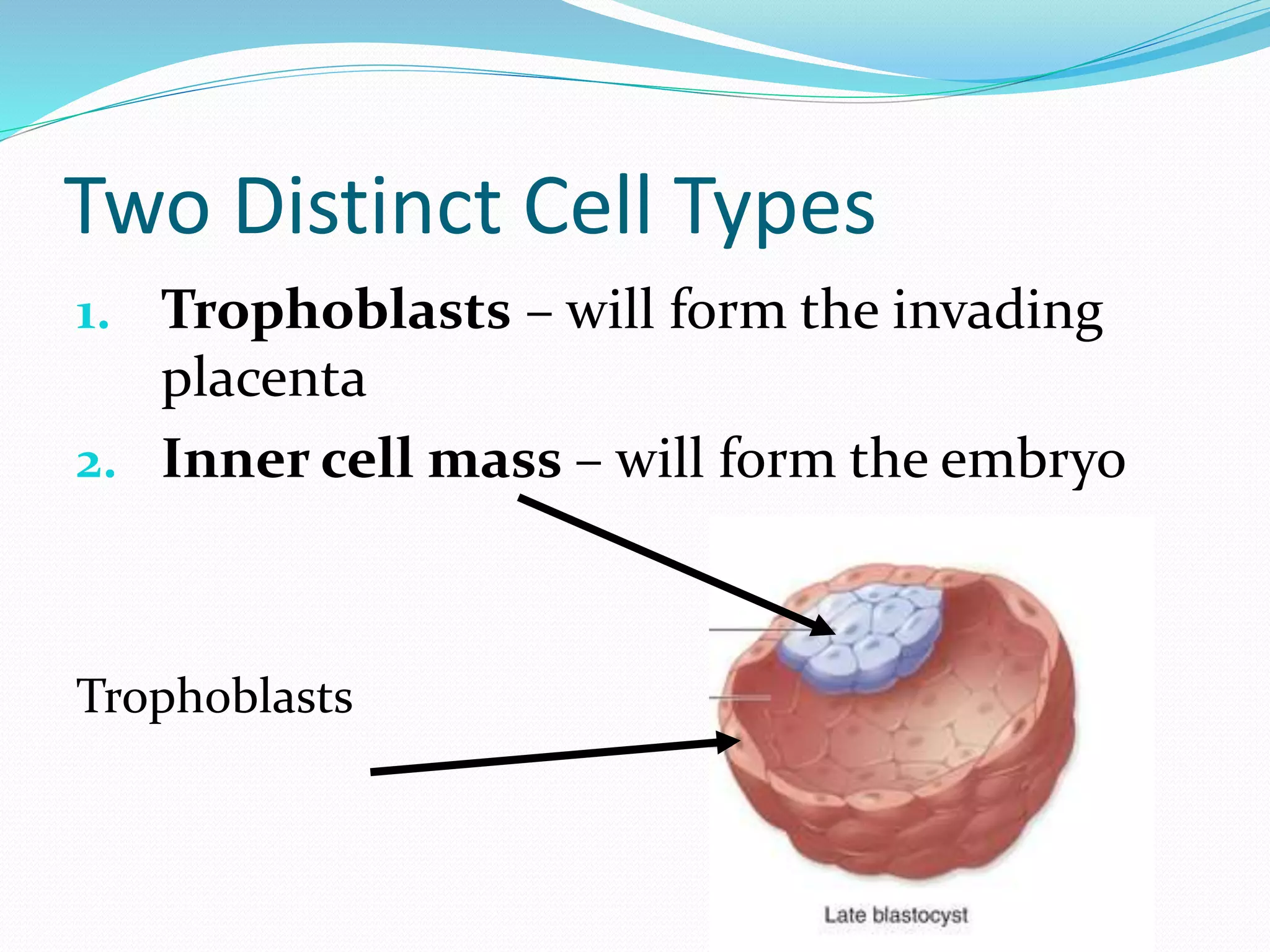
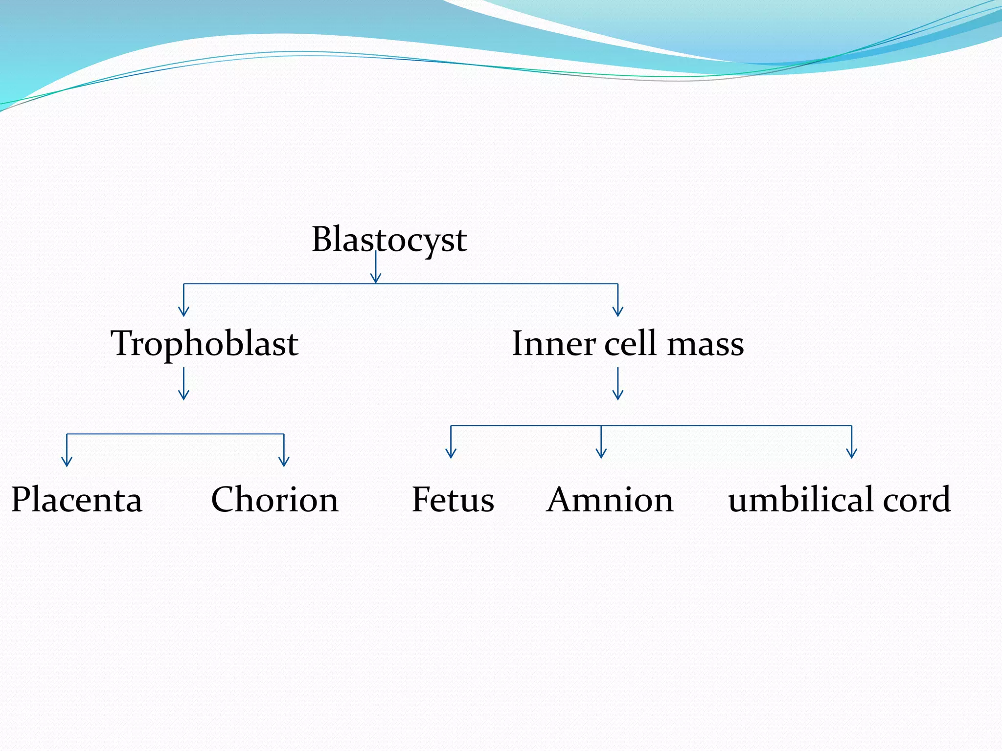
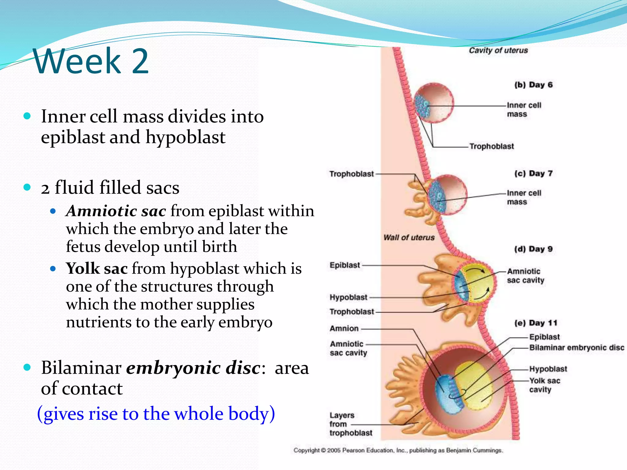
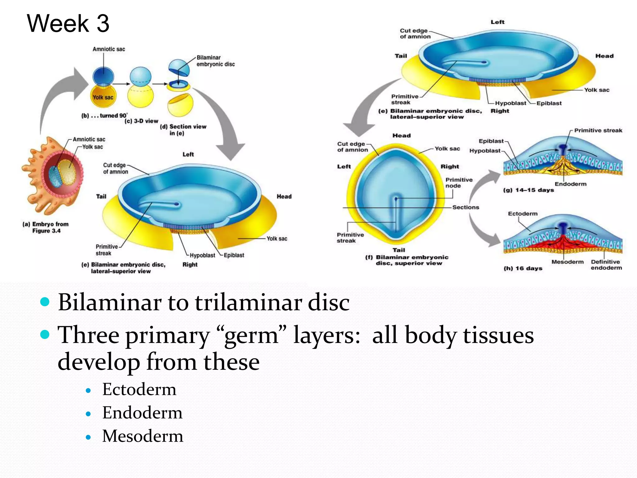
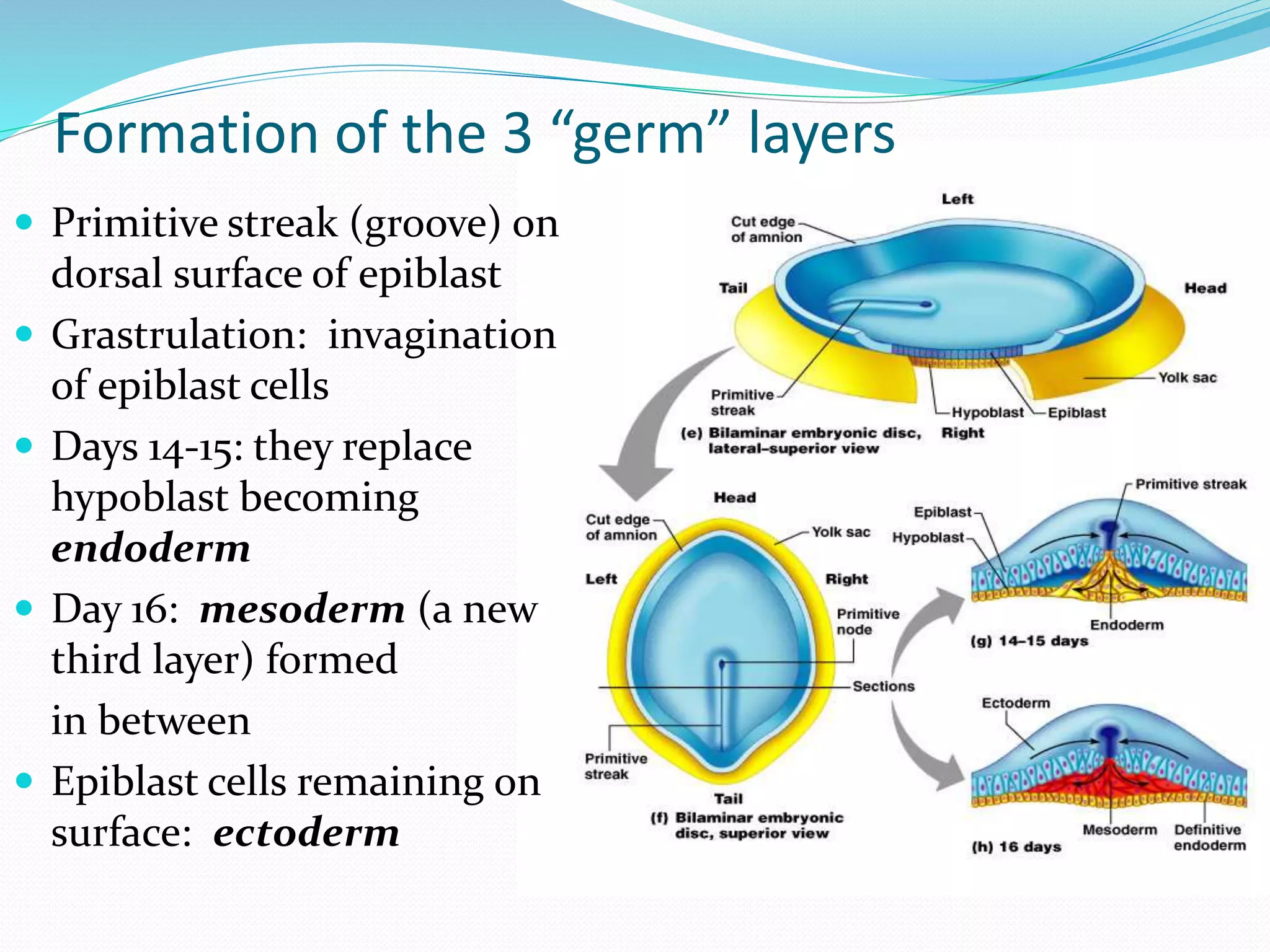
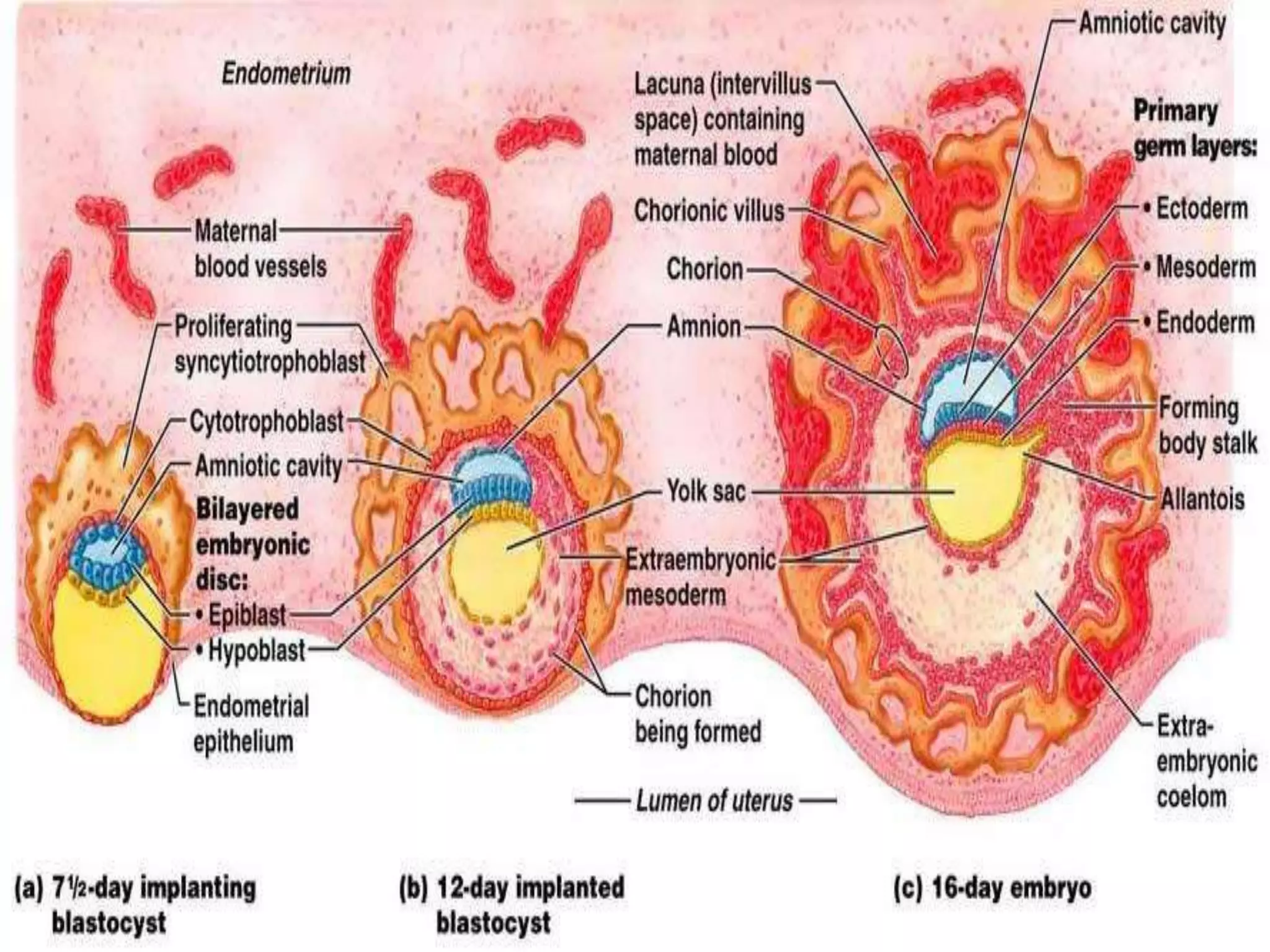
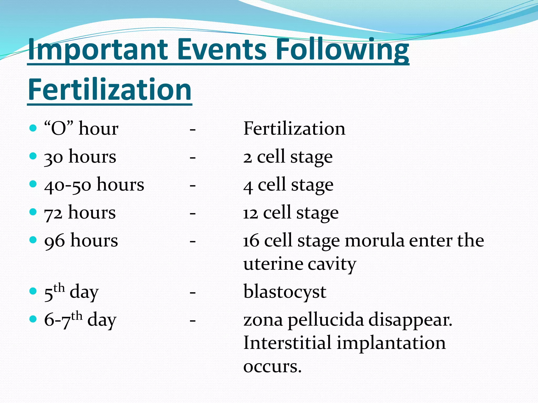
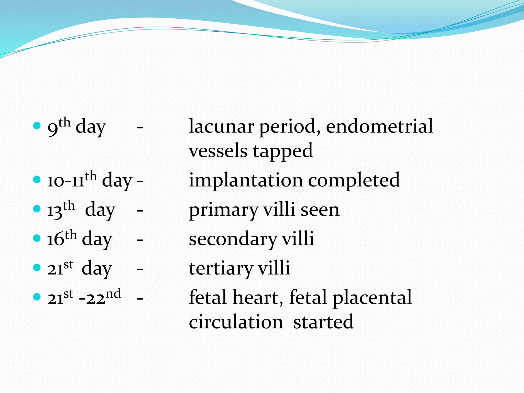
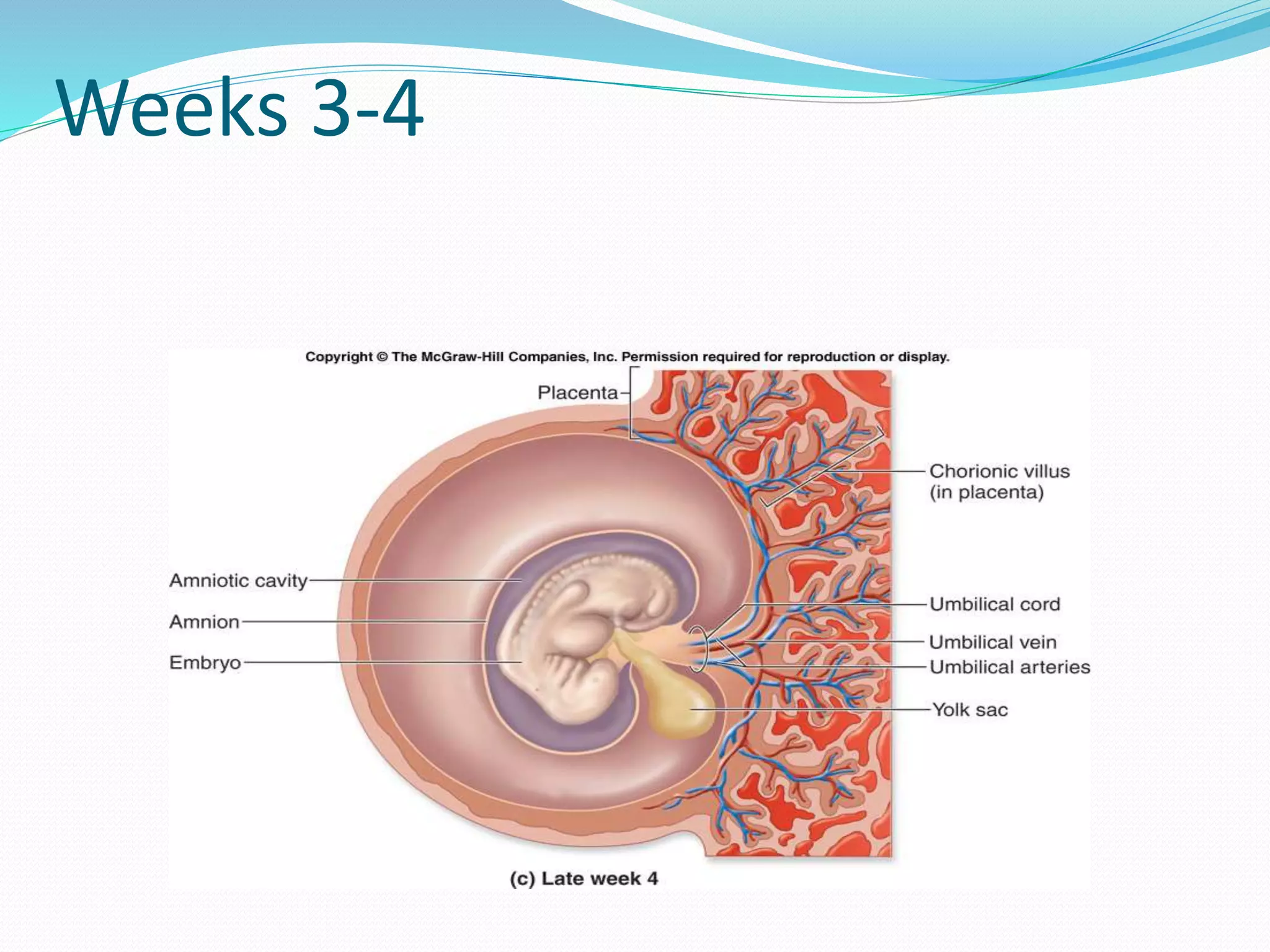
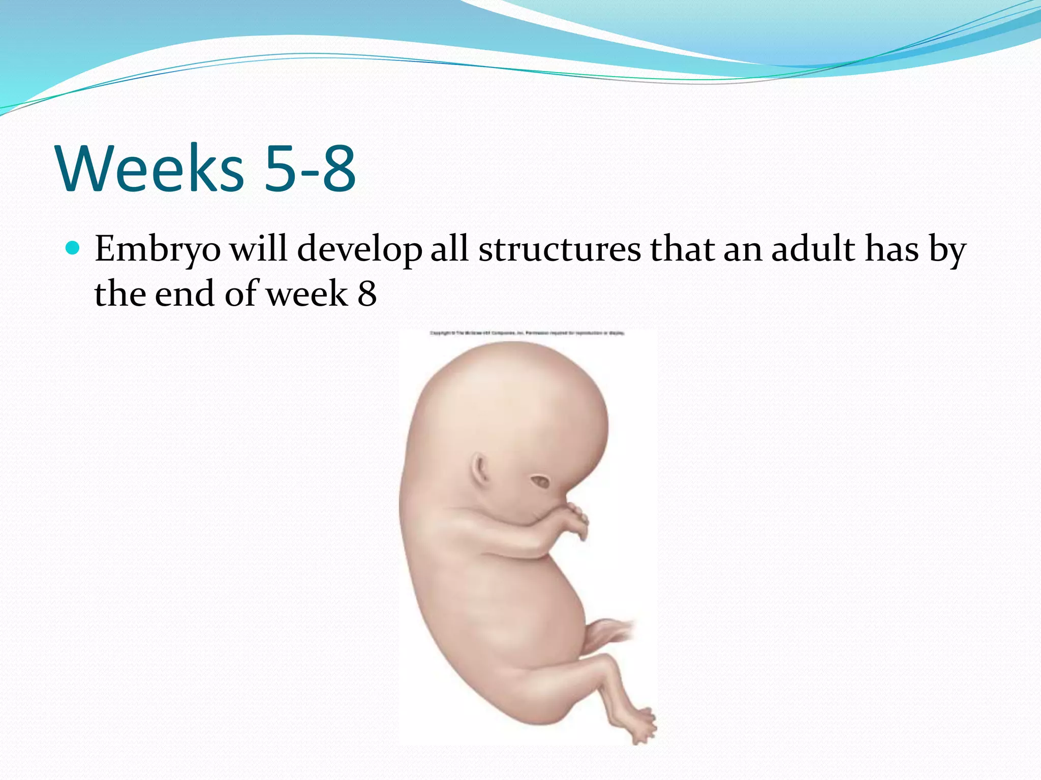
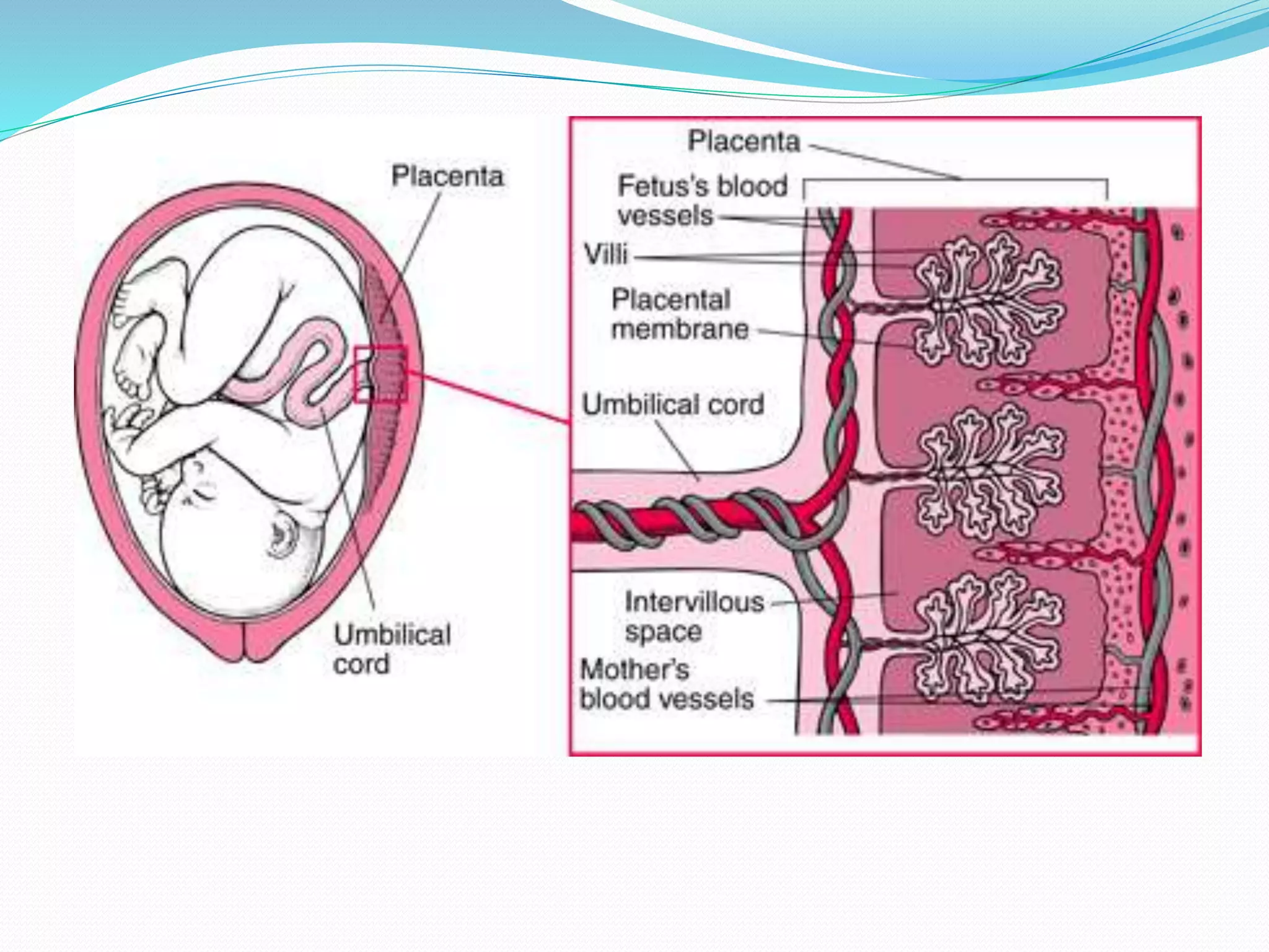
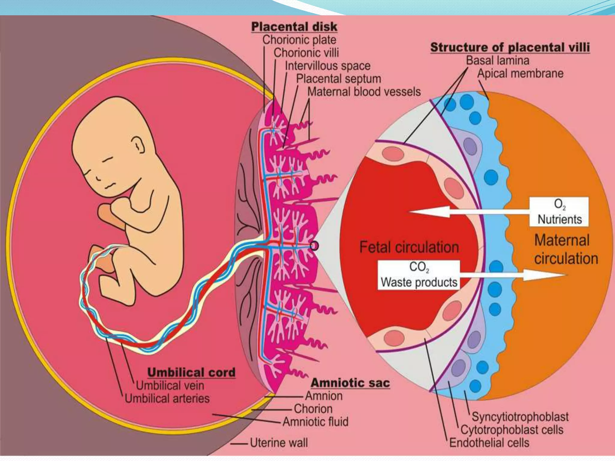
Embryology is the study of prenatal development from fertilization through the embryonic and fetal periods. During the embryonic period (first 8 weeks), the three germ layers—ectoderm, mesoderm, and endoderm—develop and give rise to all structures. The fetal period encompasses the remaining 30 weeks of development as structures and organs continue growing and maturing. Fertilization occurs when a sperm fuses with an ovum to form a zygote, which undergoes cleavage, morula, and blastocyst stages over the first week. Around day 6, implantation in the uterus occurs and the blastocyst forms an inner cell mass and trophoblast. The trophoblast develops into the pl
DEVELOPMENT OF PLACENTA,PLACENTA AT TERM , DECIDUA,PLACENTAL MEMBRANE , PLACENTAL CICULATION,PLACENTAL ENDOCRINE SYNTHESIS,ABNORMAL PLACENTA,FUNCTIONS.
ovaries, fallopian tube, component of internal genitalia, location of ovarie, boundaries of ovaries,external features of ovaries,ligaments of ovaries, support of ovaries, broad ligament, mesovarium, mesosalpinx, mesometrium, round ligament of uterus, blood supply and lymphatics of ovaries, prts of fallopian tube, blood supply of fallopian tube, ectopic pregnancy, polycystic ovaries,
In testis, the immature male germ cell (spermatogonia ) produce sperms by spermatogenesis
The spermatogonia ( sing. Spermatogonium ) present on the inside of seminiferous tubules multiply by mitotic division and increase in numbers
Each spermatogonium is diploid and contains 46 chromosomes
Some of the spermatogonia called primary spermatocytes periodically undergo meiosis.A primary spermatocyte completes the first meiotic division (reduction division) leading to formation of two equal, haploid cells called secondary spermatocyte, which have only 23 chromosomes
The secondary spermatocyte undergo the second meiotic division to produce four equal, haploid spermatids
Tooth preparation for full veneer crowns /certified fixed orthodontic course...Indian dental academy
The Indian Dental Academy is the Leader in
continuing dental education , training dentists
in all aspects of dentistry and offering a wide
range of dental certified courses in different
formats.
Indian dental academy provides dental crown &
Bridge,rotary endodontics,fixed orthodontics,
Dental implants courses.for details pls visit
www.indiandentalacademy.com ,or call
0091-9248678078
After fertilization, gestation period begins
According to development gestation period divided in to,
i. germinal period ii. embryonic period iii. fetal period
Here the first week of the germinal period is discussed
Zygote undergoes cleavage
The process of each step of cleavage is explained and shown
diagrammatically
The significance of cleavage given.
Formation of morula after the compactum of blastocytes
Pushing of embryoblast towards the animal pole and blastocoel
the formation has taken place.
Formation of blastocyst completed.
A blastocyst is ready for implantation after loosing Zonapellucida.
Implantation begins in the first week of development
Flu Vaccine Alert in Bangalore Karnatakaaddon Scans
As flu season approaches, health officials in Bangalore, Karnataka, are urging residents to get their flu vaccinations. The seasonal flu, while common, can lead to severe health complications, particularly for vulnerable populations such as young children, the elderly, and those with underlying health conditions.
Dr. Vidisha Kumari, a leading epidemiologist in Bangalore, emphasizes the importance of getting vaccinated. "The flu vaccine is our best defense against the influenza virus. It not only protects individuals but also helps prevent the spread of the virus in our communities," he says.
This year, the flu season is expected to coincide with a potential increase in other respiratory illnesses. The Karnataka Health Department has launched an awareness campaign highlighting the significance of flu vaccinations. They have set up multiple vaccination centers across Bangalore, making it convenient for residents to receive their shots.
To encourage widespread vaccination, the government is also collaborating with local schools, workplaces, and community centers to facilitate vaccination drives. Special attention is being given to ensuring that the vaccine is accessible to all, including marginalized communities who may have limited access to healthcare.
Residents are reminded that the flu vaccine is safe and effective. Common side effects are mild and may include soreness at the injection site, mild fever, or muscle aches. These side effects are generally short-lived and far less severe than the flu itself.
Healthcare providers are also stressing the importance of continuing COVID-19 precautions. Wearing masks, practicing good hand hygiene, and maintaining social distancing are still crucial, especially in crowded places.
Protect yourself and your loved ones by getting vaccinated. Together, we can help keep Bangalore healthy and safe this flu season. For more information on vaccination centers and schedules, residents can visit the Karnataka Health Department’s official website or follow their social media pages.
Stay informed, stay safe, and get your flu shot today!
Title: Sense of Smell
Presenter: Dr. Faiza, Assistant Professor of Physiology
Qualifications:
MBBS (Best Graduate, AIMC Lahore)
FCPS Physiology
ICMT, CHPE, DHPE (STMU)
MPH (GC University, Faisalabad)
MBA (Virtual University of Pakistan)
Learning Objectives:
Describe the primary categories of smells and the concept of odor blindness.
Explain the structure and location of the olfactory membrane and mucosa, including the types and roles of cells involved in olfaction.
Describe the pathway and mechanisms of olfactory signal transmission from the olfactory receptors to the brain.
Illustrate the biochemical cascade triggered by odorant binding to olfactory receptors, including the role of G-proteins and second messengers in generating an action potential.
Identify different types of olfactory disorders such as anosmia, hyposmia, hyperosmia, and dysosmia, including their potential causes.
Key Topics:
Olfactory Genes:
3% of the human genome accounts for olfactory genes.
400 genes for odorant receptors.
Olfactory Membrane:
Located in the superior part of the nasal cavity.
Medially: Folds downward along the superior septum.
Laterally: Folds over the superior turbinate and upper surface of the middle turbinate.
Total surface area: 5-10 square centimeters.
Olfactory Mucosa:
Olfactory Cells: Bipolar nerve cells derived from the CNS (100 million), with 4-25 olfactory cilia per cell.
Sustentacular Cells: Produce mucus and maintain ionic and molecular environment.
Basal Cells: Replace worn-out olfactory cells with an average lifespan of 1-2 months.
Bowman’s Gland: Secretes mucus.
Stimulation of Olfactory Cells:
Odorant dissolves in mucus and attaches to receptors on olfactory cilia.
Involves a cascade effect through G-proteins and second messengers, leading to depolarization and action potential generation in the olfactory nerve.
Quality of a Good Odorant:
Small (3-20 Carbon atoms), volatile, water-soluble, and lipid-soluble.
Facilitated by odorant-binding proteins in mucus.
Membrane Potential and Action Potential:
Resting membrane potential: -55mV.
Action potential frequency in the olfactory nerve increases with odorant strength.
Adaptation Towards the Sense of Smell:
Rapid adaptation within the first second, with further slow adaptation.
Psychological adaptation greater than receptor adaptation, involving feedback inhibition from the central nervous system.
Primary Sensations of Smell:
Camphoraceous, Musky, Floral, Pepperminty, Ethereal, Pungent, Putrid.
Odor Detection Threshold:
Examples: Hydrogen sulfide (0.0005 ppm), Methyl-mercaptan (0.002 ppm).
Some toxic substances are odorless at lethal concentrations.
Characteristics of Smell:
Odor blindness for single substances due to lack of appropriate receptor protein.
Behavioral and emotional influences of smell.
Transmission of Olfactory Signals:
From olfactory cells to glomeruli in the olfactory bulb, involving lateral inhibition.
Primitive, less old, and new olfactory systems with different path
Report Back from SGO 2024: What’s the Latest in Cervical Cancer?bkling
Are you curious about what’s new in cervical cancer research or unsure what the findings mean? Join Dr. Emily Ko, a gynecologic oncologist at Penn Medicine, to learn about the latest updates from the Society of Gynecologic Oncology (SGO) 2024 Annual Meeting on Women’s Cancer. Dr. Ko will discuss what the research presented at the conference means for you and answer your questions about the new developments.
Prix Galien International 2024 Forum ProgramLevi Shapiro
June 20, 2024, Prix Galien International and Jerusalem Ethics Forum in ROME. Detailed agenda including panels:
- ADVANCES IN CARDIOLOGY: A NEW PARADIGM IS COMING
- WOMEN’S HEALTH: FERTILITY PRESERVATION
- WHAT’S NEW IN THE TREATMENT OF INFECTIOUS,
ONCOLOGICAL AND INFLAMMATORY SKIN DISEASES?
- ARTIFICIAL INTELLIGENCE AND ETHICS
- GENE THERAPY
- BEYOND BORDERS: GLOBAL INITIATIVES FOR DEMOCRATIZING LIFE SCIENCE TECHNOLOGIES AND PROMOTING ACCESS TO HEALTHCARE
- ETHICAL CHALLENGES IN LIFE SCIENCES
- Prix Galien International Awards Ceremony
These lecture slides, by Dr Sidra Arshad, offer a quick overview of physiological basis of a normal electrocardiogram.
Learning objectives:
1. Define an electrocardiogram (ECG) and electrocardiography
2. Describe how dipoles generated by the heart produce the waveforms of the ECG
3. Describe the components of a normal electrocardiogram of a typical bipolar leads (limb II)
4. Differentiate between intervals and segments
5. Enlist some common indications for obtaining an ECG
Study Resources:
1. Chapter 11, Guyton and Hall Textbook of Medical Physiology, 14th edition
2. Chapter 9, Human Physiology - From Cells to Systems, Lauralee Sherwood, 9th edition
3. Chapter 29, Ganong’s Review of Medical Physiology, 26th edition
4. Electrocardiogram, StatPearls - https://www.ncbi.nlm.nih.gov/books/NBK549803/
5. ECG in Medical Practice by ABM Abdullah, 4th edition
6. ECG Basics, http://www.nataliescasebook.com/tag/e-c-g-basics
Lung Cancer: Artificial Intelligence, Synergetics, Complex System Analysis, S...Oleg Kshivets
RESULTS: Overall life span (LS) was 2252.1±1742.5 days and cumulative 5-year survival (5YS) reached 73.2%, 10 years – 64.8%, 20 years – 42.5%. 513 LCP lived more than 5 years (LS=3124.6±1525.6 days), 148 LCP – more than 10 years (LS=5054.4±1504.1 days).199 LCP died because of LC (LS=562.7±374.5 days). 5YS of LCP after bi/lobectomies was significantly superior in comparison with LCP after pneumonectomies (78.1% vs.63.7%, P=0.00001 by log-rank test). AT significantly improved 5YS (66.3% vs. 34.8%) (P=0.00000 by log-rank test) only for LCP with N1-2. Cox modeling displayed that 5YS of LCP significantly depended on: phase transition (PT) early-invasive LC in terms of synergetics, PT N0—N12, cell ratio factors (ratio between cancer cells- CC and blood cells subpopulations), G1-3, histology, glucose, AT, blood cell circuit, prothrombin index, heparin tolerance, recalcification time (P=0.000-0.038). Neural networks, genetic algorithm selection and bootstrap simulation revealed relationships between 5YS and PT early-invasive LC (rank=1), PT N0—N12 (rank=2), thrombocytes/CC (3), erythrocytes/CC (4), eosinophils/CC (5), healthy cells/CC (6), lymphocytes/CC (7), segmented neutrophils/CC (8), stick neutrophils/CC (9), monocytes/CC (10); leucocytes/CC (11). Correct prediction of 5YS was 100% by neural networks computing (area under ROC curve=1.0; error=0.0).
CONCLUSIONS: 5YS of LCP after radical procedures significantly depended on: 1) PT early-invasive cancer; 2) PT N0--N12; 3) cell ratio factors; 4) blood cell circuit; 5) biochemical factors; 6) hemostasis system; 7) AT; 8) LC characteristics; 9) LC cell dynamics; 10) surgery type: lobectomy/pneumonectomy; 11) anthropometric data. Optimal diagnosis and treatment strategies for LC are: 1) screening and early detection of LC; 2) availability of experienced thoracic surgeons because of complexity of radical procedures; 3) aggressive en block surgery and adequate lymph node dissection for completeness; 4) precise prediction; 5) adjuvant chemoimmunoradiotherapy for LCP with unfavorable prognosis.
micro teaching on communication m.sc nursing.pdfAnurag Sharma
Microteaching is a unique model of practice teaching. It is a viable instrument for the. desired change in the teaching behavior or the behavior potential which, in specified types of real. classroom situations, tends to facilitate the achievement of specified types of objectives.
Ozempic: Preoperative Management of Patients on GLP-1 Receptor Agonists Saeid Safari
Preoperative Management of Patients on GLP-1 Receptor Agonists like Ozempic and Semiglutide
ASA GUIDELINE
NYSORA Guideline
2 Case Reports of Gastric Ultrasound
TEST BANK for Operations Management, 14th Edition by William J. Stevenson, Ve...kevinkariuki227
TEST BANK for Operations Management, 14th Edition by William J. Stevenson, Verified Chapters 1 - 19, Complete Newest Version.pdf
Tom Selleck Health: A Comprehensive Look at the Iconic Actor’s Wellness Journeygreendigital
Tom Selleck, an enduring figure in Hollywood. has captivated audiences for decades with his rugged charm, iconic moustache. and memorable roles in television and film. From his breakout role as Thomas Magnum in Magnum P.I. to his current portrayal of Frank Reagan in Blue Bloods. Selleck's career has spanned over 50 years. But beyond his professional achievements. fans have often been curious about Tom Selleck Health. especially as he has aged in the public eye.
Follow us on: Pinterest
Introduction
Many have been interested in Tom Selleck health. not only because of his enduring presence on screen but also because of the challenges. and lifestyle choices he has faced and made over the years. This article delves into the various aspects of Tom Selleck health. exploring his fitness regimen, diet, mental health. and the challenges he has encountered as he ages. We'll look at how he maintains his well-being. the health issues he has faced, and his approach to ageing .
Early Life and Career
Childhood and Athletic Beginnings
Tom Selleck was born on January 29, 1945, in Detroit, Michigan, and grew up in Sherman Oaks, California. From an early age, he was involved in sports, particularly basketball. which played a significant role in his physical development. His athletic pursuits continued into college. where he attended the University of Southern California (USC) on a basketball scholarship. This early involvement in sports laid a strong foundation for his physical health and disciplined lifestyle.
Transition to Acting
Selleck's transition from an athlete to an actor came with its physical demands. His first significant role in "Magnum P.I." required him to perform various stunts and maintain a fit appearance. This role, which he played from 1980 to 1988. necessitated a rigorous fitness routine to meet the show's demands. setting the stage for his long-term commitment to health and wellness.
Fitness Regimen
Workout Routine
Tom Selleck health and fitness regimen has evolved. adapting to his changing roles and age. During his "Magnum, P.I." days. Selleck's workouts were intense and focused on building and maintaining muscle mass. His routine included weightlifting, cardiovascular exercises. and specific training for the stunts he performed on the show.
Selleck adjusted his fitness routine as he aged to suit his body's needs. Today, his workouts focus on maintaining flexibility, strength, and cardiovascular health. He incorporates low-impact exercises such as swimming, walking, and light weightlifting. This balanced approach helps him stay fit without putting undue strain on his joints and muscles.
Importance of Flexibility and Mobility
In recent years, Selleck has emphasized the importance of flexibility and mobility in his fitness regimen. Understanding the natural decline in muscle mass and joint flexibility with age. he includes stretching and yoga in his routine. These practices help prevent injuries, improve posture, and maintain mobilit
2. What is embryology?
The study of developmental events that
occur during the prenatal stage .
The branch of biology and medicine
concerned with the study of embryos
and their development.
3. Embryonic period vs. Fetal period
Embryonic – first 8 weeks Development of the
three primary germ layers give rise to all structures and
Basic body plan takes shape
Fetal period – remaining 30 weeks. Structures
and organs continue to grow and develop.
8 weeks
5. Stages of Development
1. Fertilization
2. Cleavage
3. Gastrulation Embryogenesis
4.Organogenesis
5.Maturation
6. Fertilization
The process of fusion or union of the
spermatozoon with the mature ovum is known as
conception / fertilizaiton/impregnantation.
Which produced the fertilized single mono-
nucleated cell called the zygote.
8. 2. Gastrulation : is a phase early in the
embryonic development of most animals/human
being, during which the single-layered blastula is
reorganized into a trilaminar ("three-layered")
structure known as the gastrula. These three germ
layers are known as the ectoderm, mesoderm, and
endoderm.
3. Organogenesis: The production and
development of the organs of an animal or plant.
9. How fertilization occurs????
Following ovulation, the ovum, which is about 0.15 mm in
diameter is picked up by the tubal fimbriae and is moved along by
the cilia and by peristaltic movement of the tube.
At the time the cervix under the influence of estrogen, secretes a
flow of alkaline mucus that deposited in the vagina, only
thousands capacitated spermatozoa enter the uterine tube while
300-500 reach the ovum, and remainder are destroyed by the acid
medium of the vagina.
It takes about 1 hour for sperm to reach the site.
10. The sperm release the enzyme, Hylluronidase
which allows penetration of the zona pellucida and
the cell membrane surrounding the ovum.
Many sperm are needed for this to take place but
only one will enter the ovum.
After this the membrane is sealed to prevent entry
of any further sperm and the nucluei of the two
cell fuse.
11. The sperm and ovum contribute half the
complement of chromosomes to make a
total of 46.
The sperm and ovum is known as the male
and female gametes and the fertilized ovum
as the zygote
13. Normal site for Conception
The most common site of conception is the
ampullary part (Ampulla ) of the fallopian
tube which is the widest part located closed
to the ovary
14. Usual Time for Conception
Neither sperm nor ovum can survive longer than 2-3
days and the fertilization is most likely to occur when
intercourse takes place not more than 48 hours before
and 24 hours after ovulation.
So the conception will take place about 14 days before
the next period is due.
The sex of the new individual at the time of
conception is determined by sex chromosomes.
15. Every human cell contains 46 chromosomes,
which are made up of 44 autosome chromosome
and 2 sex chromosomes.
The sex chromosome are X and Y .
Woman have no Y chromosome and male has Y
chromosome (male 44+X+Y) (female 44+X+X).
Therefor e, sex of child is always determined by
father
16.
17. Morula
1. After fertilization, the Zygote divides into 2 cells
(blastomere) (mitosis division)in about 30 hours
after fertilization.
2. The blastomeres continue to divide by binary
division through 4, 8, and 16 cell stage until a
cluster of cells is formed– Morula, resemblibg
a mulberry
18. The morula after spending about 3 days(72 hours)
in the uterine tube enters the uterine cavity
through the narrow uterine ostium (1mm) on the
4th day .
21. 4. Blastocyst
Morula, once entering the uterine cavity, floats
freely(next 2 days) and is covered by endometrial
fluid and mucus.
This fluid is absorbed through the canaliculi of the
zona pellucida and Morula begins to accumulate
fluid and forms a cavity between its cells.
Once cavity appears, it is now called a blastocyst.
23. Blastocyst
The zona pellucida
becomes stretched, thinned
and gradually disappear
soon prior to implantation.
The cell of the outer cell
mass forms the wall of the
blastocyst and is known as
trophoblast.
The inner cell mass is
concerned with the
development of the
embryo.
24. Two Distinct Cell Types
1. Trophoblasts – will form the invading
placenta
2. Inner cell mass – will form the embryo
Trophoblasts
26. hCG is produced
hCG is human chorionic gonadotropin
hormone
It is produced by the trophoblasts starting on day 6
hCG causes endometrium of uterus to grow and
proliferate
hCG prevents the menstrual cycle from occuring
This is why a female misses her periods when she
is pregnant
28. Implantation occurs about 6th day after
fertilization (approx the 20th day of a regular
menstrual cycle).
As the zona pellucida surrounding the blastocyst
disappear, there is increased adhesiveness of the
trophoblastic cells to the endometrium.
The trophoblastic cells invade the stromal cells
lying between the secretary endometrial glands by
histolytic action of the blastocyst bringing about
deeper penetration in the decidua.
29. The syncytial cells ( multinucleate cell) ultimately
penetrate the endothelial coat of the maternal
decidual capillaries establishing communication with
the syncytial lacunar system
Thus maternal blood circulates through the syncytial
lacunae providing nourishment to the blastocyst.
Maternal blood vassels
30. Further penetration is arrested by the maternal
immunological blocking factor. The point of entry of
the blastocyst into the decidual is sealed by a fibrin
coat and later by epithelium
The process of implantation is completed by the 10th
or 11th day of fertilization.
31. This type of deeper penetration of the human
blastocyst is called interstitial implantation and the
blastocyst is covered on all sides by the endometrium
(decidual).
Occasionally, there may be increased blood flow into
the lacunar space and cause bleeding. It is called
implantation bleeding and This corresponds approx to
13th day after fertilization (at about the expected day of
period).
This may produce confusion in determination of the
EDD
32.
33. The outer cell mass of the blastocyst form the
trophoblast layer.
The inner cell mass on the 8th day differentiates into
bilaminar germ disc consisting of a dorsal ectodermal
layer of the tall columnar cells and a ventral
endodermal layer of flat polyhydral cells.
The bilaminar germ disc is connected to the
trophoblast by a mesenchymal connecting stalk called
the body stalk which later forms the umblical cord.
34. Formation of Amniotic cavity and
Yolk sac
Two cavities appear on either side of the bilaminar
embryonic disc.
The dorsal cavity between the ectoderm and
trophoblastic layers lined by mesenchyme(connective
tissue) is called the amniotic cavity.
The cavity on the ventral aspect lined by the primitive
mesenchyme on the outside and the endodermal layer
of the germ cell disc on the inside is called the yolk sac
35. Week 2
Inner cell mass divides into
epiblast and hypoblast
2 fluid filled sacs
Amniotic sac from epiblast within
which the embryo and later the
fetus develop until birth
Yolk sac from hypoblast which is
one of the structures through
which the mother supplies
nutrients to the early embryo
Bilaminar embryonic disc: area
of contact
(gives rise to the whole body)
36. Bilaminar to trilaminar disc
Three primary “germ” layers: all body tissues
develop from these
Ectoderm
Endoderm
Mesoderm
Week 3
37. Formation of the 3 “germ” layers
Primitive streak (groove) on
dorsal surface of epiblast
Grastrulation: invagination
of epiblast cells
Days 14-15: they replace
hypoblast becoming
endoderm
Day 16: mesoderm (a new
third layer) formed
in between
Epiblast cells remaining on
surface: ectoderm
53. Trophoblast
Small projection begins to appear all over
the surface of the blastocyst, becoming most
prolific at the area of contact. These
trophoblastic cells differentiate into 3 layer.
1. The outer syncytiotrophoblast
(syncytium): it erodes the walls of the blood
vessels of the decidua, making the nutrient
in the maternal blood accessible to the
developing fetus.
54. 2. The inner cytotrophoblast: it’s a single
layer cells which produces a hormone HCG. This is
responsible for informing the corpus luteum that a
pregnancy has begun and corpus luteum
continues to produce estrogen and progesterone,
which maintain the integrity of the decidua.
3. In the inner aspect: a layer of mesoderm or
primitive mesenchyme, which consists of loose
connective tissue.
There is similar tissue in the inner cell mass and
the two are continuous at the point where they join
in the body stalk. (fig 3.9 1st picture from book
tuitui)
55.
56.
57. The trophoblast is responsible for the formation of
the placenta and fetal membrane and sub-serve
the important functions of attachment of the fetus
to maternal tissues, providing nutrition,
oxygenation, and clearing of the fetal metabolic
wastes and producing hormones, thus ensuring
growth and development of the fetus.
58. Decidua
The endometrial lining of the uterus is
called decidua during pregnancy and it is
shed after delivery.
Progesterone from the corpus luteum and
placenta maintain decidua during
pregnancy. It is called the decidual reaction.
59. Well developed Decidua consists
of 3 layers.
1. Superficial compact layer: consists of
compact mass of decidual cells, gland, ducts and
dilated capillaries. The greater part of the surface
epithelium is either thinned out or lost.
2. Intermediate spongy layer (cavernous
or functional layer): contains dilated uterine
glands, decidual cells and blood vessesls. Placenta
separation occurs in this layer.
60. 3. The basal layer: containing the basal
portion of the glands and is opposed to the
uterine muscle. Regeneration of the mucous
coat occurs from this layer following
parturition.
After the implantation of the blastocyst into the
compact layer of the decidua, the different
portions of the decidua are renamed as:
61. 1. Decidua basalis or
serotina: The portion of
the decidua in contact
with the base of the
blastocyst.
2. Decidua capsularies or
reflexa: The thin
superficial compact layer
covering the blastocyst.
3. Decidua vera or
parietalis: the rest of
the decidua lining the
uterine cavity outside the
site of implantation.
62. The ovum bulges into the
uterine cavity. The space
between the decidua
capsularies and decidua vera
is called the decidual space.
It progressively diminishes
as the ovum enlarges in
pregnancy
Until at 16 weeks, the space
is obliterate because of the
fusion of the decidua
capsularis with the decidua
vera.
At term this fused layer is
gradually attenuated and its
constituent layers can not
be identified.
The decidua basalis
becomes the maternal
portion of the placenta.
63. Functions of Decidua
1. To provide place for implantation of the
fertilized ovum.
2. To provide nutrition to the growing ovum.
3. Provide a protective action.
4. Provides the basal plate of the placenta.
64. Important Events Following
Fertilization
“O” hour - Fertilization
30 hours - 2 cell stage
40-50 hours - 4 cell stage
72 hours - 12 cell stage
96 hours - 16 cell stage morula enter the
uterine cavity
5th day - blastocyst
6-7th day - zona pellucida disappear.
Interstitial implantation
occurs.
65. 9th day - lacunar period, endometrial
vessels tapped
10-11th day - implantation completed
13th day - primary villi seen
16th day - secondary villi
21st day - tertiary villi
21st -22nd - fetal heart, fetal placental
circulation started
66.
67. The prenatal development of the fetus
can be divided into 3 phases.
1. Ovular period or germinal period (It
lasts from the stage of fertilization upto 2 weeks after
ovulation)
The embryonic period (first 8 weeks
Development of the three primary germ layers give rise
to all structures and Basic body plan takes shape )
Fetal period (remaining 30 weeks. Structures
and organs continue to grow and develop)
68. Fetal Length
In the earlier weeks, it is expressed as the
measurement from the vertex to the coccyx
(crown rump length) while after mid
pregnancy (20 weeks onwards), the
measurement of the fetus is determined
from the vertex to the heel (crown heel
length).
69. Age of the Fetus
The length is more reliable criterion than
the weight to calculate the age of the fetus.
Haase’s rule is employed in calculating the
age of fetus from its length in centimeters,
divided by 5.
70. 1. First lunar month: Fertilization to
2weeks of embryonic growth
Implantation is complete.
Primary chorionic villi forming
Embryo develops into two cell layers (bilaminar
embryonic disks)
Amniotic cavity appears.
Note: lunar month is a period that is measured based on the
movement of the moon. It is roughly 28 days and it constitutes
a month. In that context, pregnancy is 10.
71. 2. Second lunar month: 3 to 6
weeks of embryonic growth
At the end of 6 weeks of growth, the embryo is
approximatey 1.2 cm long.
Arms and leg buds are visible; arm buds are more
developed with finger ridges beginning to appear.
Rudiments of the eyes, ears and nose appear.
Primitive intestinal tract is developing.
Primitive cardiovascular system is functioning.
Neural tube, which forms the brain and spinal cord
closes by the 4th week.
72. 3. Third lunar month: 7 to 10
weeks of growth
The middle of this period (8 weeks)marks the end of the
embryonic period and beginning of the fetal period.
At the end of 10 weeks of growth, the fetus is 6.1 cm from
crown to rump and weigh 14 gm.
Appearance of external genitalia.
By the middle of this month, all major organs, systems have
formed.
The heart has formed four chambers (by 7th week)
The fetus assumes a human appearance
Bone ossification begins
Rudimentary kidney begins to secrete urine.
73. 4. Fourth lunar month: 11 to 14
weeks old fetus
At the end of 14 weeks of growth, the fetus is
12 cm crown to rump length and 110 gm.
Head erect; lower extremities well
developed.
Hard palate and nasal septum have fused.
External genitalia of male and female can
now be differentiated.
Eyelids are sealed
74. 5.Fifth lunar month: 15 to 18
weeks old fetus
At the end of 18 weeks of growth, the fetus is 16 cm
crown rump length and 320 gm.
Ossification of fetal skeleton can be seen on X-ray.
Ears stand out from head.
Fetus makes sucking motions and swallows
amniotic fluid.
Fetal movement may be felt by the mother (end of
month)
75. 6. Sixth lunar month: 19-22 weeks
old fetus
At the end of 22 weeks of
growth, the fetus
is 21 cm crown rump length
and 630 gm.
Vernix caseosa covers the skin.
Head and body hair (lanugo)
visible.
Skin is wrinkled and red.
Brown fat(adipose tissue), an
important site of heat
production, is present in neck
and sternal area.
76. 7. Seventh lunar months: 23-26
weeks old fetus
At the end of 26 weeks of
growth, the fetus is 25 cm
crown to rump length and
1000 gm.
Fingernails present.
Lean body
Eyes partially open:
eyelashes present.
Bronchioles are present:
primitive alveoli (terminal
sacs) are forming
Skin begins to thicken on
hands and feet
Startle reflex present,
grasp reflex is strong.
77. 8. Eighth lunar month: 27-30 weeks
old fetus
At the end of 30 weeks
of growth, the fetus is
28 cm crown rump
length and 1,700 gm.
Eyes open
Ample hair on head:
lanugo begins to fade
Skin slightly wrinkled
Toe nails presents
Testes in inguinal
canal, begin descent to
scrotal sac
Surfactant coat in
much of the alveolar
epithelium
78. 9. Ninth lunar months: 31-34
weeks of old fetus
At the end of 34 weeks of growth, the fetus
is about 32 cm crown –rump length and
2,500 gm
Fingernails reach finger tips
Skin pink and smooth
Testes in scrotal sac
79. 10. Tenth lunar month: 35 to 38
weeks old fetus
End of 38 weeks of growth, fetus is about 36 cm
crown –rump length and 3400 gm.
Ample of subcutaneous fat present
Lanugo is decreasing
Toe nails reach upto toe tips
Testes in scrotum
Vernix caseosa mainly on the back
Breasts are firm.