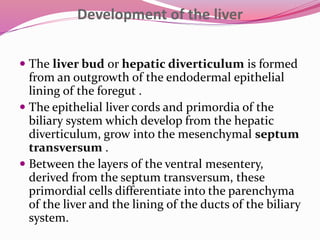
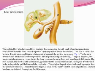
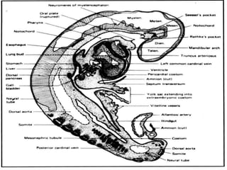
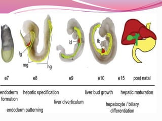
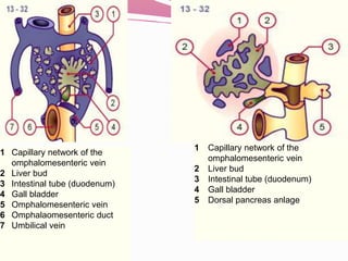
The liver develops from the endoderm layer of the foregut as a bud called the hepatic diverticulum. This bud grows into the mesenchyme and its cells differentiate into the liver parenchyma and biliary system ducts. The gallbladder also develops from the hepatic diverticulum. After birth, the umbilical vein and ductus venosus regress and the liver takes over metabolic functions from the fetal liver.
2. Development of the liver
The liver bud or hepatic diverticulum is formed
from an outgrowth of the endodermal epithelial
lining of the foregut .
The epithelial liver cords and primordia of the
biliary system which develop from the hepatic
diverticulum, grow into the mesenchymal septum
transversum .
Between the layers of the ventral mesentery,
derived from the septum transversum, these
primordial cells differentiate into the parenchyma
of the liver and the lining of the ducts of the biliary
system.
4. Early liver morphogenesis
Liver is derived from endoderm.
It forms from a diverticulum (bud) which
branches out from the primitive gut.
The pancreas develops dorsally, while the liver
bud develops ventrally.
The liver metabolises nutrients absorbed from
the
gut. Therefore first organ to receive intake.
It removes toxic compounds which are
absorbed
by modifying them so they are soluble.
5. The gallbladder, bile ducts, and liver begin to develop during the 4th week of embryogenesis as a
ventral bud from the most caudal aspect of the foregut (the future duodenum). This bud is called the
hepatic diverticulum, and it grows between the layers of the ventral mesentery (Fig 1). The hepatic
diverticulum has two distinct components: pars hepatica and pars cystica (1). The pars hepatica, the
most cranial component, gives rise to the liver, common hepatic duct, and intrahepatic bile ducts. The
pars cystica, the most caudal component, gives rise to the cystic diverticulum. The cystic diverticulum
is the anlage of the gallbladder and the cystic duct. The original hepatic diverticulum elongates to form
the common bile duct. These structures begin as solid cords, but by the 8th week of gestation, a lumen
has been established throughout the biliary tract.
Liver development
6. Gross Anatomy
The liver is divided) into four
lobes: the right (the largest lobe),
left, quadrate and caudate
lobes.
Supplied with blood via the protal
vein and hepatic artery.
Blood carried away by the hepatic
vein.
It is connected to the diaphragm
and abdomainal walls by five
ligaments.
Gall Bladder
Muscular bag for the storage,
concentration, acidification and
delivery of bile to small intestine
The liver is the only human organ that
has the remarkable property of self-
regeneration. If a part of the liver is
removed, the remaining parts can grow
back to its original size and shape.
7. Fetal blood supply
In the growing fetus, a major source of blood to
the liver is the umbilical vein which supplies
nutrients to the growing fetus. The umbilical vein
enters the abdomen at the umbilicus, and passes
upward along the free margin of the falciform
ligament of the liver to the inferior surface of the
liver. There it joins with the left branch of the
portal vein. The ductus venosus carries blood from
the left portal vein to the left hepatic vein and then
to the inferior vena cava, allowing placental blood
to bypass the liver.
8.
9. Fetal blood supply
In the fetus, the liver develops throughout normal
gestation, and does not perform the normal
filtration of the infant liver. The liver does not
perform digestive processes because the fetus does
not consume meals directly, but receives
nourishment from the mother via the placenta.
The fetal liver releases some blood stem cells that
migrate to the fetal thymus, so initially the
lymphocytes, called T-cells, are created from fetal
liver stem cells. Once the fetus is delivered, the
formation of blood stem cells in infants shifts to
the red bone marrow.
10. The liver and biliarysystem develop from the hepatic diverticulum
•The distal end of the hepatic divertivculum proliferates rapidly in
the septum transversum to form the hepatocytes and intra-hepatic
biliary system
•The proximal part in the ventral mesogastrium does not proliferate
rapidly, becomes relatively narrow and forms the extrahepatic biliary
system. A secondary outgrowth from it forms the gall bladder and
cystic duct during the 4thweek (day 24)
•The vitelline and umbilical veins, disrupted by growth of hepatic
cells in the septum transversum, form the liver sinusoids
•The septum transversum mesoderm surrounding the hepatic cords
forms the Kuppfer cells and haemopoietic cells
11. Developemental stages
After birth, the umbilical vein and ductus venosus are
completely obliterated in two to five days; the former
becomes the ligamentum teres and the latter becomes
the ligamentum venosum. In the disease state of cirrhosis
and portal hypertension, the umbilical vein can open up
again.
During childhood-At birth the liver comprises roughly
4% of body weight and is at average 120g. Over the course
of development it will increase to 1,4–1,6 kg but will only
take up 2.5–3.5% of body weight.
12.
13. Cardiac mesoderm is necessary
for liver formation
In mid 1960’s Le Douarin developed a
model using cultured fragments of tissue
from chick embryos.
Piece of primitive gut (endoderm) cannot
develop into liver by itself.
Requires interaction with cardiac mesoderm to
produce glycogen storing hepatocytes.
A physical barrier between the two
14. The liver and biliarysystem develop
from the hepatic diverticulum
•The distal end proliferates rapidly in
the septum transversumto form the
hepatocytes and intra-hepatic
biliarysystem
•The proximal part in the ventral
mesogastriumproliferate slowly,
becomes relatively narrow and forms
the extrahepatic biliarysystem.
•A secondary outgrowth from it forms
the gall bladderand cysticductduring
the 4thweek (day 24)
17. Synthesis Protein metabolism
Synthesis of amino acids
Carbohydrate metabolism
Gluconeogenesis
Glycogenolysis
Glycogenesis
Lipid metabolism
Cholesterol synthesis
Lipogenesis
Production of coagulation factors I, II, V, VII, IX, X and XI,
and protein C, protein S and antithrombin
Main site of red blood cell production
Produces insulin-like growth factor 1 (IGF-1), a polypeptide
protein – anabolic effects
Production of trombopoetin
18. Breakdown
Breaks down insulin and other hormones
Breaks down hemoglobin
Breaks down or modifies toxic substances
(methylation) → sometimes results in toxication
Converts ammonia to urea
19. Other functions
Produces albumin, the major osmolar component of
blood serum
Synthesizes angiotensinogen, the hormone
responsible for raising blood pressure when activated
by renin (enzyme released when the kidney senses low
blood pressure)
20. 1
2
3
4
5
6
7
Capillary network of the
omphalomesenteric vein
Liver bud
Intestinal tube (duodenum)
Gall bladder
Omphalomesenteric vein
Omphalaomesenteric duct
Umbilical vein
1
2
3
4
5
Capillary network of the
omphalomesenteric vein
Liver bud
Intestinal tube (duodenum)
Gall bladder
Dorsal pancreas anlage
21. Summary
Liver develops from ventral endoderm
Initially, hepatoblasts with limited liver function are
generated (this requires GATA and HNF3).
This process depends on interaction between endoderm
and cardiac mesoderm as this produces inducing factors
such as Hex.
Later, hepatoblasts differentiate into hepatocytes. This
requires further interaction with the septum transversum
(which provides Bmp4).
In the perinatal stages more functions are acquired by
hepatocytes and this is driven by hormones which initiate
transcription of many liver specific genes.
22. Some questions
1.In child bone marrow is not developed then where is
is formed?
what do you mean by De novo in childs liver disease?
What is difference between “centrilobular” and
“periportal”.
Editor's Notes
- Glukoniozdinisis (the synthesis of glucose from certain amino acids, lactate or glycerol). Note that humans and some other mammals cannot synthesize glucose from glycerol.
-3. (the breakdown of glycogen into glucose)
-4. (the formation of glycogen from glucose)(muscle tissues can also do this)
-I (fibrinogen), II (prothrombin)
-in the first trimester fetus
-Thrombopoietin is a glycoprotein hormone that regulates the production of platelets by the bone marrow.
-2. metabolites that are added to bile as pigment (bilirubin and biliverdin)
3. when the metabolite is more toxic than its precursor. Preferably, the toxins are conjugated to avail excretion in bile or urine.