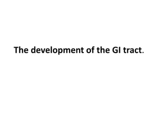
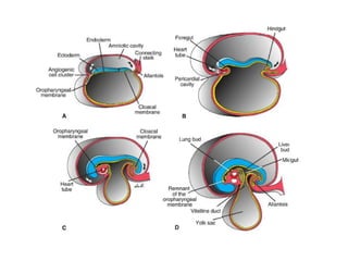
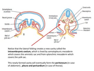
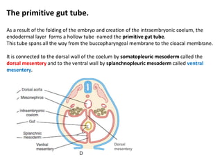
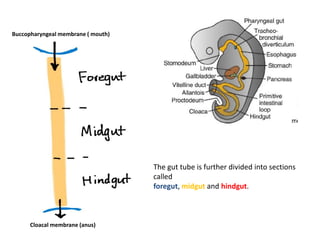
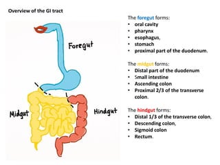
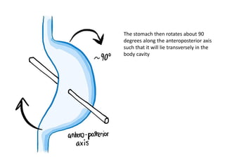
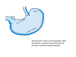
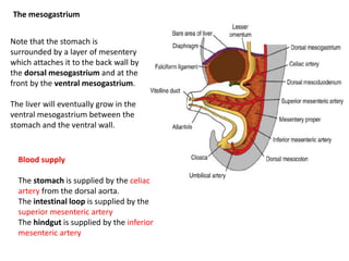
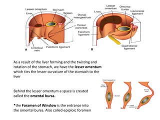
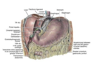
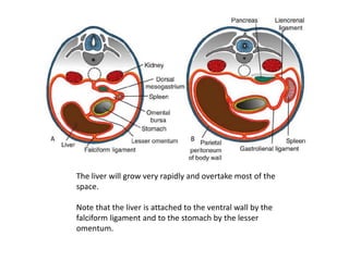
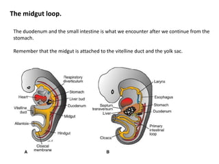
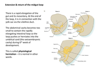
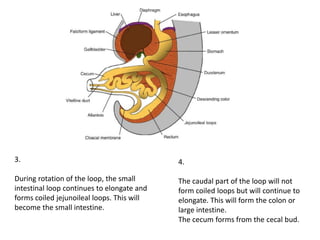
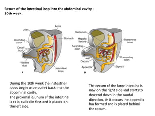
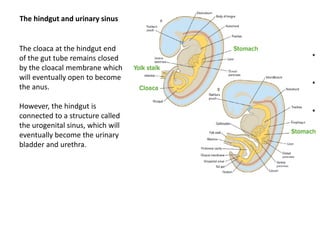
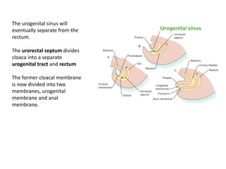
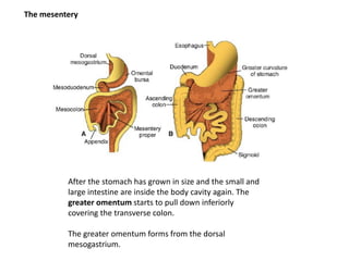
Embryology part 5
Jón Kolbeinn Guðmundsson
The development of the GI tract.
Folding of the embryo
As we already know, the folding of the embryo occurs soon after
gastrulation. The folding occurs laterally and cephalocaudally.
The amniotic cavity starts to enfold almost the entire embryo, except the
yolk sac and it’s associated vitelline duct which are for the time being
lying outside of the amniotic cavity
Notice that the lateral folding creates a new cavity called the
intraembryonic coelum, which is lined by somatopleuric mesoderm
which covers the amniotic sac and from splanchnic mesoderm which
covers the yolk sac.
This newly formed cavity will eventually form the peritoneum (in case
of abdomen) , pleura and pericardium (in case of thorax).
The primitive gut tube.
As a result of the folding of the embryo and creation of the intraembryonic coelum, the
endodermal layer forms a hollow tube named the primitive gut tube.
This tube spans all the way from the buccopharyngeal membrane to the cloacal membrane.
It is connected to the dorsal wall of the coelum by somatopleuric mesoderm called the
dorsal mesentery and to the ventral wall by splanchnopleuric mesoderm called ventral
mesentery.
Buccopharyngeal membrane ( mouth)
Cloacal membrane (anus)
The gut tube is further divided into sections
called
foregut, midgut and hindgut.
The foregut forms:
• oral cavity
• pharynx
• esophagus,
• stomach
• proximal part of the duodenum.
The midgut forms:
• Distal part of the duodenum
• Small intestine
• Ascending colon
• Proximal 2/3 of the transverse
colon.
The hindgut forms:
• Distal 1/3 of the transverse colon,
• Descending colon,
• Sigmoid colon
• Rectum.
Overview of the GI tract
The development of the stomach
Topic 19
The stomach starts to form as a
swelling/dilation in the forgut region.
The swelling is greater towards the dorsal
wall. The bulge towards the back is called the
Greater curvature and the smaller one on the
ventral side is called the Lesser curvature.
Once the curvatures have been established.
The stomach twists around its longitudinal
axis, such that the greater curvature is now
on the left side.
The stomach then rotates about 90
degrees along the anteroposterior axis
such that it will lie transversely in the
body cavity
pylorus
fundus
cardia
The stomach is now in its final position. With
the greater curvature facing inferiorly and
the lesser curvature facing superiorly.
The mesogastrium
Note that the stomach is
surrounded by a layer of mesentery
which attaches it to the back wall by
the dorsal mesogastrium and at the
front by the ventral mesogastrium.
The liver will eventually grow in the
ventral mesogastrium between the
stomach and the ventral wall.
Blood supply
The stomach is supplied by the celiac
artery from the dorsal aorta.
The intestinal loop is supplied by the
superior mesenteric artery
The hindgut is supplied by the inferior
mesenteric artery
As a result of the liver forming and the twisting and
rotation of the stomach, we have the lesser omentum
which ties the lesser curvature of the stomach to the
liver
Behind the lesser omentum a space is created
called the omental bursa.
*the Foramen of Winslow is the entrance into
the omental bursa. Also called epiploic foramen
The liver will grow very rapidly and overtake most of the
space.
Note that the liver is attached to the ventral wall by the
falciform ligament and to the stomach by the lesser
omentum.
The development of the small
intestine & large intestine
Topic 20 & 21
The midgut loop.
The duodenum and the small intestine is what we encounter after we continue from the
stomach.
Remember that the midgut is attached to the vitelline duct and the yolk sac.
Extension & return of the midgut loop
There is a rapid elongation of the
gut and its mesentery. At the end of
the loop, it is in connection with the
yolk sac via the vitelline duct.
The abdominal cavity becomes too
small to contain the rapidly
elongating intestinal loop so the
loop pushes or herniates into the
umbilical cord (the extraembryonic
cavity) during 6th week of
development.
This is called physiological
herniation – it is normal in other
words.
1.
The intestinal loop has now a cephalic limb
and a caudal limb, being supplied by the
superior mesenteric artery.
2.
The caudal limb rotates counterclockwise
over to the right side (270 deg).
A cecal bud starts to form.
Note that the loop is attached to
mesentery which will rotate with it.
3.
During rotation of the loop, the small
intestinal loop continues to elongate and
forms coiled jejunoileal loops. This will
become the small intestine.
4.
The caudal part of the loop will not
form coiled loops but will continue to
elongate. This will form the colon or
large intestine.
The cecum forms from the cecal bud.
Return of the intestinal loop into the abdominal cavity –
10th week
During the 10th week the instestinal
loops begin to be pulled back into the
abdominal cavity.
The proximal jejunum of the intestinal
loop is pulled in first and is placed on
the left side.
The cecum of the large intestine is
now on the right side and starts to
descend down in the caudal
direction. As it occurs the appendix
has formed and is placed behind
the cecum.
The cloaca at the hindgut end
of the gut tube remains closed
by the cloacal membrane which
will eventually open to become
the anus.
However, the hindgut is
connected to a structure called
the urogenital sinus, which will
eventually become the urinary
bladder and urethra.
The hindgut and urinary sinus
The urogenital sinus will
eventually separate from the
rectum.
The urorectal septum divides
cloaca into a separate
urogenital tract and rectum
The former cloacal membrane
is now divided into two
membranes, urogenital
membrane and anal
membrane.
The mesentery
After the stomach has grown in size and the small and
large intestine are inside the body cavity again. The
greater omentum starts to pull down inferiorly
covering the transverse colon.
The greater omentum forms from the dorsal
mesogastrium.
Eventually the greater omentum fuses with the mesentery of the
transverse colon.
Once the colon has placed itself with the ascending part on the right and the
descending part on the left, their mesentery (mesocolon) fuses with the
mesentery of the posterior wall. This makes the ascending and descending
colon, along with the rectum stuck to the back wall.
The transverse mesocolon however does not fuse with the dorsal wall and is
freely movable.

Embryology part 5

  • 1.
    Embryology part 5 JónKolbeinn Guðmundsson
  • 2.
    The development ofthe GI tract.
  • 3.
    Folding of theembryo As we already know, the folding of the embryo occurs soon after gastrulation. The folding occurs laterally and cephalocaudally. The amniotic cavity starts to enfold almost the entire embryo, except the yolk sac and it’s associated vitelline duct which are for the time being lying outside of the amniotic cavity
  • 6.
    Notice that thelateral folding creates a new cavity called the intraembryonic coelum, which is lined by somatopleuric mesoderm which covers the amniotic sac and from splanchnic mesoderm which covers the yolk sac. This newly formed cavity will eventually form the peritoneum (in case of abdomen) , pleura and pericardium (in case of thorax).
  • 7.
    The primitive guttube. As a result of the folding of the embryo and creation of the intraembryonic coelum, the endodermal layer forms a hollow tube named the primitive gut tube. This tube spans all the way from the buccopharyngeal membrane to the cloacal membrane. It is connected to the dorsal wall of the coelum by somatopleuric mesoderm called the dorsal mesentery and to the ventral wall by splanchnopleuric mesoderm called ventral mesentery.
  • 8.
    Buccopharyngeal membrane (mouth) Cloacal membrane (anus) The gut tube is further divided into sections called foregut, midgut and hindgut.
  • 9.
    The foregut forms: •oral cavity • pharynx • esophagus, • stomach • proximal part of the duodenum. The midgut forms: • Distal part of the duodenum • Small intestine • Ascending colon • Proximal 2/3 of the transverse colon. The hindgut forms: • Distal 1/3 of the transverse colon, • Descending colon, • Sigmoid colon • Rectum. Overview of the GI tract
  • 10.
    The development ofthe stomach Topic 19
  • 11.
    The stomach startsto form as a swelling/dilation in the forgut region.
  • 12.
    The swelling isgreater towards the dorsal wall. The bulge towards the back is called the Greater curvature and the smaller one on the ventral side is called the Lesser curvature.
  • 13.
    Once the curvatureshave been established. The stomach twists around its longitudinal axis, such that the greater curvature is now on the left side.
  • 14.
    The stomach thenrotates about 90 degrees along the anteroposterior axis such that it will lie transversely in the body cavity
  • 15.
    pylorus fundus cardia The stomach isnow in its final position. With the greater curvature facing inferiorly and the lesser curvature facing superiorly.
  • 16.
    The mesogastrium Note thatthe stomach is surrounded by a layer of mesentery which attaches it to the back wall by the dorsal mesogastrium and at the front by the ventral mesogastrium. The liver will eventually grow in the ventral mesogastrium between the stomach and the ventral wall. Blood supply The stomach is supplied by the celiac artery from the dorsal aorta. The intestinal loop is supplied by the superior mesenteric artery The hindgut is supplied by the inferior mesenteric artery
  • 17.
    As a resultof the liver forming and the twisting and rotation of the stomach, we have the lesser omentum which ties the lesser curvature of the stomach to the liver Behind the lesser omentum a space is created called the omental bursa. *the Foramen of Winslow is the entrance into the omental bursa. Also called epiploic foramen
  • 19.
    The liver willgrow very rapidly and overtake most of the space. Note that the liver is attached to the ventral wall by the falciform ligament and to the stomach by the lesser omentum.
  • 20.
    The development ofthe small intestine & large intestine Topic 20 & 21
  • 21.
    The midgut loop. Theduodenum and the small intestine is what we encounter after we continue from the stomach. Remember that the midgut is attached to the vitelline duct and the yolk sac.
  • 22.
    Extension & returnof the midgut loop There is a rapid elongation of the gut and its mesentery. At the end of the loop, it is in connection with the yolk sac via the vitelline duct. The abdominal cavity becomes too small to contain the rapidly elongating intestinal loop so the loop pushes or herniates into the umbilical cord (the extraembryonic cavity) during 6th week of development. This is called physiological herniation – it is normal in other words.
  • 23.
    1. The intestinal loophas now a cephalic limb and a caudal limb, being supplied by the superior mesenteric artery. 2. The caudal limb rotates counterclockwise over to the right side (270 deg). A cecal bud starts to form. Note that the loop is attached to mesentery which will rotate with it.
  • 24.
    3. During rotation ofthe loop, the small intestinal loop continues to elongate and forms coiled jejunoileal loops. This will become the small intestine. 4. The caudal part of the loop will not form coiled loops but will continue to elongate. This will form the colon or large intestine. The cecum forms from the cecal bud.
  • 25.
    Return of theintestinal loop into the abdominal cavity – 10th week During the 10th week the instestinal loops begin to be pulled back into the abdominal cavity. The proximal jejunum of the intestinal loop is pulled in first and is placed on the left side. The cecum of the large intestine is now on the right side and starts to descend down in the caudal direction. As it occurs the appendix has formed and is placed behind the cecum.
  • 26.
    The cloaca atthe hindgut end of the gut tube remains closed by the cloacal membrane which will eventually open to become the anus. However, the hindgut is connected to a structure called the urogenital sinus, which will eventually become the urinary bladder and urethra. The hindgut and urinary sinus
  • 27.
    The urogenital sinuswill eventually separate from the rectum. The urorectal septum divides cloaca into a separate urogenital tract and rectum The former cloacal membrane is now divided into two membranes, urogenital membrane and anal membrane.
  • 28.
    The mesentery After thestomach has grown in size and the small and large intestine are inside the body cavity again. The greater omentum starts to pull down inferiorly covering the transverse colon. The greater omentum forms from the dorsal mesogastrium.
  • 29.
    Eventually the greateromentum fuses with the mesentery of the transverse colon.
  • 30.
    Once the colonhas placed itself with the ascending part on the right and the descending part on the left, their mesentery (mesocolon) fuses with the mesentery of the posterior wall. This makes the ascending and descending colon, along with the rectum stuck to the back wall. The transverse mesocolon however does not fuse with the dorsal wall and is freely movable.