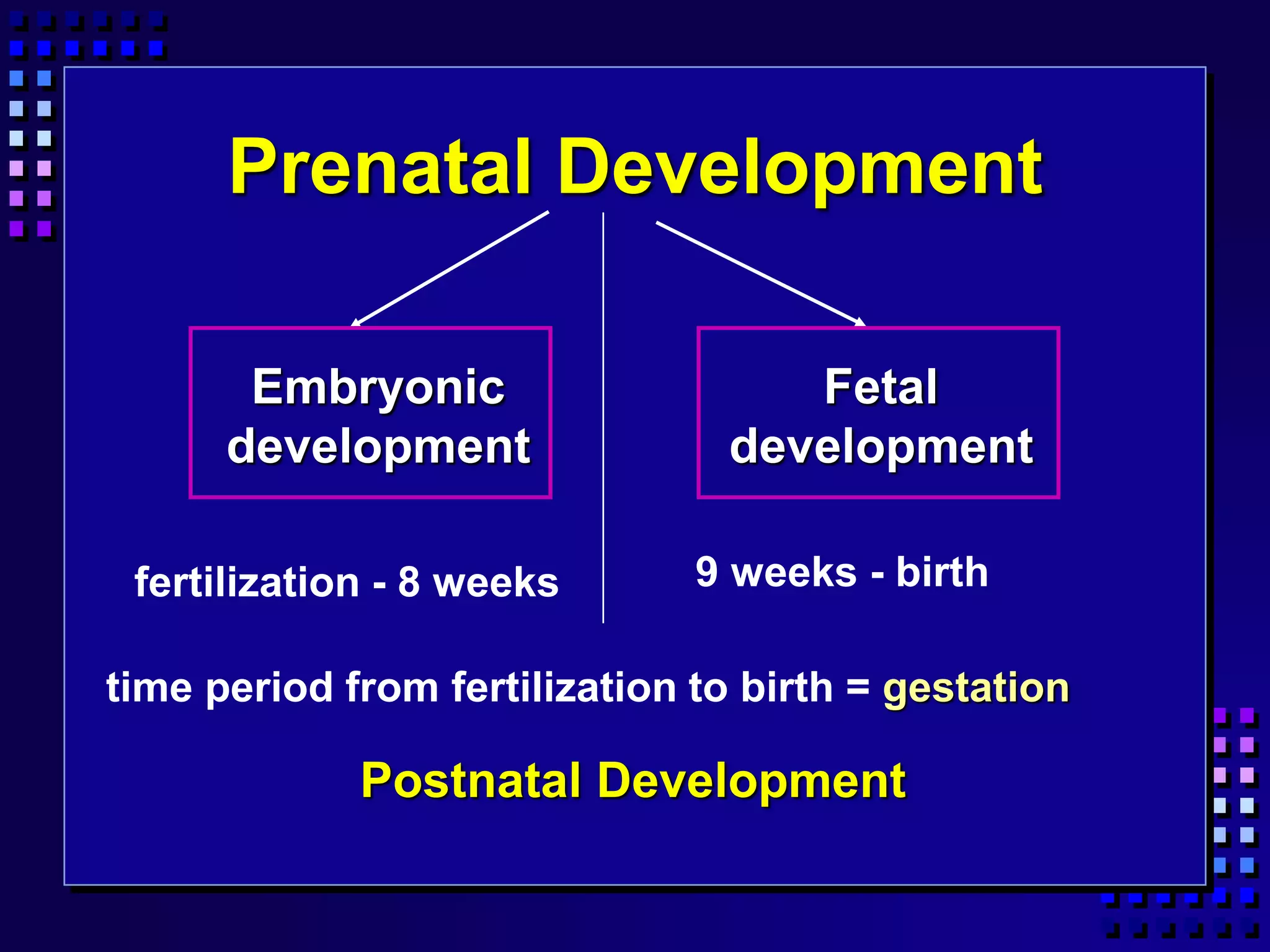
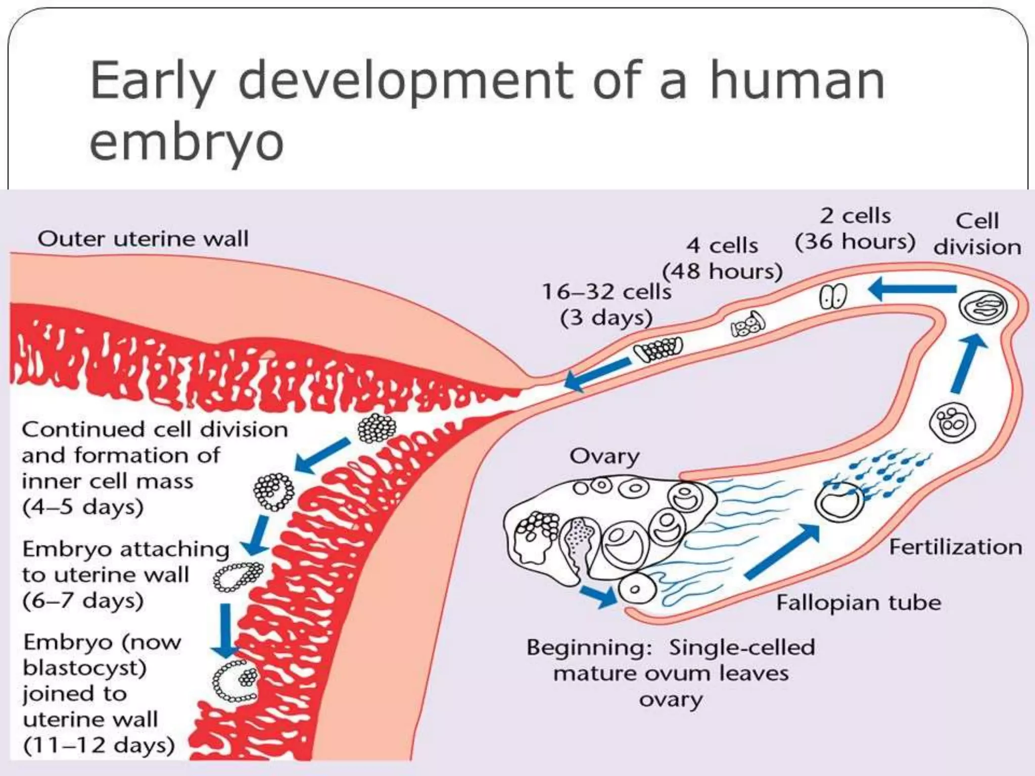
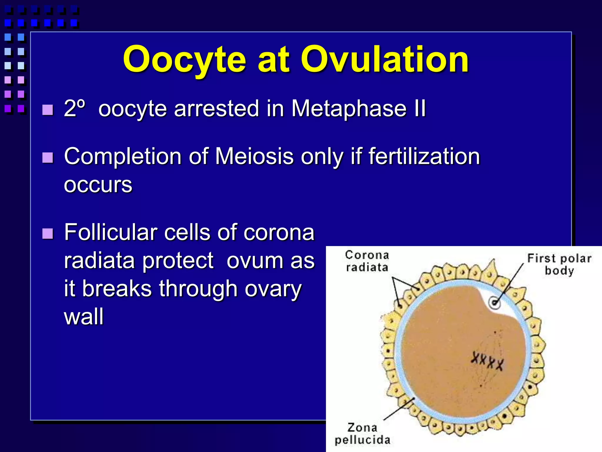
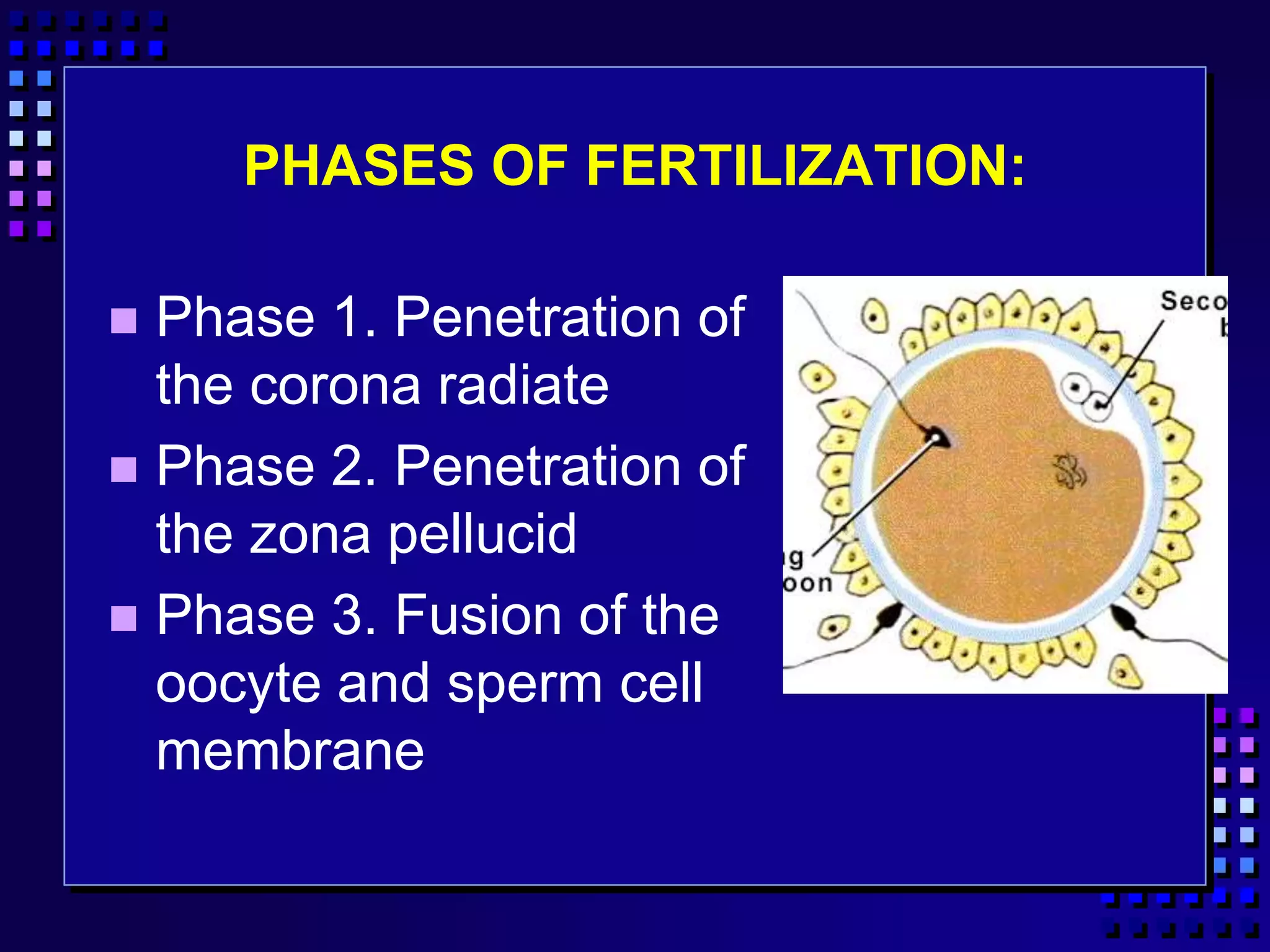
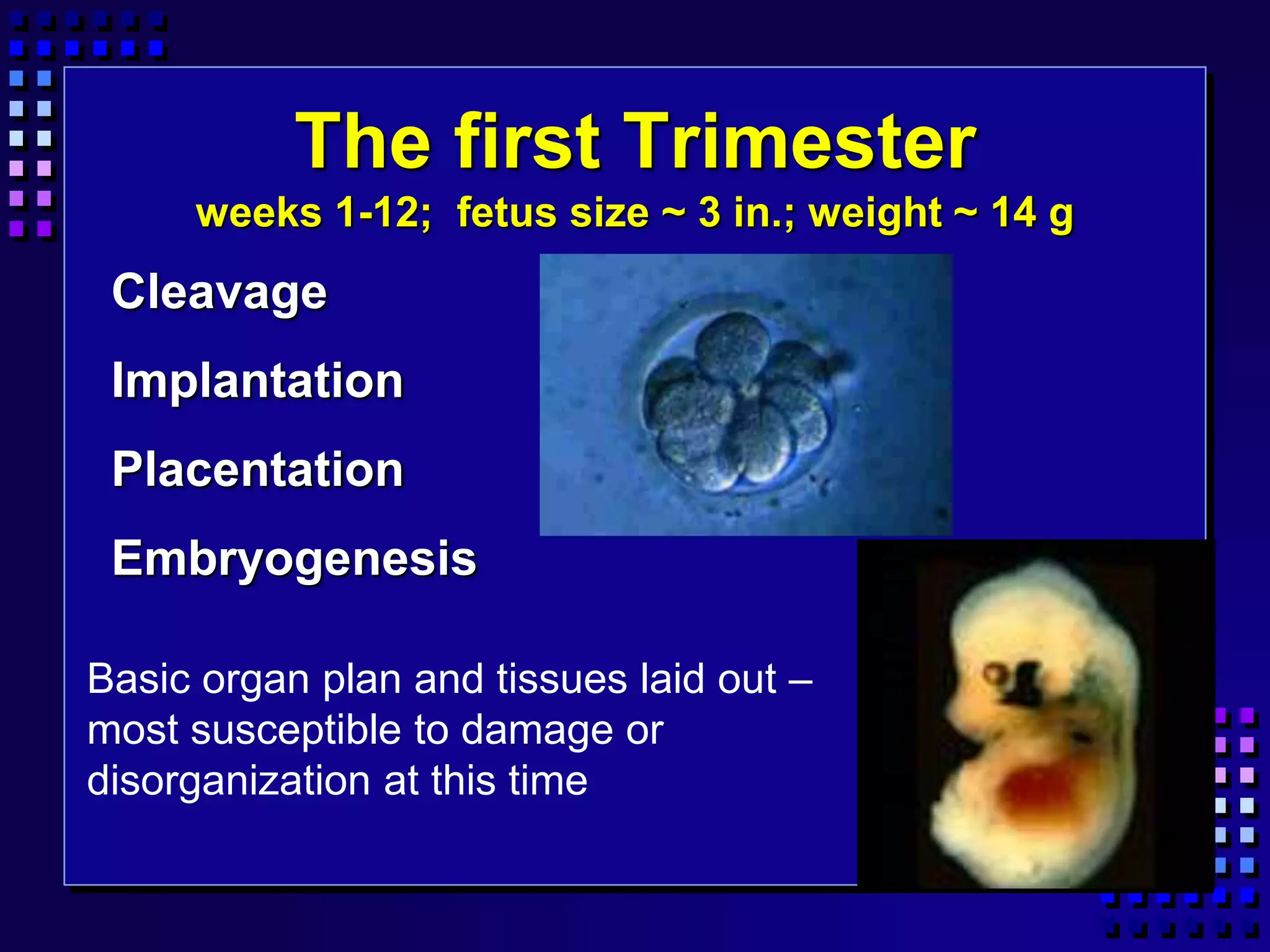
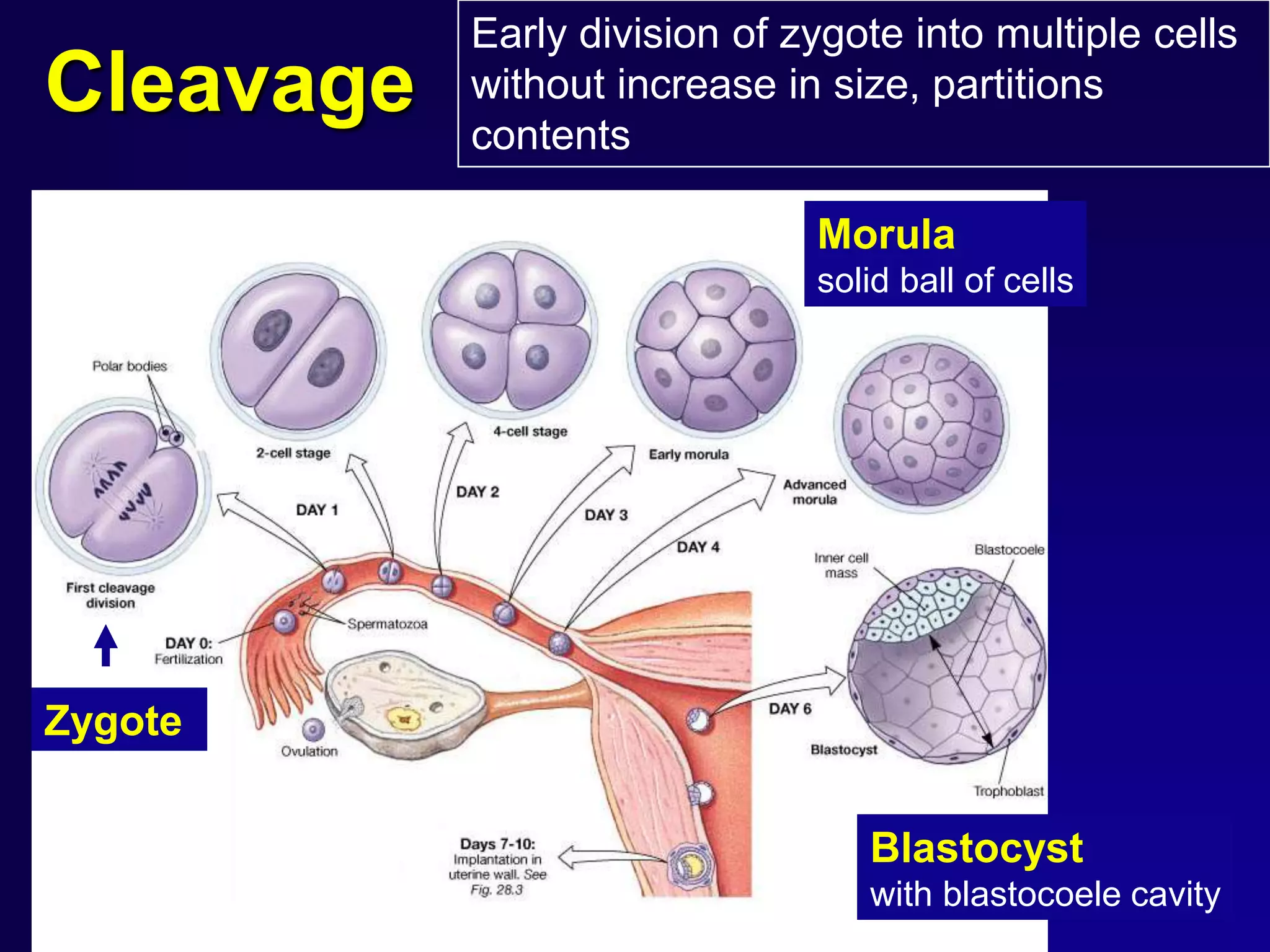
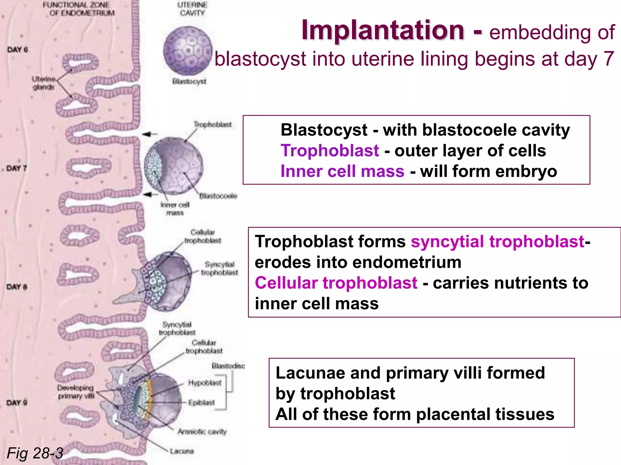
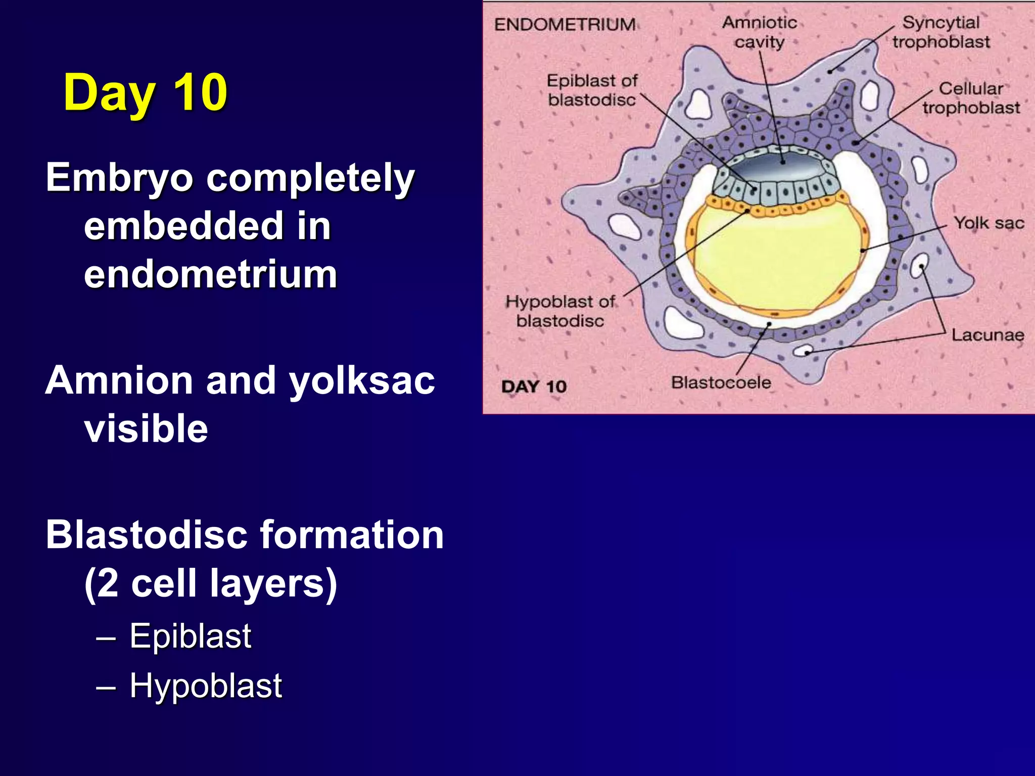
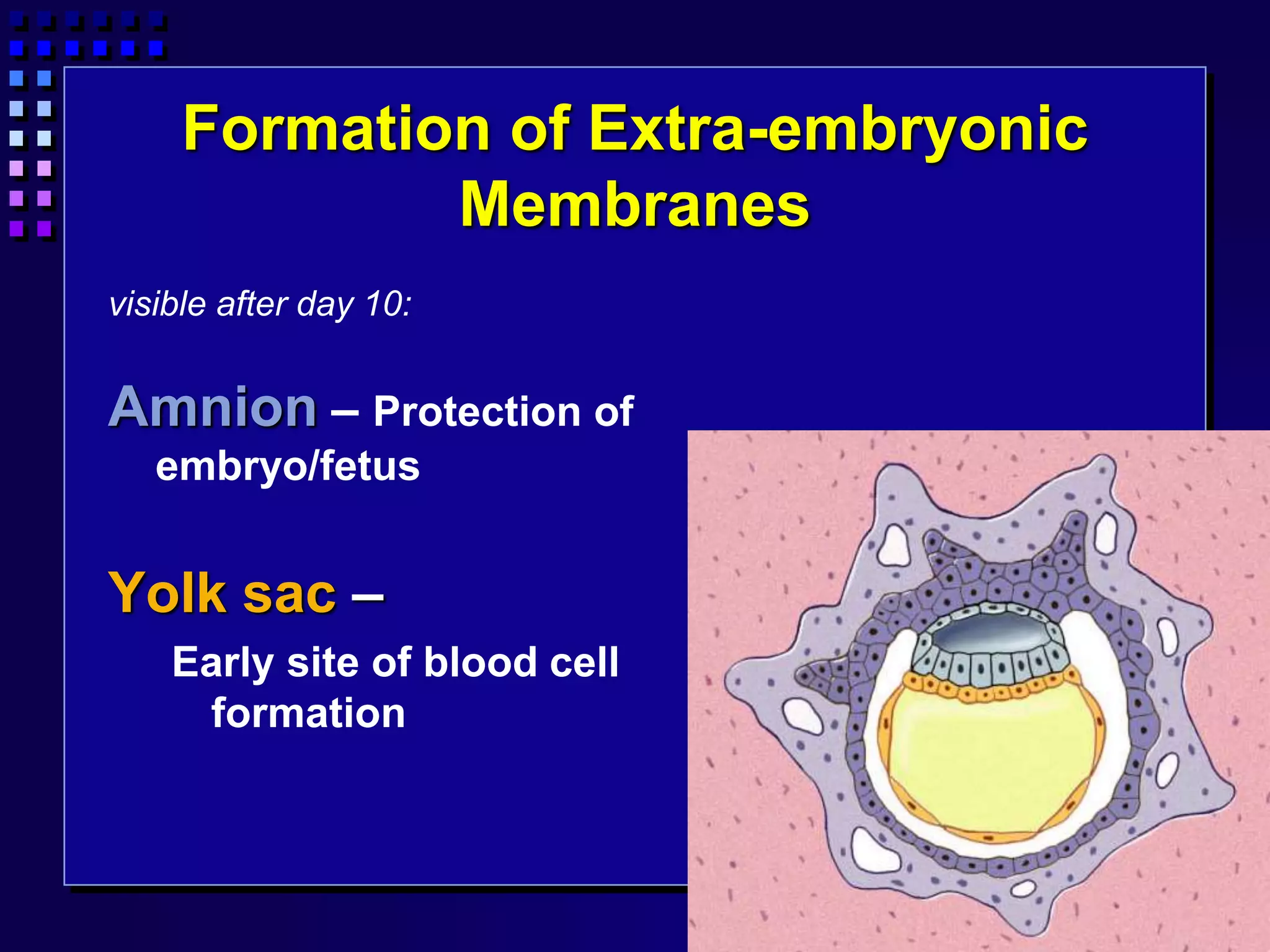
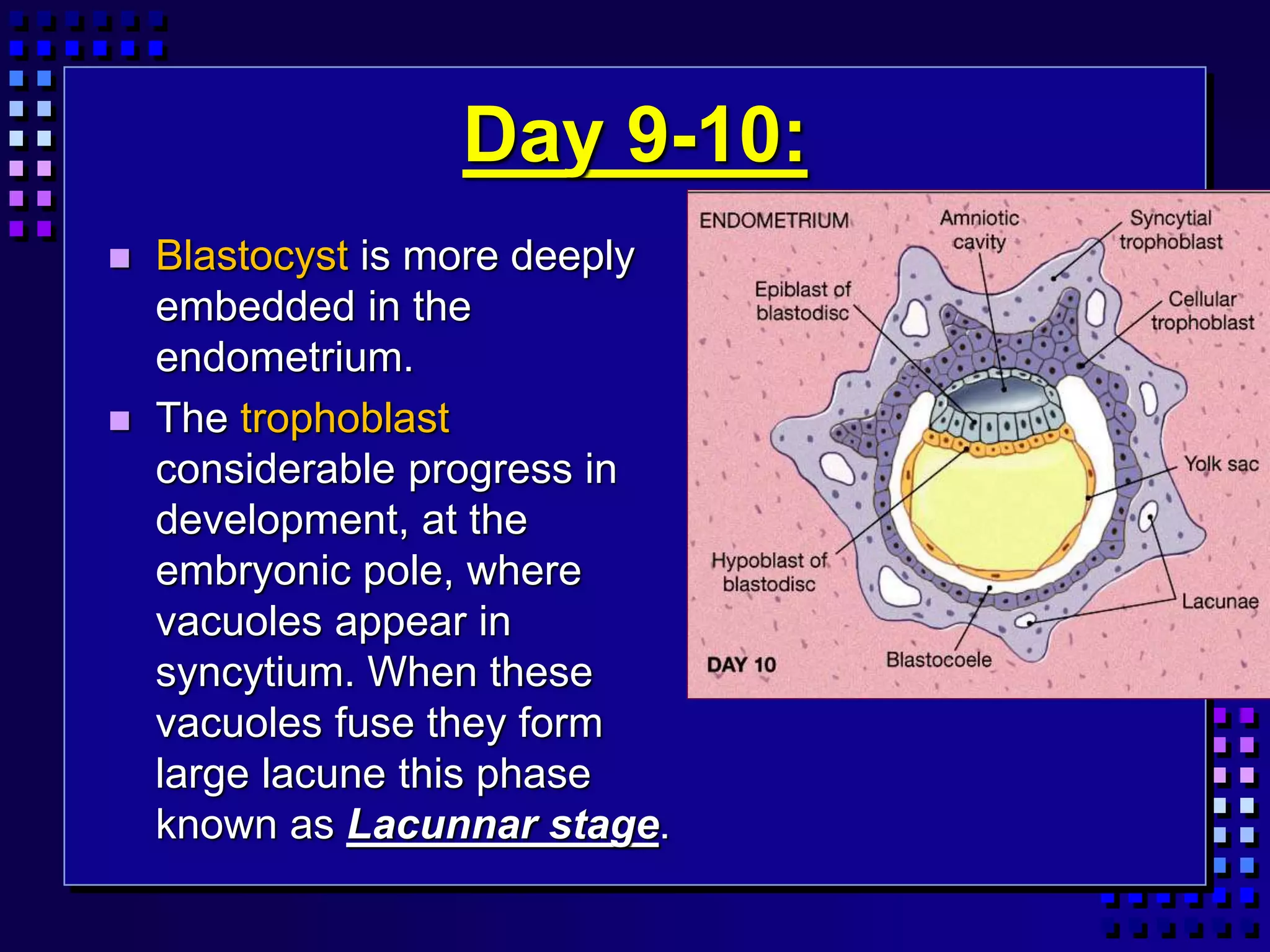
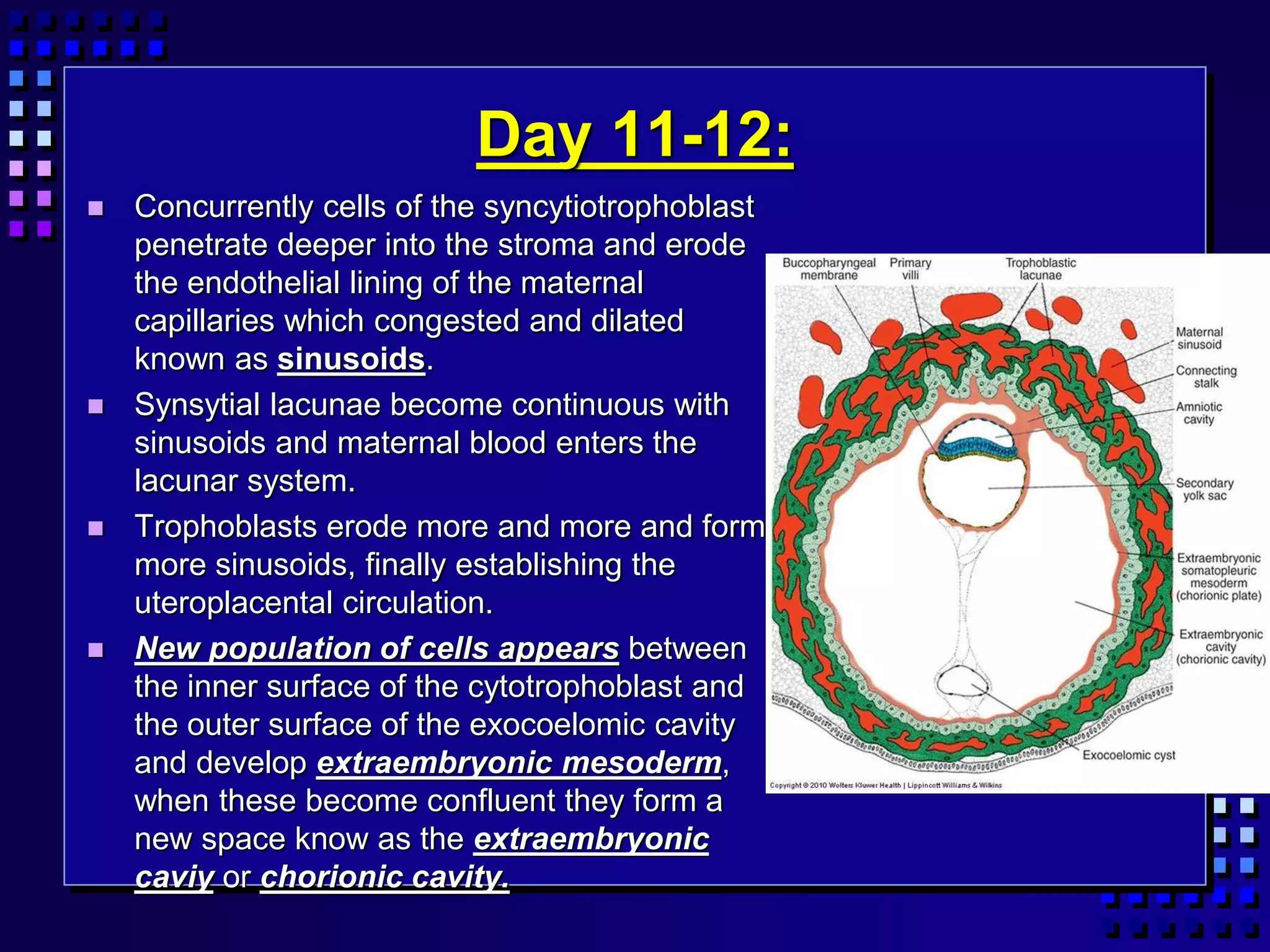
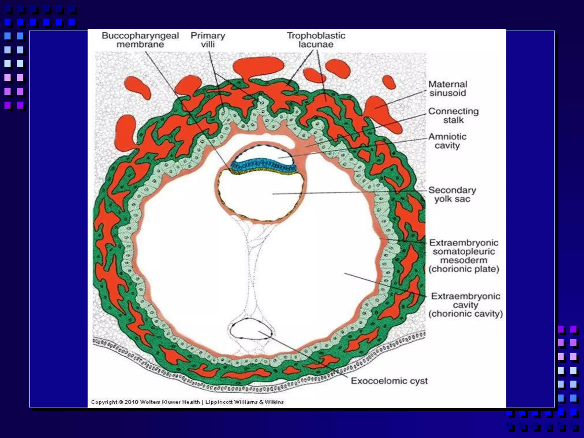
Human Development
Muhammad Ishaque Mujeeb Rehman
Lecturer
Department of Eastern Medicine
Faculty of Pharmacy & Health Sciences
University of Balochistan
Prenatal Development
Embryonic
development
fertilization - 8 weeks
Fetal
development
9 weeks - birth
Postnatal Development
time period from fertilization to birth = gestation
Oocyte at Ovulation
 2º oocyte arrested in Metaphase II
 Completion of Meiosis only if fertilization
occurs
 Follicular cells of corona
radiata protect ovum as
it breaks through ovary
wall
Fertilization
Taking place in ampula
Viability of gametes:
– Oocyte 12-24 h
– Sperm 12-48 h
Acrosome contains hyaluronidase
 acrosomal reaction breaks down intercellular
cement between adjacent follicle cells
Single sperm fuses with oocyte
fusion of sperm and oocyte pronuclei
Fertilization:
 Only 1% sperm reach to target. (300-500) out of 2 to
6 million. The trip from cervix to oviduct (ampulla of
uterine tube) may occur rapid as 30 minutes and may
delay to 6 days.
 Spermatozoa are not able to fertilize the oocyte
immediately upon arrival in the female genital tract
but must undergo
1. Capacitation:
2. The acrosome reaction:
Capacitation and
Acrosome reaction:
1 CAPACITATION:
 It is removal of the glycoprotein coat covering the external
surface of the sperm by the result of secretion of the uterine
tube. Capacitation helps sperm to easily penetrate corona
radiata.

2. THE ACROSOME REACTION:
 This reaction occur after binding to the zona pellucid, this
reaction enzyme (acrosin & trypsin-like substance) release by
acrosome to penetrate the zona pellucid.
PHASES OF FERTILIZATION:
 Phase 1. Penetration of
the corona radiate
 Phase 2. Penetration of
the zona pellucid
 Phase 3. Fusion of the
oocyte and sperm cell
membrane
The first Trimester
weeks 1-12; fetus size ~ 3 in.; weight ~ 14 g
Cleavage
Implantation
Placentation
Embryogenesis
Basic organ plan and tissues laid out –
most susceptible to damage or
disorganization at this time
CLEAVAGE:
 Once the zygote has reached the two-cell stage, it undergoes a
series of mitotic division, increase in the number of cells. These
cells, which become smaller with each cleavage division are
known as blastomeres. Until the eight-cell stage they form a
loosely arranged clump, after the third cleavage blastomeres
maximize their contact with each other forming the compact ball
of cells. After 3 days of fertilization cells of the compacted
embryo divide again to form a 16-cell morulla (Mulberry)
 Inner cells of the morulla constitute the inner cell mass and
surrounding cells composed the outer cell mass. The inner cell
mass gives rise to tissue of the embryo proper, and the outer
cell mass form the trophoblast, which later contributes to the
placenta.
Blastocyst
with blastocoele cavity
Morula
solid ball of cells
Cleavage
Early division of zygote into multiple cells
without increase in size, partitions
contents
Zygote
BLASTOCYT FORMATION:
 After enters of morulla in uterine cavity, fluid begins
to penetrate through the zona pellucid into the
intercellular spaces of the inner cell mass. Gradually
the intercellular space becomes confluent, and finally
a single cavity forms blastocele. At this time embryo
is a blastocyste, Cell of inner cells mass now called
the embryoblast are at one pole and those of the
outer cell mass (trophoblast) flatten and form the
epithelial wall of the blastocyst.
Blastocyst - with blastocoele cavity
Trophoblast - outer layer of cells
Inner cell mass - will form embryo
Lacunae and primary villi formed
by trophoblast
All of these form placental tissues
Implantation - embedding of
blastocyst into uterine lining begins at day 7
Fig 28-3
Trophoblast forms syncytial trophoblast-
erodes into endometrium
Cellular trophoblast - carries nutrients to
inner cell mass
Day 10
Embryo completely
embedded in
endometrium
Amnion and yolksac
visible
Blastodisc formation
(2 cell layers)
– Epiblast
– Hypoblast
Formation of Extra-embryonic
Membranes
visible after day 10:
Amnion – Protection of
embryo/fetus
Yolk sac –
Early site of blood cell
formation
Day 9-10:
 Blastocyst is more deeply
embedded in the
endometrium.
 The trophoblast
considerable progress in
development, at the
embryonic pole, where
vacuoles appear in
syncytium. When these
vacuoles fuse they form
large lacune this phase
known as Lacunnar stage.
Day 11-12:
 Concurrently cells of the syncytiotrophoblast
penetrate deeper into the stroma and erode
the endothelial lining of the maternal
capillaries which congested and dilated
known as sinusoids.
 Synsytial lacunae become continuous with
sinusoids and maternal blood enters the
lacunar system.
 Trophoblasts erode more and more and form
more sinusoids, finally establishing the
uteroplacental circulation.
 New population of cells appears between
the inner surface of the cytotrophoblast and
the outer surface of the exocoelomic cavity
and develop extraembryonic mesoderm,
when these become confluent they form a
new space know as the extraembryonic
caviy or chorionic cavity.
Day 13-14:
 Trophoblast form the primary villi with the
syncytiotrophoblast.
 In the main time the hypoblast produces additional cells that
migrate along the inside of the exocoelomic membrane.
These cells proliferate and gradually form a new cavity within
the exocoelomic cavity. This cavity is known as the
secondary yolk sac or definitive yolk sac.
 Exocoelomic cavity or primitive yolk sac pinched off and
this portions are represented by exocoelomic cysts.
Which are found in the extraembryonic coelom or
chorionic cavity.
 The lining inside of the cytotrophoblast is known as
chorionic plate.
 The only place where extraembryonic mesoderm
traverses the chorionic cavity is in the connecting stalk.
The stalk become the umbilical cord./
1st and 2nd week of human development

1st and 2nd week of human development

  • 1.
    Human Development Muhammad IshaqueMujeeb Rehman Lecturer Department of Eastern Medicine Faculty of Pharmacy & Health Sciences University of Balochistan
  • 2.
    Prenatal Development Embryonic development fertilization -8 weeks Fetal development 9 weeks - birth Postnatal Development time period from fertilization to birth = gestation
  • 4.
    Oocyte at Ovulation 2º oocyte arrested in Metaphase II  Completion of Meiosis only if fertilization occurs  Follicular cells of corona radiata protect ovum as it breaks through ovary wall
  • 5.
    Fertilization Taking place inampula Viability of gametes: – Oocyte 12-24 h – Sperm 12-48 h Acrosome contains hyaluronidase  acrosomal reaction breaks down intercellular cement between adjacent follicle cells Single sperm fuses with oocyte fusion of sperm and oocyte pronuclei
  • 6.
    Fertilization:  Only 1%sperm reach to target. (300-500) out of 2 to 6 million. The trip from cervix to oviduct (ampulla of uterine tube) may occur rapid as 30 minutes and may delay to 6 days.  Spermatozoa are not able to fertilize the oocyte immediately upon arrival in the female genital tract but must undergo 1. Capacitation: 2. The acrosome reaction:
  • 7.
    Capacitation and Acrosome reaction: 1CAPACITATION:  It is removal of the glycoprotein coat covering the external surface of the sperm by the result of secretion of the uterine tube. Capacitation helps sperm to easily penetrate corona radiata.  2. THE ACROSOME REACTION:  This reaction occur after binding to the zona pellucid, this reaction enzyme (acrosin & trypsin-like substance) release by acrosome to penetrate the zona pellucid.
  • 8.
    PHASES OF FERTILIZATION: Phase 1. Penetration of the corona radiate  Phase 2. Penetration of the zona pellucid  Phase 3. Fusion of the oocyte and sperm cell membrane
  • 11.
    The first Trimester weeks1-12; fetus size ~ 3 in.; weight ~ 14 g Cleavage Implantation Placentation Embryogenesis Basic organ plan and tissues laid out – most susceptible to damage or disorganization at this time
  • 12.
    CLEAVAGE:  Once thezygote has reached the two-cell stage, it undergoes a series of mitotic division, increase in the number of cells. These cells, which become smaller with each cleavage division are known as blastomeres. Until the eight-cell stage they form a loosely arranged clump, after the third cleavage blastomeres maximize their contact with each other forming the compact ball of cells. After 3 days of fertilization cells of the compacted embryo divide again to form a 16-cell morulla (Mulberry)  Inner cells of the morulla constitute the inner cell mass and surrounding cells composed the outer cell mass. The inner cell mass gives rise to tissue of the embryo proper, and the outer cell mass form the trophoblast, which later contributes to the placenta.
  • 13.
    Blastocyst with blastocoele cavity Morula solidball of cells Cleavage Early division of zygote into multiple cells without increase in size, partitions contents Zygote
  • 14.
    BLASTOCYT FORMATION:  Afterenters of morulla in uterine cavity, fluid begins to penetrate through the zona pellucid into the intercellular spaces of the inner cell mass. Gradually the intercellular space becomes confluent, and finally a single cavity forms blastocele. At this time embryo is a blastocyste, Cell of inner cells mass now called the embryoblast are at one pole and those of the outer cell mass (trophoblast) flatten and form the epithelial wall of the blastocyst.
  • 15.
    Blastocyst - withblastocoele cavity Trophoblast - outer layer of cells Inner cell mass - will form embryo Lacunae and primary villi formed by trophoblast All of these form placental tissues Implantation - embedding of blastocyst into uterine lining begins at day 7 Fig 28-3 Trophoblast forms syncytial trophoblast- erodes into endometrium Cellular trophoblast - carries nutrients to inner cell mass
  • 16.
    Day 10 Embryo completely embeddedin endometrium Amnion and yolksac visible Blastodisc formation (2 cell layers) – Epiblast – Hypoblast
  • 17.
    Formation of Extra-embryonic Membranes visibleafter day 10: Amnion – Protection of embryo/fetus Yolk sac – Early site of blood cell formation
  • 18.
    Day 9-10:  Blastocystis more deeply embedded in the endometrium.  The trophoblast considerable progress in development, at the embryonic pole, where vacuoles appear in syncytium. When these vacuoles fuse they form large lacune this phase known as Lacunnar stage.
  • 19.
    Day 11-12:  Concurrentlycells of the syncytiotrophoblast penetrate deeper into the stroma and erode the endothelial lining of the maternal capillaries which congested and dilated known as sinusoids.  Synsytial lacunae become continuous with sinusoids and maternal blood enters the lacunar system.  Trophoblasts erode more and more and form more sinusoids, finally establishing the uteroplacental circulation.  New population of cells appears between the inner surface of the cytotrophoblast and the outer surface of the exocoelomic cavity and develop extraembryonic mesoderm, when these become confluent they form a new space know as the extraembryonic caviy or chorionic cavity.
  • 20.
    Day 13-14:  Trophoblastform the primary villi with the syncytiotrophoblast.  In the main time the hypoblast produces additional cells that migrate along the inside of the exocoelomic membrane. These cells proliferate and gradually form a new cavity within the exocoelomic cavity. This cavity is known as the secondary yolk sac or definitive yolk sac.
  • 22.
     Exocoelomic cavityor primitive yolk sac pinched off and this portions are represented by exocoelomic cysts. Which are found in the extraembryonic coelom or chorionic cavity.  The lining inside of the cytotrophoblast is known as chorionic plate.  The only place where extraembryonic mesoderm traverses the chorionic cavity is in the connecting stalk. The stalk become the umbilical cord./