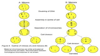
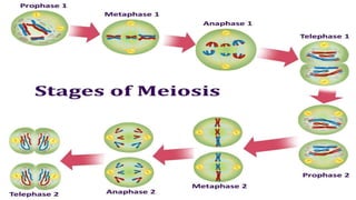
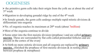
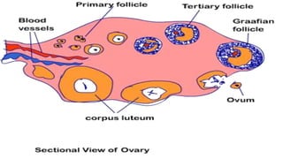
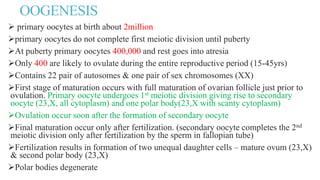
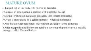
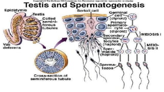
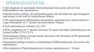
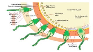
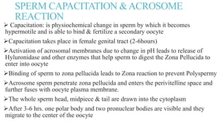
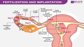
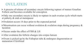
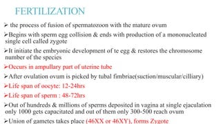
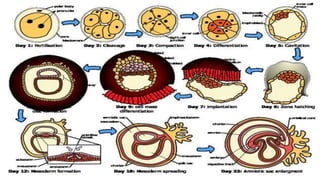
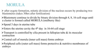
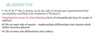
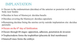
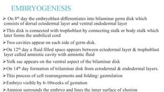
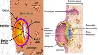
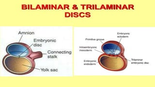
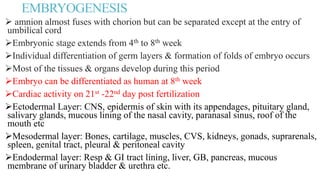
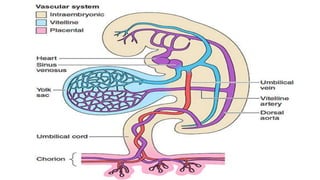
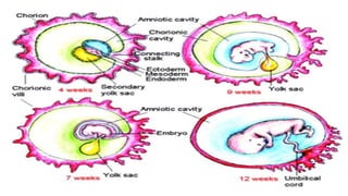
FERTILIZATION &
EMBRYOLOGY
By: Maj Saminder Malik
MSc (N) Obs & Gyn
GAMETOGENESIS
The process involved in the maturation of two highly specialized cells
(spermatozoon & ovum) before they unite to form zygote.
Oogenesis : process of maturation of ovum
 Spermatogenesis: process of maturation of spermatids
OOGENESIS
 the primitive germ cells take their origin from the yolk sac at about the end of
3rd week
Migration to developing gonadal ridge by end of the 4th week
In female gonads, the germ cells undergo multiple rapid mitotic divisions &
differentiate into oogonia
No. of oogonia reaches its maximum at 20th week (about 7million)
Most of the oogonia continue to divide
Some enter into the first meiotic division (prophase ) and are called primary
oocytes. These are surrounded by flat cells called primordial follicles and are
present in the cortex of the ovary
At birth no more mitotic division and all oogonia are replaced by primary
oocytes . (finished the prophase of first meiotic division & in resting phase
between prophase & metaphase)
OOGENESIS
 primary oocytes at birth about 2million
primary oocytes do not complete first meiotic division until puberty
At puberty primary oocytes 400,000 and rest goes into atresia
Only 400 are likely to ovulate during the entire reproductive period (15-45yrs)
Contains 22 pair of autosomes & one pair of sex chromosomes (XX)
First stage of maturation occurs with full maturation of ovarian follicle just prior to
ovulation. Primary oocyte undergoes 1st meiotic division giving rise to secondary
oocyte (23,X, all cytoplasm) and one polar body(23,X with scanty cytoplasm)
Ovulation occur soon after the formation of secondary oocyte
Final maturation occur only after fertilization. (secondary oocyte completes the 2nd
meiotic division only after fertilization by the sperm in fallopian tube)
Fertilization results in formation of two unequal daughter cells – mature ovum (23,X)
& second polar body (23,X)
Polar bodies degenerate
MATURE OVUM
 Largest cell in the body, 130 micron in diameter
Consists of cytoplasm & a nucleus with nucleolus (23,X)
During fertilization nucleus is converted into female pronucleus
Ovum is surrounded by a cell membrane – vitelline membrane
Also has an outer transparent mucoprotein envelope – zona pellucida
After escape from follicle ovum retains a covering of granulosa cells radially
arranged called Corona Radiata
SPERMATOGENESIS
 Development of spermatids from primordial male germ cells & their
differentiation into spermatozoa
Shortly before puberty, the primordial germ cells develop into spermatogonia
and remain in the wall of seminiferous tubules
The spermatogonia differentiate into primary spermatocytes which remain in
stage of prophase of 1st meiotic division (about 16days)
Each spermatocyte contains (22, XY)
With completion of 1st meiotic division, 02 equal secondary spermatocytes are
formed (either 23,X/ 23,Y).
Immediately follows second meiotic division with formation of 04 spermatids
(two each 23,X & 23,Y)
Spermtids undergo extensive morphological differentiation & conversion into
spermatozoa
Development of Spermatogonium to mature spermatozoon = 61days
SPERM CAPACITATION &ACROSOME
REACTION
 Capacitation: is physiochemical change in sperm by which it becomes
hypermotile and is able to bind & fertilize a secondary oocyte
Capacitation takes place in female genital tract (2-6hours)
Activation of acrosomal membranes due to change in pH leads to release of
Hyluronidase and other enzymes that help sperm to digest the Zona Pellucida to
enter into oocyte
Binding of sperm to zona pellucida leads to Zona reaction to prevent Polyspermy
Acrosome sperm penetrate zona pellucida and enters the perivitelline space and
further fuses with oocyte plasma membrane.
The whole sperm head, midpiece & tail are drawn into the cytoplasm
After 3-6 hrs. one polar body and two pronuclear bodies are visible and they
migrate to the center of the oocyte
OVULATION
 A process of release of secondary oocyte following rupture of mature Graafian
Follicle and becomes available for conception
Only one secondary oocyte is likely to rupture in each ovarian cycle which starts
at puberty & ends at menopause
Ovulation occurs 14 days prior to the expected period
Menstruation can occur without ovulation & ovulation stops during pregnancy &
lactation
Occurs under the effect of FSH & LH
After ovulation the follicle changes into corpus luteum
Ovum is picked up by the Fallopian tube & undergoes degeneration or
maturation if fertilized
FERTILIZATION
 the process of fusion of spermatozoon with the mature ovum
Begins with sperm egg collision & ends with production of a mononucleated
single cell called zygote
It initiate the embryonic development of te egg & restores the chromosome
number of the species
Occurs in ampullary part of uterine tube
After ovulation ovum is picked by tubal fimbriae(suction/muscular/cilliary)
Life span of oocyte: 12-24hrs
Life span of sperm : 48-72hrs
Out of hundreds & millions of sperms deposited in vagina at single ejaculation
only 1000 gets capacitated and out of them only 300-500 reach ovum
Union of gametes takes place (46XX or 46XY), forms Zygote
MORULA
 after zygote formation, mitotic division of the nucleus occurs by producing two
blastomeres (takes 30hrs after fertilization)
Blastomere continue to divide by binary division through 4, 8, 16-cell stage until
a cluster is formed called MORULA (mulberry like)
Spend 3 days in uterine tube
Enters the uterine cavity the 4th day in 16-64 cell stage
Transport is controlled by cilia present in fallopian tube & its muscular
contraction
Central cell of morula (inner cell mass) forms embryo
Peripheral cells (outer cell mass) forms protective & nutritive membranes of
embryo
BLASTOCYST
 On 4th & 5th day in uterine cavity the cells of morula gets separated and fluid
accumulation resulting in the formation of blastocyst
Implantation occurs by Zona Hatching (lysis of zona pellucida layer & escape of
embryo)
Cells on outer side of morula – trophectoderm differentiates into chorion which
further becomes placenta
Cells on inner side differentiates into embryo
IMPLANTATION
 Occurs in the endometrium (decidua) of the anterior or posterior wall of the
body near fundus
Decidua in base of blastocyst: decidua basalis
Decidua covering the blastocyst: decidua capsularis
Remaining decidua lining the uterine cavity outside implantation site: decidua
parietalis
Occurs on the 6th day of fertilization
Occurs through 04 stages: apposition, adhesion, penetration & invasion
Trophectoderm forms the trophoblast (placenta & fetal membranes)
Innercell mass forms the embryo
EMBRYOGENESIS
 On 8th day the embryoblast differentiates into bilaminar germ disk which
consists of dorsal ectodermal layer and ventral endodermal layer
This disk is connected with trophoblast by connecting stalk or body stalk which
later forms the umbilical cord
Two cavities appear on each side of germ disk.
On 12th day a fluid filled space appears between ectodermal layer & trophoblast
layer called amniotic cavity with amniotic fluid
Yolk sac appears on the ventral aspect of the bilaminar disk
On 14th day formation of trilaminar disk from ectodermal & endodermal layers.
This process of cell rearrangements and folding: gastrulation
Embryo visible by 6-10weeks of gestation
Amnion surrounds the embryo and lines the inner surface of chorion
EMBRYOGENESIS
 amnion almost fuses with chorion but can be separated except at the entry of
umbilical cord
Embryonic stage extends from 4th to 8th week
Individual differentiation of germ layers & formation of folds of embryo occurs
Most of the tissues & organs develop during this period
Embryo can be differentiated as human at 8th week
Cardiac activity on 21st -22nd day post fertilization
Ectodermal Layer: CNS, epidermis of skin with its appendages, pituitary gland,
salivary glands, mucous lining of the nasal cavity, paranasal sinus, roof of the
mouth etc
Mesodermal layer: Bones, cartilage, muscles, CVS, kidneys, gonads, suprarenals,
spleen, genital tract, pleural & peritoneal cavity
Endodermal layer: Resp & GI tract lining, liver, GB, pancreas, mucous
membrane of urinary bladder & urethra etc.
Embryology & fertilization

Embryology & fertilization

  • 1.
    FERTILIZATION & EMBRYOLOGY By: MajSaminder Malik MSc (N) Obs & Gyn
  • 2.
    GAMETOGENESIS The process involvedin the maturation of two highly specialized cells (spermatozoon & ovum) before they unite to form zygote. Oogenesis : process of maturation of ovum  Spermatogenesis: process of maturation of spermatids
  • 5.
    OOGENESIS  the primitivegerm cells take their origin from the yolk sac at about the end of 3rd week Migration to developing gonadal ridge by end of the 4th week In female gonads, the germ cells undergo multiple rapid mitotic divisions & differentiate into oogonia No. of oogonia reaches its maximum at 20th week (about 7million) Most of the oogonia continue to divide Some enter into the first meiotic division (prophase ) and are called primary oocytes. These are surrounded by flat cells called primordial follicles and are present in the cortex of the ovary At birth no more mitotic division and all oogonia are replaced by primary oocytes . (finished the prophase of first meiotic division & in resting phase between prophase & metaphase)
  • 7.
    OOGENESIS  primary oocytesat birth about 2million primary oocytes do not complete first meiotic division until puberty At puberty primary oocytes 400,000 and rest goes into atresia Only 400 are likely to ovulate during the entire reproductive period (15-45yrs) Contains 22 pair of autosomes & one pair of sex chromosomes (XX) First stage of maturation occurs with full maturation of ovarian follicle just prior to ovulation. Primary oocyte undergoes 1st meiotic division giving rise to secondary oocyte (23,X, all cytoplasm) and one polar body(23,X with scanty cytoplasm) Ovulation occur soon after the formation of secondary oocyte Final maturation occur only after fertilization. (secondary oocyte completes the 2nd meiotic division only after fertilization by the sperm in fallopian tube) Fertilization results in formation of two unequal daughter cells – mature ovum (23,X) & second polar body (23,X) Polar bodies degenerate
  • 8.
    MATURE OVUM  Largestcell in the body, 130 micron in diameter Consists of cytoplasm & a nucleus with nucleolus (23,X) During fertilization nucleus is converted into female pronucleus Ovum is surrounded by a cell membrane – vitelline membrane Also has an outer transparent mucoprotein envelope – zona pellucida After escape from follicle ovum retains a covering of granulosa cells radially arranged called Corona Radiata
  • 11.
    SPERMATOGENESIS  Development ofspermatids from primordial male germ cells & their differentiation into spermatozoa Shortly before puberty, the primordial germ cells develop into spermatogonia and remain in the wall of seminiferous tubules The spermatogonia differentiate into primary spermatocytes which remain in stage of prophase of 1st meiotic division (about 16days) Each spermatocyte contains (22, XY) With completion of 1st meiotic division, 02 equal secondary spermatocytes are formed (either 23,X/ 23,Y). Immediately follows second meiotic division with formation of 04 spermatids (two each 23,X & 23,Y) Spermtids undergo extensive morphological differentiation & conversion into spermatozoa Development of Spermatogonium to mature spermatozoon = 61days
  • 13.
    SPERM CAPACITATION &ACROSOME REACTION Capacitation: is physiochemical change in sperm by which it becomes hypermotile and is able to bind & fertilize a secondary oocyte Capacitation takes place in female genital tract (2-6hours) Activation of acrosomal membranes due to change in pH leads to release of Hyluronidase and other enzymes that help sperm to digest the Zona Pellucida to enter into oocyte Binding of sperm to zona pellucida leads to Zona reaction to prevent Polyspermy Acrosome sperm penetrate zona pellucida and enters the perivitelline space and further fuses with oocyte plasma membrane. The whole sperm head, midpiece & tail are drawn into the cytoplasm After 3-6 hrs. one polar body and two pronuclear bodies are visible and they migrate to the center of the oocyte
  • 15.
    OVULATION  A processof release of secondary oocyte following rupture of mature Graafian Follicle and becomes available for conception Only one secondary oocyte is likely to rupture in each ovarian cycle which starts at puberty & ends at menopause Ovulation occurs 14 days prior to the expected period Menstruation can occur without ovulation & ovulation stops during pregnancy & lactation Occurs under the effect of FSH & LH After ovulation the follicle changes into corpus luteum Ovum is picked up by the Fallopian tube & undergoes degeneration or maturation if fertilized
  • 16.
    FERTILIZATION  the processof fusion of spermatozoon with the mature ovum Begins with sperm egg collision & ends with production of a mononucleated single cell called zygote It initiate the embryonic development of te egg & restores the chromosome number of the species Occurs in ampullary part of uterine tube After ovulation ovum is picked by tubal fimbriae(suction/muscular/cilliary) Life span of oocyte: 12-24hrs Life span of sperm : 48-72hrs Out of hundreds & millions of sperms deposited in vagina at single ejaculation only 1000 gets capacitated and out of them only 300-500 reach ovum Union of gametes takes place (46XX or 46XY), forms Zygote
  • 18.
    MORULA  after zygoteformation, mitotic division of the nucleus occurs by producing two blastomeres (takes 30hrs after fertilization) Blastomere continue to divide by binary division through 4, 8, 16-cell stage until a cluster is formed called MORULA (mulberry like) Spend 3 days in uterine tube Enters the uterine cavity the 4th day in 16-64 cell stage Transport is controlled by cilia present in fallopian tube & its muscular contraction Central cell of morula (inner cell mass) forms embryo Peripheral cells (outer cell mass) forms protective & nutritive membranes of embryo
  • 19.
    BLASTOCYST  On 4th& 5th day in uterine cavity the cells of morula gets separated and fluid accumulation resulting in the formation of blastocyst Implantation occurs by Zona Hatching (lysis of zona pellucida layer & escape of embryo) Cells on outer side of morula – trophectoderm differentiates into chorion which further becomes placenta Cells on inner side differentiates into embryo
  • 21.
    IMPLANTATION  Occurs inthe endometrium (decidua) of the anterior or posterior wall of the body near fundus Decidua in base of blastocyst: decidua basalis Decidua covering the blastocyst: decidua capsularis Remaining decidua lining the uterine cavity outside implantation site: decidua parietalis Occurs on the 6th day of fertilization Occurs through 04 stages: apposition, adhesion, penetration & invasion Trophectoderm forms the trophoblast (placenta & fetal membranes) Innercell mass forms the embryo
  • 23.
    EMBRYOGENESIS  On 8thday the embryoblast differentiates into bilaminar germ disk which consists of dorsal ectodermal layer and ventral endodermal layer This disk is connected with trophoblast by connecting stalk or body stalk which later forms the umbilical cord Two cavities appear on each side of germ disk. On 12th day a fluid filled space appears between ectodermal layer & trophoblast layer called amniotic cavity with amniotic fluid Yolk sac appears on the ventral aspect of the bilaminar disk On 14th day formation of trilaminar disk from ectodermal & endodermal layers. This process of cell rearrangements and folding: gastrulation Embryo visible by 6-10weeks of gestation Amnion surrounds the embryo and lines the inner surface of chorion
  • 26.
    EMBRYOGENESIS  amnion almostfuses with chorion but can be separated except at the entry of umbilical cord Embryonic stage extends from 4th to 8th week Individual differentiation of germ layers & formation of folds of embryo occurs Most of the tissues & organs develop during this period Embryo can be differentiated as human at 8th week Cardiac activity on 21st -22nd day post fertilization Ectodermal Layer: CNS, epidermis of skin with its appendages, pituitary gland, salivary glands, mucous lining of the nasal cavity, paranasal sinus, roof of the mouth etc Mesodermal layer: Bones, cartilage, muscles, CVS, kidneys, gonads, suprarenals, spleen, genital tract, pleural & peritoneal cavity Endodermal layer: Resp & GI tract lining, liver, GB, pancreas, mucous membrane of urinary bladder & urethra etc.