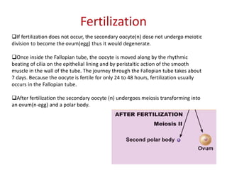
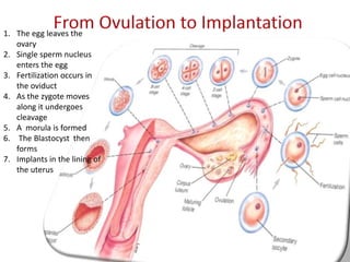
1. Oogenesis begins before birth as oogonia undergo mitosis to form primary oocytes, which begin but halt meiosis. At puberty, one oocyte resumes meiosis to become the ovum.
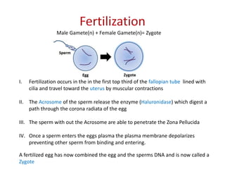
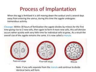
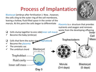
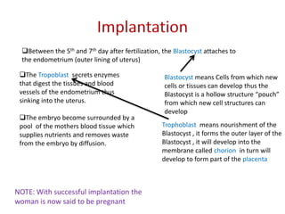
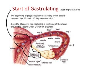
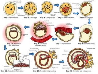
2. Fertilization occurs when a sperm penetrates the ovum in the fallopian tube. The zygote then undergoes cleavage and forms a blastocyst, which implants in the uterus.
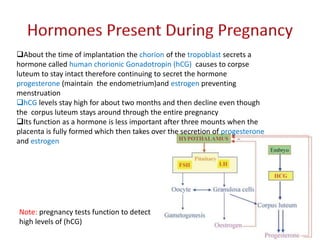
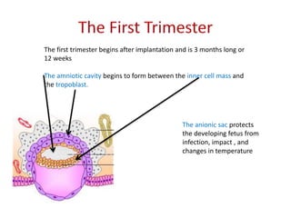
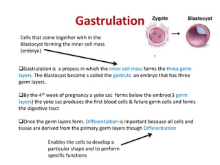
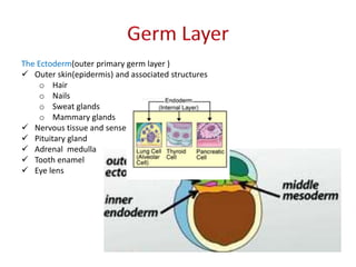
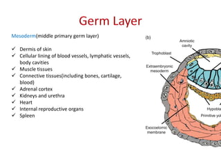
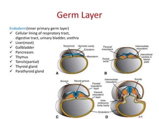
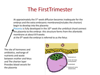
3. If implantation is successful, the blastocyst develops into an embryo with three germ layers—ectoderm, mesoderm, and endoderm—over the course of the first trimester. Concurrently, the placenta forms to support fetal development.