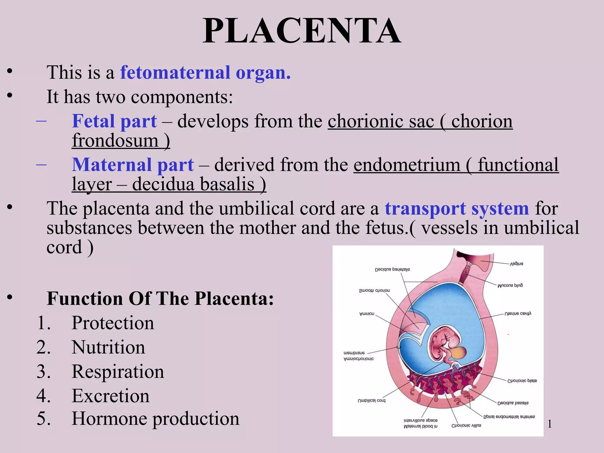
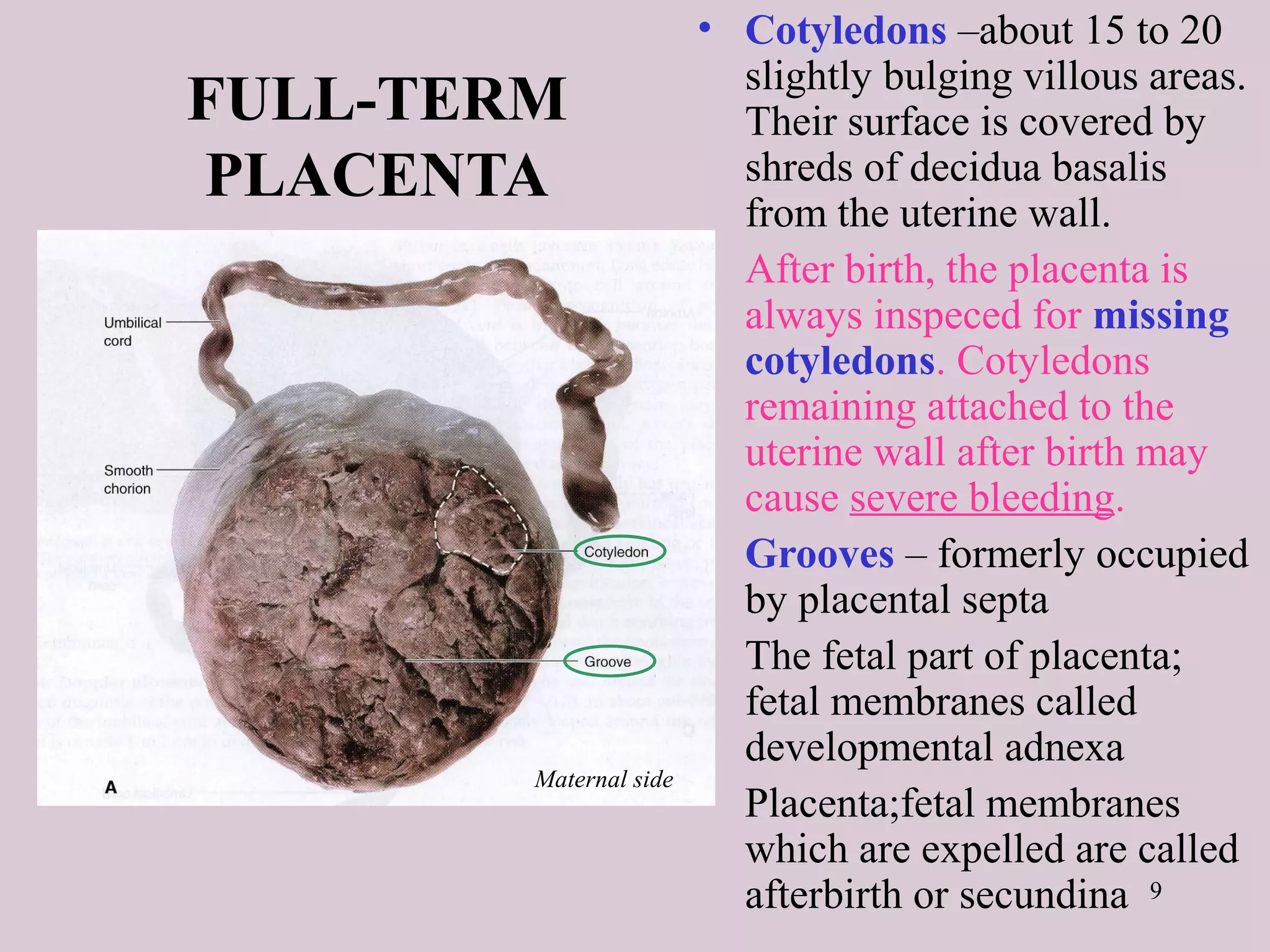
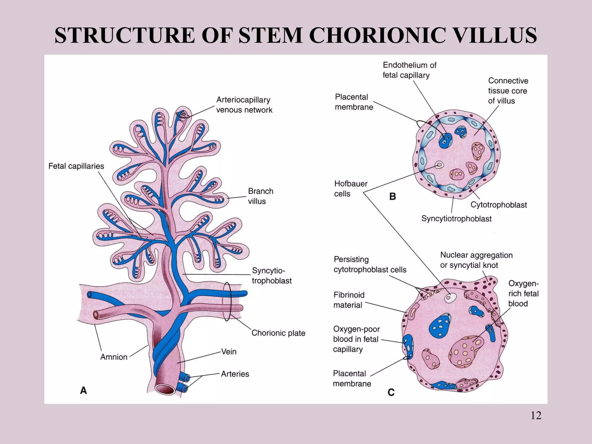
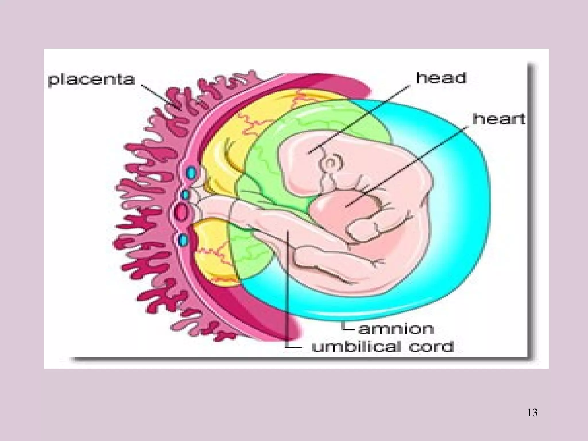
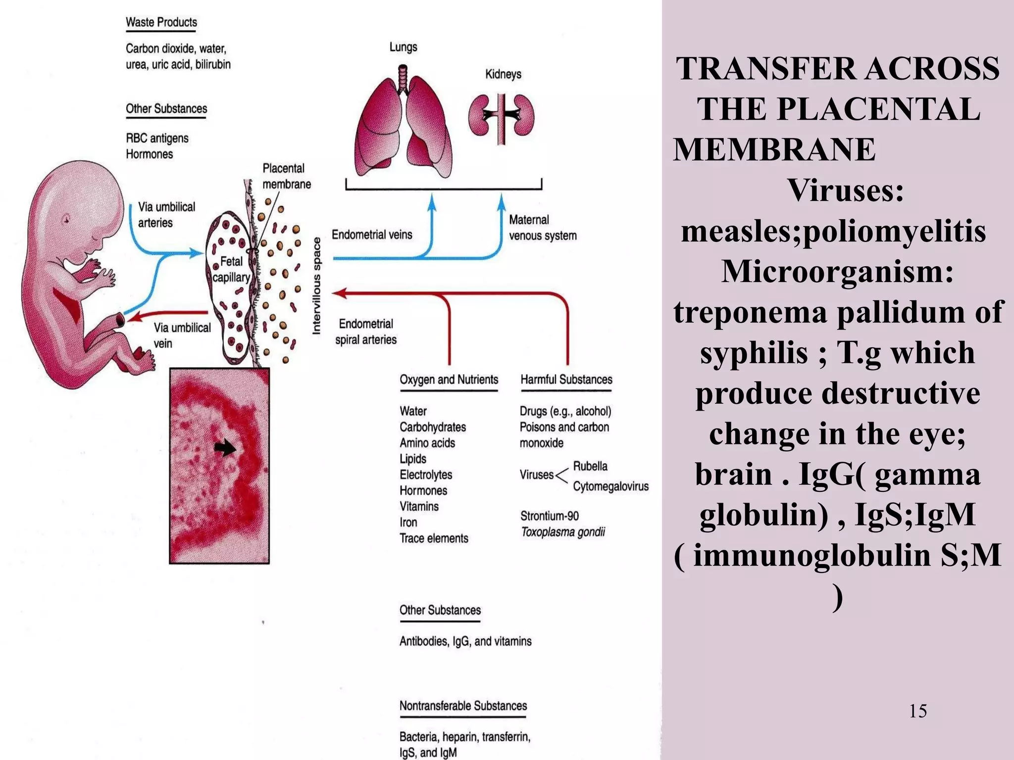
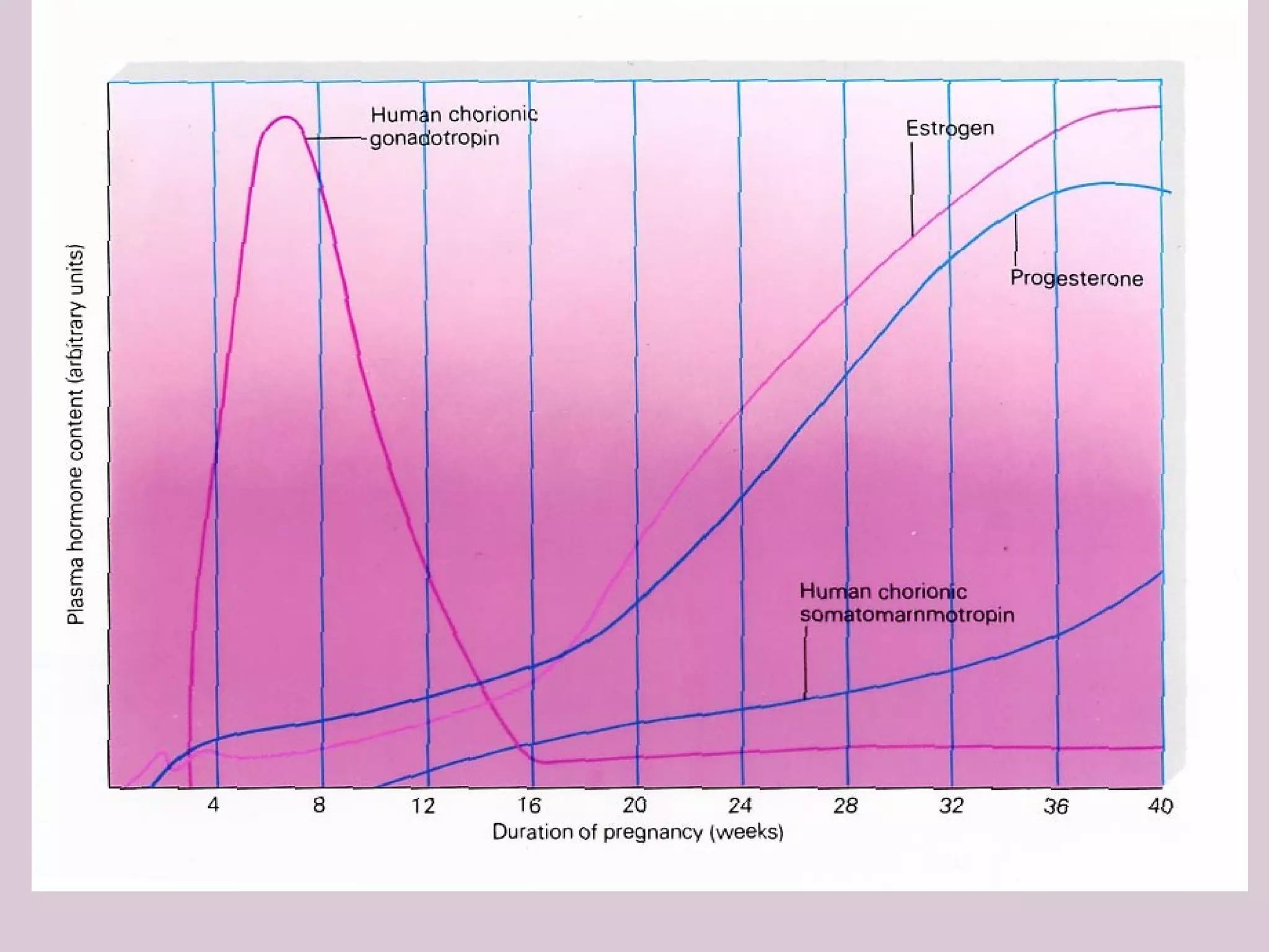
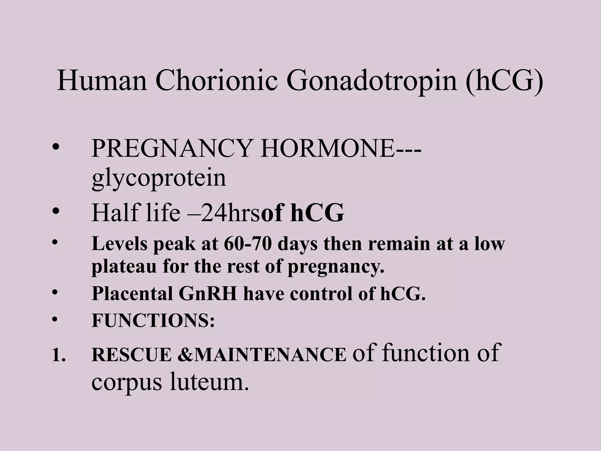
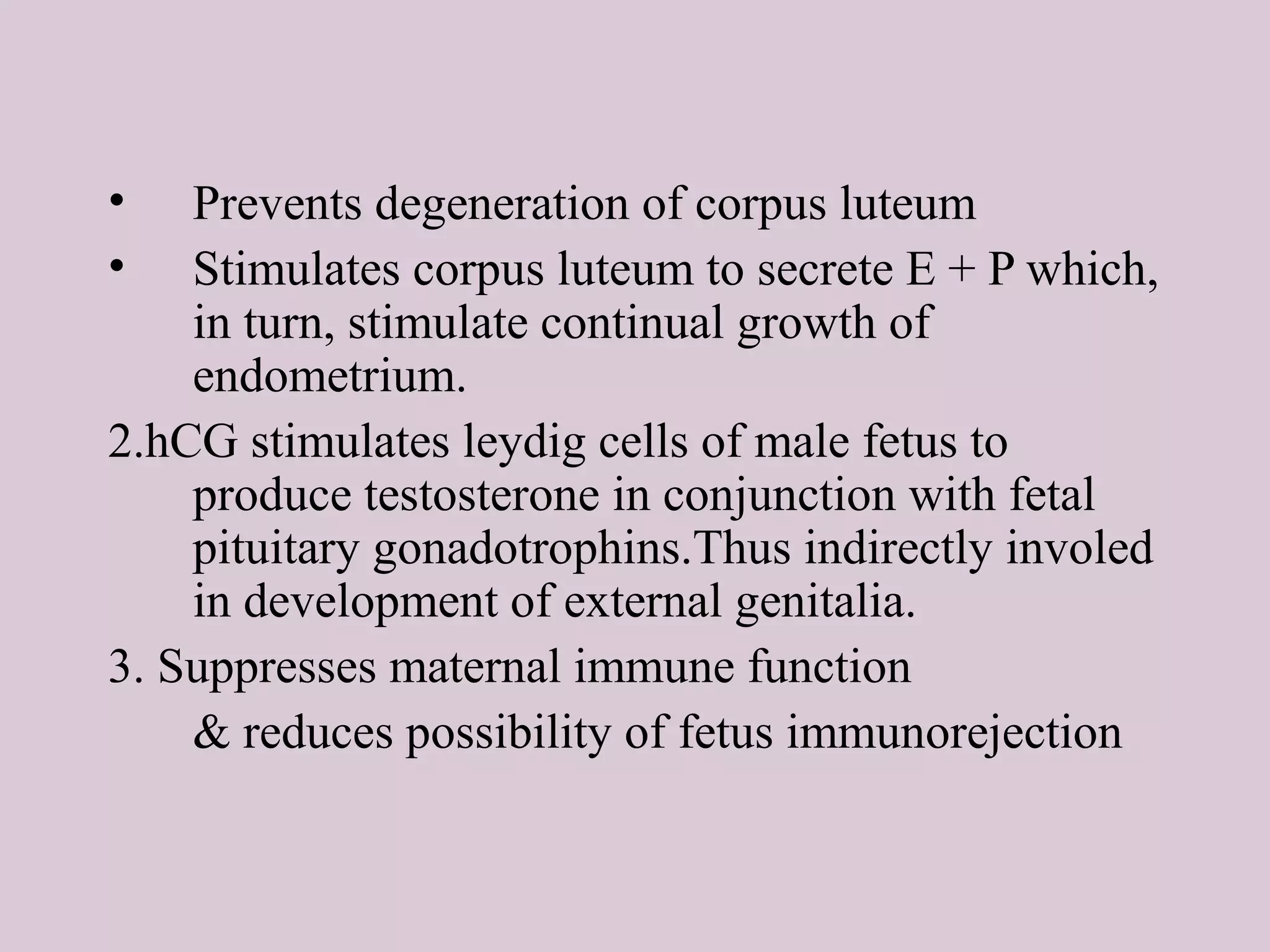
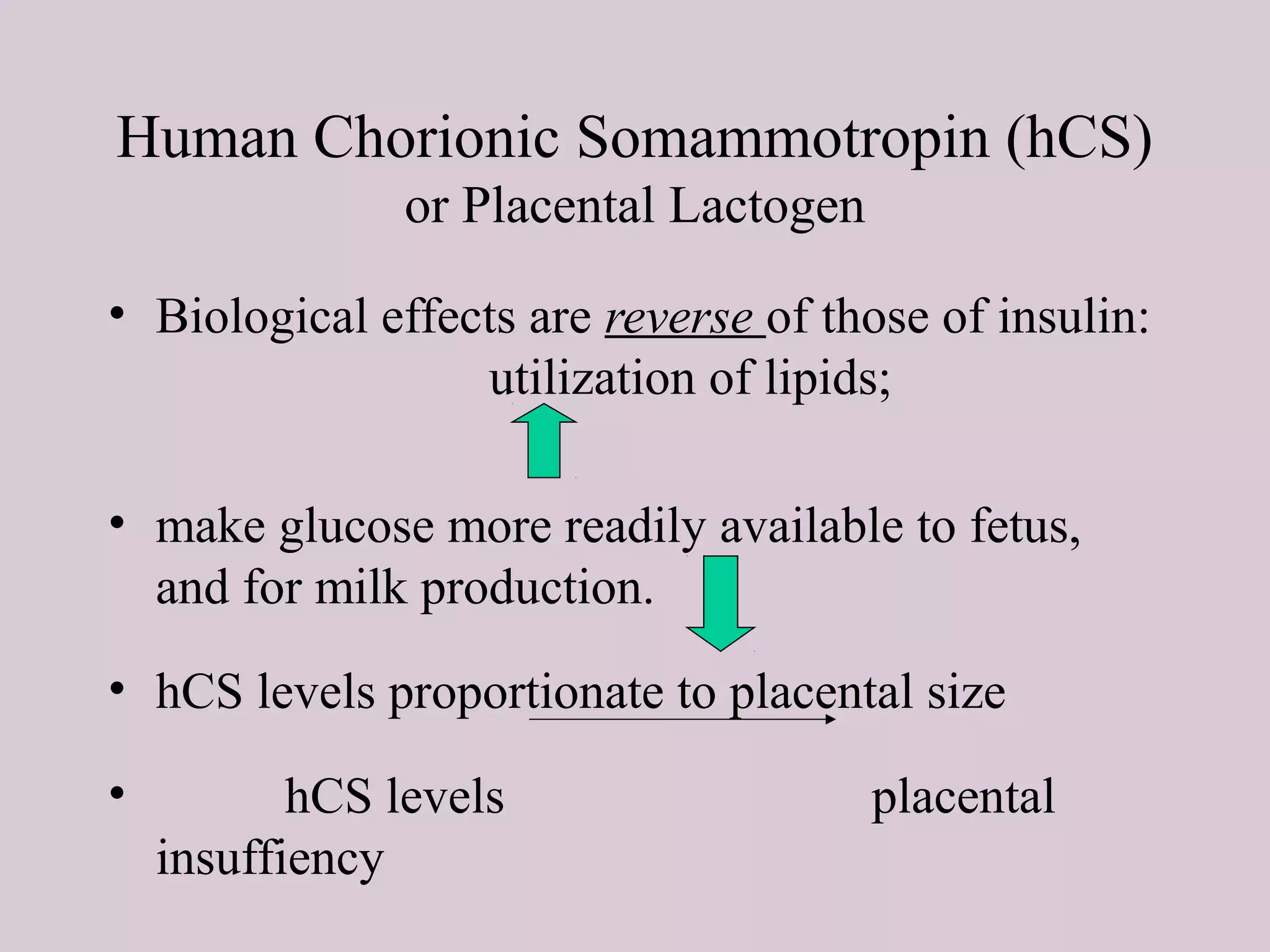
1
PLACENTA
• This is a fetomaternal organ.
• It has two components:
– Fetal part – develops from the chorionic sac ( chorion
frondosum )
– Maternal part – derived from the endometrium ( functional
layer – decidua basalis )
• The placenta and the umbilical cord are a transport system for
substances between the mother and the fetus.( vessels in umbilical
cord )
• Function Of The Placenta:
1. Protection
2. Nutrition
3. Respiration
4. Excretion
5. Hormone production
2
Further Development of Chorionic
Villi
Early in the 3rd
week,
mesenchyme growth into the
primary villi forming a core of
mesenchymal tissue. Thus the
Secondary Chorionic Villi are
formed over the entire surface of
the chorionic sac.
Some mesenchymal cells in the
secondary villi differentiate into
capillaries and blood cells
forming the Tertiary Chorionic
Villi.
The capillaries in the villi fuse to
form arteriocapillary networks.
The previous formed
arteriocapillary networks
become connected with the
embryonic heart through
vessels which are formed in the
mesenchyme of the chorion and
connecting stalk.
By the end of the 3rd
week,
embryonic blood begins to flow
through the capillaries in the
chorionic villi.
Oxygen & nutrients in the
maternal blood in the intervillous
space diffuse through the walls
of the villi and enter the
embryo’s blood.
Carbon dioxide & waste
products diffuse from blood in
the fetal capillaries through the
wall of the chorionic villi into the
6
DECIDUA
• This is the endometrium
of the gravid (pregnant)
uterus.
• It has four parts:
– Decidua basalis: it
forms the maternal
part of the placenta
– Decidua capsularis:
it covers the
conceptus
– Decidua parietalis:
the rest of the
endometrium
– Decidua reflexa:
– Junction between
capsularis &
parietalis.
7
DEVELOPMENT OF PLACENTA
• Until the beginning of the 8th
week, the entire chorionic
sac is covered with villi.
• After that, as the sac grows,
only the part that is
associated with Decidua
basalis retain its villi.
• Villi of Decidua capsularis
compressed by the
developing sac.
• Thus, two types of chorion
are formed:
– Chorion frondosum
(villous chorion)
– Chorion laeve – bare
(smooth) chorion
– About 18 weeks old, it
covers 15-30% of the
decidua and weights
about 1 6 of fetus
8
DEVELOPMENT OF
PLACENTA
• The villous
chorion ( increase
in number, enlarge
and branch ) will
form the fetal part
of the placenta.
• The decidua
basalis will form
the maternal part
of the placenta.
• The placenta will
grow rapidly.
• By the end of the
4th
month, the
decidua basalis is
almost entirely
replaced by the
fetal part of the
placenta.
9
FULL-TERM
PLACENTA
• Cotyledons –about 15 to 20
slightly bulging villous areas.
Their surface is covered by
shreds of decidua basalis
from the uterine wall.
• After birth, the placenta is
always inspeced for missing
cotyledons. Cotyledons
remaining attached to the
uterine wall after birth may
cause severe bleeding.
• Grooves – formerly occupied
by placental septa
• The fetal part of placenta;
fetal membranes called
developmental adnexa
• Placenta;fetal membranes
which are expelled are called
afterbirth or secundina
Maternal side
10
FULL-TERM PLACENTA
( Discoid shape -500- 600 gm- Diameter 15-20 cm
– Thickness of 2-3 cm)
• Fetal surface:
• This side is smooth
and shiny. It is
covered by amnion.
• The umbilical cord is
attached close to the
center of the placenta.
• The umbilical vessels
radiate from the
umbilical cord.
• They branch on the
fetal surface to form
chorionic vessels.
• They enter the
chorionic villi to form
arteriocapillary-
venous system.
Fetal side
11
PLACENTAL CIRCULATION 80 to 100
each cotyledon - inflow
12
STRUCTURE OF STEM CHORIONIC VILLUS
13
14
PLACENTAL MEMBRANE
knot –syncytiotrophoblast
–Toward end of pregnancy –
phagocytic cells
• This is a composite structure
that consists of the extrafetal
tissues separating the fetal
blood from the maternal
blood.
• It has four layers:
– Syncytiotrophoblast
– Cytotrophoblast
– Connective tissue of
villus
– Endothelium of fetal
capillaries
• After the 20th
week, the
cytotrophoblastic cells
disappear and the placental
membrane consists only of
three layers.
15
TRANSFER ACROSS
THE PLACENTAL
MEMBRANE
Viruses:
measles;poliomyelitis
Microorganism:
treponema pallidum of
syphilis ; T.g which
produce destructive
change in the eye;
brain . IgG( gamma
globulin) , IgS;IgM
( immunoglobulin S;M
)
Placental endocrine synthesis
• The syncytiotrophoblast synthesizes protein
&steroid hormones
• The protein homones
• 1- human chorionic gonadotropin
• 2- h.c. somatomammotropin
• 3-h.c. thyrotropin
• 4-h.c. corticotropin
• The steroid hormones
• Progesterone & Estrogens
17
18
19
Third trimester
bleeding is the common
sign of these anomalies
20
Biscoid
placenta
Bipartita
Horseshoe
When villi persist on the entire
surface of the chorionic sac ,a
thin layer of placenta attaches to
a large area of the uterus …… it
is a membranous placenta.
22
23
24
25
26
27
29
PLACENTAL FUNCTION
• Transfer of nutrients and waste products bn
the mother & fetus.
RESPIRATORY
EXCRETORY
NUTRITIVE
• Produces or metabolizes the hormones &
enzymes necessary to maintain the
pregnancy.
PLACENTAL FUNCTION
• BARRIER FUNCTION
• IMMUNOLOGICAL FUNCTION
Transfer function
• Transport is facilitated by the close
approximation of maternal and fetal
vascular systems within the placenta.
• It is important to recognize that there
normally is no mixing of fetal and maternal
blood within the placenta.
• Respiratory function—Intake of o2 &
output of co2 takes place by simple
diffusion.o2 supply to fetus rate of
5ml/kg/min & this achieved with cord flow
of 165-330ml/min.
• Excretory function—waste products urea,
uric acid,creatinine are excreted to maternal
blood by simple diffusion.
NUTRITIVE FUNCTION
• Glucose is the major energy substrate
provided to the placenta and fetus. It is
transported across the placenta by facilitated
diffusion via hexose transporters
• Although the fetus receives large amounts of
intact glucose, a large amount is oxidized
within the placenta to lactate, which is used
for fetal energy production.
• Amino acid concentrations in fetal blood
are higher than in maternal blood.
Amino acids are therefore transported
to the fetus by active transport .
• LIPIDS—TG`s & FA directly
transported from mother to fetus in early
pregnancy but synthesized in fetus later
in pregnancy. Thus, fetal fat has got
dual origin.
• Water & electrolytes—Na,K+,Cl- by
simple diffusion.Ca,Ph,iron by active
transport.
• BARRIER FUNCTION:-Protective
barrier to the fetus against noxious
agents circulating in maternal blood.
(High MW >500daltons.
IMMUNOLOGICAL
FUNCTION
• Fetus & placenta contain paternally determined
antigens,foreign to the mother . Inspite of this
,no evidence of graft rejection. Probably:
1. Fibrinoid & sialomucin coating of trophoblast
may suppress the troblastic antigen.
2. Placental hormones ,steroids, HCG have got
weak immunosuppressive effect, may be
responsible for producing sialomucin.
3.Nitabuch`s layer which intervenes bn
decidua basalis &cytotrophoblast probably
inactivates the antigenic property of tissue.
4.There is little HLA & blood group antigens on
trophoblast surface. so antigenic stimulus is
poor.
5. Production of block antibodies by mother
,protects fetus from rejection.
• ENDOCRINE—hormones secreted
internally.
• HORMONE--Any organic chemical that is
secreted by a gland into the circulatory
system and is transported to some target
organ. The target may be either peripheral
tissue (such as muscle or other gland) or
brain.
Fetal, placental & maternal compartments
form an integrated hormonal unit
The feto-placental-maternal (FPM) unit
creates the
Endocrine Environment
that maintains and drives the processes of
pregnancy and pre-natal development.
PLACENTAL HORMONES
• Human Chorionic
Gonadotropin (hCG
• Human Chorionic
Somammotropin (hCS)
or Placental Lactogen(hPL)
OTHER HORMONES
• Chorionic
Adrenocorticotropin
• Chorionic thyrotropin
• Relaxin
• PTH-rP
• hGH-V
• Estrogen (E)
• Progesterone (P)
• HYPOTHALAMIC-LIKE
RELEASING HORMONES
• GnRH
• CRH
• cTRH
• GH-RH
• PLACENTAL PEPTIDE
HORMONES
• Neuropeptide-Y
• Inhibin & Activin
• ANP
To understand the FPM one should know:
1. The major hormones involved:
hCGn
Progesterone
Estrogen
Human Chorionic Somatomammotropin (hCS)
(placental lactogen)
2. How the FPM compartments work together
to produce the steroid hormones
3. The transfer of hormones between
the FPM compartments.
Human Chorionic Gonadotropin (hCG)
• PREGNANCY HORMONE---
glycoprotein
• Half life –24hrsof hCG
• Levels peak at 60-70 days then remain at a low
plateau for the rest of pregnancy.
• Placental GnRH have control of hCG.
• FUNCTIONS:
1. RESCUE &MAINTENANCE of function of
corpus luteum.
• Prevents degeneration of corpus luteum
• Stimulates corpus luteum to secrete E + P which,
in turn, stimulate continual growth of
endometrium.
2.hCG stimulates leydig cells of male fetus to
produce testosterone in conjunction with fetal
pituitary gonadotrophins.Thus indirectly involed
in development of external genitalia.
3. Suppresses maternal immune function
& reduces possibility of fetus immunorejection
Human Chorionic Somammotropin (hCS)
or Placental Lactogen
• Structure similar to growth hormone
• Produced by the placenta
• Levels throughout pregnancy
• Large amounts in maternal blood but
DO NOT reach the fetus
Human Chorionic Somammotropin (hCS)
or Placental Lactogen
• Biological effects are reverse of those of insulin:
utilization of lipids;
• make glucose more readily available to fetus,
and for milk production.
• hCS levels proportionate to placental size
• hCS levels placental
insuffiency
Estrogen (E)
• FORMS-estriol,estradiol &estrone .
• Estriol most important .
• Levels increase throughout pregnancy
• 90% produced by placenta.
(syncytiotrophoblast)
• Placental production is transferred to both
maternal and fetal compartments
• Two of the principle effects of placental
estrogens are:
• Stimulate growth of the myometrium and
antagonize the myometrial-suppressing activity of
progesterone. In many species, the high levels of
estrogen in late gestation induces myometrial
oxytocin receptors, thereby preparing the uterus
for parturition.
• Stimulate mammary gland development.
Estrogens are one in a battery of hormones
necessary for both ductal and alveolar growth in
the mammary gland.
Progesterone (P)
• Levels increase throughout pregnancy
• 80-90% is produced by placenta and
secreted to both fetus and mother
• Progestins, including progesterone, have two
major roles during pregnancy:
• Support of the endometrium to provide an
environment conducive to fetal survival. If the
endometrium is deprived of progestins, the
pregnancy will inevitably be terminated.
• Suppression of contractility in uterine smooth
muscle, which, if unchecked, would clearly be a
disaster. This is often called the "progesterone
block" on the myometrium. Toward the end of
gestation, this myometrial-quieting effect is
antagonized by rising levels of estrogens, thereby
facilitating parturition.
• Progesterone and other progestins also
potently inhibit secretion of the pituitary
gonadotropins luteinizing hormone and
follicle stimulating hormone. This effect
almost always prevents ovulation from
occuring during pregnancy
FETAL ADRENAL GLAND
Adrenal Gland Development
• Adrenal Cortex
– Vital to organism survival
– Begins to develop at 4th
week of embryonic life
– Functional around 10th
to 12th
week of embryonic life
– Enzymes necessary for biosynthesis of adrenocortical
hormones do not develop simultaneously
– hCG may have a role in stimulating Adrenocortical
development
• Adrenal Medulla
– Originates from nervous system
– Ganglia of Autonomic Nervous System
Fetal Adrenal Cortex Function
– Adrenal Cortex
• Zona Glomerulosa
– Has enzymes to convert Pregnenalone to:
» Progesterone
» Deoxycorticosterone
» Corticosterone
» Aldosterone
• Zona Fasciculata
– Converts Pregnenalone and Progesterone to 17OH-Pre
and 17OH-Pro
– 17OH-Pro is converted to cortisol (major glucocorticoid)
• Zona Reticularis
– Converts 17OH-Pre into DHEA and Androstenedione
(androgens)
THE END

PLACENTA

  • 1.
    1 PLACENTA • This isa fetomaternal organ. • It has two components: – Fetal part – develops from the chorionic sac ( chorion frondosum ) – Maternal part – derived from the endometrium ( functional layer – decidua basalis ) • The placenta and the umbilical cord are a transport system for substances between the mother and the fetus.( vessels in umbilical cord ) • Function Of The Placenta: 1. Protection 2. Nutrition 3. Respiration 4. Excretion 5. Hormone production
  • 2.
  • 3.
    Further Development ofChorionic Villi Early in the 3rd week, mesenchyme growth into the primary villi forming a core of mesenchymal tissue. Thus the Secondary Chorionic Villi are formed over the entire surface of the chorionic sac. Some mesenchymal cells in the secondary villi differentiate into capillaries and blood cells forming the Tertiary Chorionic Villi. The capillaries in the villi fuse to form arteriocapillary networks.
  • 4.
    The previous formed arteriocapillarynetworks become connected with the embryonic heart through vessels which are formed in the mesenchyme of the chorion and connecting stalk. By the end of the 3rd week, embryonic blood begins to flow through the capillaries in the chorionic villi. Oxygen & nutrients in the maternal blood in the intervillous space diffuse through the walls of the villi and enter the embryo’s blood. Carbon dioxide & waste products diffuse from blood in the fetal capillaries through the wall of the chorionic villi into the
  • 6.
    6 DECIDUA • This isthe endometrium of the gravid (pregnant) uterus. • It has four parts: – Decidua basalis: it forms the maternal part of the placenta – Decidua capsularis: it covers the conceptus – Decidua parietalis: the rest of the endometrium – Decidua reflexa: – Junction between capsularis & parietalis.
  • 7.
    7 DEVELOPMENT OF PLACENTA •Until the beginning of the 8th week, the entire chorionic sac is covered with villi. • After that, as the sac grows, only the part that is associated with Decidua basalis retain its villi. • Villi of Decidua capsularis compressed by the developing sac. • Thus, two types of chorion are formed: – Chorion frondosum (villous chorion) – Chorion laeve – bare (smooth) chorion – About 18 weeks old, it covers 15-30% of the decidua and weights about 1 6 of fetus
  • 8.
    8 DEVELOPMENT OF PLACENTA • Thevillous chorion ( increase in number, enlarge and branch ) will form the fetal part of the placenta. • The decidua basalis will form the maternal part of the placenta. • The placenta will grow rapidly. • By the end of the 4th month, the decidua basalis is almost entirely replaced by the fetal part of the placenta.
  • 9.
    9 FULL-TERM PLACENTA • Cotyledons –about15 to 20 slightly bulging villous areas. Their surface is covered by shreds of decidua basalis from the uterine wall. • After birth, the placenta is always inspeced for missing cotyledons. Cotyledons remaining attached to the uterine wall after birth may cause severe bleeding. • Grooves – formerly occupied by placental septa • The fetal part of placenta; fetal membranes called developmental adnexa • Placenta;fetal membranes which are expelled are called afterbirth or secundina Maternal side
  • 10.
    10 FULL-TERM PLACENTA ( Discoidshape -500- 600 gm- Diameter 15-20 cm – Thickness of 2-3 cm) • Fetal surface: • This side is smooth and shiny. It is covered by amnion. • The umbilical cord is attached close to the center of the placenta. • The umbilical vessels radiate from the umbilical cord. • They branch on the fetal surface to form chorionic vessels. • They enter the chorionic villi to form arteriocapillary- venous system. Fetal side
  • 11.
    11 PLACENTAL CIRCULATION 80to 100 each cotyledon - inflow
  • 12.
    12 STRUCTURE OF STEMCHORIONIC VILLUS
  • 13.
  • 14.
    14 PLACENTAL MEMBRANE knot –syncytiotrophoblast –Towardend of pregnancy – phagocytic cells • This is a composite structure that consists of the extrafetal tissues separating the fetal blood from the maternal blood. • It has four layers: – Syncytiotrophoblast – Cytotrophoblast – Connective tissue of villus – Endothelium of fetal capillaries • After the 20th week, the cytotrophoblastic cells disappear and the placental membrane consists only of three layers.
  • 15.
    15 TRANSFER ACROSS THE PLACENTAL MEMBRANE Viruses: measles;poliomyelitis Microorganism: treponemapallidum of syphilis ; T.g which produce destructive change in the eye; brain . IgG( gamma globulin) , IgS;IgM ( immunoglobulin S;M )
  • 16.
    Placental endocrine synthesis •The syncytiotrophoblast synthesizes protein &steroid hormones • The protein homones • 1- human chorionic gonadotropin • 2- h.c. somatomammotropin • 3-h.c. thyrotropin • 4-h.c. corticotropin • The steroid hormones • Progesterone & Estrogens
  • 17.
  • 18.
  • 19.
    19 Third trimester bleeding isthe common sign of these anomalies
  • 20.
  • 21.
    When villi persiston the entire surface of the chorionic sac ,a thin layer of placenta attaches to a large area of the uterus …… it is a membranous placenta.
  • 22.
  • 23.
  • 24.
  • 25.
  • 26.
  • 27.
  • 29.
  • 30.
    PLACENTAL FUNCTION • Transferof nutrients and waste products bn the mother & fetus. RESPIRATORY EXCRETORY NUTRITIVE • Produces or metabolizes the hormones & enzymes necessary to maintain the pregnancy.
  • 31.
    PLACENTAL FUNCTION • BARRIERFUNCTION • IMMUNOLOGICAL FUNCTION
  • 32.
    Transfer function • Transportis facilitated by the close approximation of maternal and fetal vascular systems within the placenta. • It is important to recognize that there normally is no mixing of fetal and maternal blood within the placenta.
  • 33.
    • Respiratory function—Intakeof o2 & output of co2 takes place by simple diffusion.o2 supply to fetus rate of 5ml/kg/min & this achieved with cord flow of 165-330ml/min. • Excretory function—waste products urea, uric acid,creatinine are excreted to maternal blood by simple diffusion.
  • 35.
    NUTRITIVE FUNCTION • Glucoseis the major energy substrate provided to the placenta and fetus. It is transported across the placenta by facilitated diffusion via hexose transporters • Although the fetus receives large amounts of intact glucose, a large amount is oxidized within the placenta to lactate, which is used for fetal energy production.
  • 36.
    • Amino acidconcentrations in fetal blood are higher than in maternal blood. Amino acids are therefore transported to the fetus by active transport . • LIPIDS—TG`s & FA directly transported from mother to fetus in early pregnancy but synthesized in fetus later in pregnancy. Thus, fetal fat has got dual origin.
  • 37.
    • Water &electrolytes—Na,K+,Cl- by simple diffusion.Ca,Ph,iron by active transport. • BARRIER FUNCTION:-Protective barrier to the fetus against noxious agents circulating in maternal blood. (High MW >500daltons.
  • 38.
    IMMUNOLOGICAL FUNCTION • Fetus &placenta contain paternally determined antigens,foreign to the mother . Inspite of this ,no evidence of graft rejection. Probably: 1. Fibrinoid & sialomucin coating of trophoblast may suppress the troblastic antigen. 2. Placental hormones ,steroids, HCG have got weak immunosuppressive effect, may be responsible for producing sialomucin.
  • 39.
    3.Nitabuch`s layer whichintervenes bn decidua basalis &cytotrophoblast probably inactivates the antigenic property of tissue. 4.There is little HLA & blood group antigens on trophoblast surface. so antigenic stimulus is poor. 5. Production of block antibodies by mother ,protects fetus from rejection.
  • 40.
    • ENDOCRINE—hormones secreted internally. •HORMONE--Any organic chemical that is secreted by a gland into the circulatory system and is transported to some target organ. The target may be either peripheral tissue (such as muscle or other gland) or brain.
  • 41.
    Fetal, placental &maternal compartments form an integrated hormonal unit The feto-placental-maternal (FPM) unit creates the Endocrine Environment that maintains and drives the processes of pregnancy and pre-natal development.
  • 42.
    PLACENTAL HORMONES • HumanChorionic Gonadotropin (hCG • Human Chorionic Somammotropin (hCS) or Placental Lactogen(hPL) OTHER HORMONES • Chorionic Adrenocorticotropin • Chorionic thyrotropin • Relaxin • PTH-rP • hGH-V • Estrogen (E) • Progesterone (P) • HYPOTHALAMIC-LIKE RELEASING HORMONES • GnRH • CRH • cTRH • GH-RH • PLACENTAL PEPTIDE HORMONES • Neuropeptide-Y • Inhibin & Activin • ANP
  • 43.
    To understand theFPM one should know: 1. The major hormones involved: hCGn Progesterone Estrogen Human Chorionic Somatomammotropin (hCS) (placental lactogen) 2. How the FPM compartments work together to produce the steroid hormones 3. The transfer of hormones between the FPM compartments.
  • 45.
    Human Chorionic Gonadotropin(hCG) • PREGNANCY HORMONE--- glycoprotein • Half life –24hrsof hCG • Levels peak at 60-70 days then remain at a low plateau for the rest of pregnancy. • Placental GnRH have control of hCG. • FUNCTIONS: 1. RESCUE &MAINTENANCE of function of corpus luteum.
  • 46.
    • Prevents degenerationof corpus luteum • Stimulates corpus luteum to secrete E + P which, in turn, stimulate continual growth of endometrium. 2.hCG stimulates leydig cells of male fetus to produce testosterone in conjunction with fetal pituitary gonadotrophins.Thus indirectly involed in development of external genitalia. 3. Suppresses maternal immune function & reduces possibility of fetus immunorejection
  • 47.
    Human Chorionic Somammotropin(hCS) or Placental Lactogen • Structure similar to growth hormone • Produced by the placenta • Levels throughout pregnancy • Large amounts in maternal blood but DO NOT reach the fetus
  • 48.
    Human Chorionic Somammotropin(hCS) or Placental Lactogen • Biological effects are reverse of those of insulin: utilization of lipids; • make glucose more readily available to fetus, and for milk production. • hCS levels proportionate to placental size • hCS levels placental insuffiency
  • 49.
    Estrogen (E) • FORMS-estriol,estradiol&estrone . • Estriol most important . • Levels increase throughout pregnancy • 90% produced by placenta. (syncytiotrophoblast) • Placental production is transferred to both maternal and fetal compartments
  • 51.
    • Two ofthe principle effects of placental estrogens are: • Stimulate growth of the myometrium and antagonize the myometrial-suppressing activity of progesterone. In many species, the high levels of estrogen in late gestation induces myometrial oxytocin receptors, thereby preparing the uterus for parturition. • Stimulate mammary gland development. Estrogens are one in a battery of hormones necessary for both ductal and alveolar growth in the mammary gland.
  • 52.
    Progesterone (P) • Levelsincrease throughout pregnancy • 80-90% is produced by placenta and secreted to both fetus and mother
  • 53.
    • Progestins, includingprogesterone, have two major roles during pregnancy: • Support of the endometrium to provide an environment conducive to fetal survival. If the endometrium is deprived of progestins, the pregnancy will inevitably be terminated. • Suppression of contractility in uterine smooth muscle, which, if unchecked, would clearly be a disaster. This is often called the "progesterone block" on the myometrium. Toward the end of gestation, this myometrial-quieting effect is antagonized by rising levels of estrogens, thereby facilitating parturition.
  • 54.
    • Progesterone andother progestins also potently inhibit secretion of the pituitary gonadotropins luteinizing hormone and follicle stimulating hormone. This effect almost always prevents ovulation from occuring during pregnancy
  • 55.
  • 57.
    Adrenal Gland Development •Adrenal Cortex – Vital to organism survival – Begins to develop at 4th week of embryonic life – Functional around 10th to 12th week of embryonic life – Enzymes necessary for biosynthesis of adrenocortical hormones do not develop simultaneously – hCG may have a role in stimulating Adrenocortical development • Adrenal Medulla – Originates from nervous system – Ganglia of Autonomic Nervous System
  • 58.
    Fetal Adrenal CortexFunction – Adrenal Cortex • Zona Glomerulosa – Has enzymes to convert Pregnenalone to: » Progesterone » Deoxycorticosterone » Corticosterone » Aldosterone • Zona Fasciculata – Converts Pregnenalone and Progesterone to 17OH-Pre and 17OH-Pro – 17OH-Pro is converted to cortisol (major glucocorticoid) • Zona Reticularis – Converts 17OH-Pre into DHEA and Androstenedione (androgens)
  • 59.

Editor's Notes

  • #56 FETAL ADRENAL GLAND