Downloaded 232 times

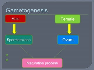
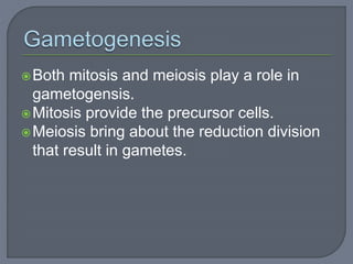
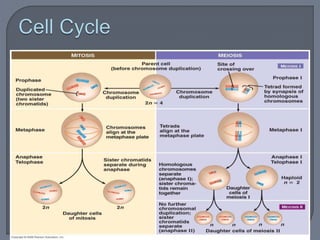
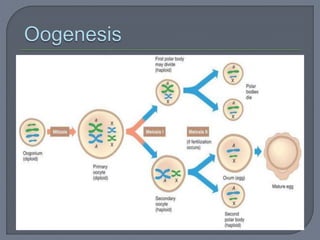
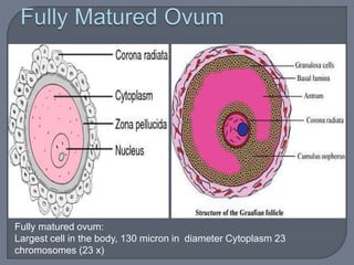
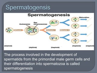
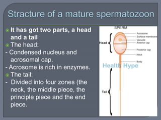
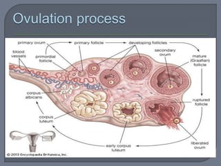


The document summarizes conception and fertilization. It describes the maturation process of both male and female gametes. Spermatogenesis produces sperm from male germ cells, while oogenesis results in a mature ovum. Ovulation releases a secondary oocyte from the ovaries. Fertilization occurs when a sperm fuses with the ovum in the fallopian tubes, restoring the chromosome number and initiating embryonic development. This forms a zygote containing a mix of paternal and maternal genetic material.


