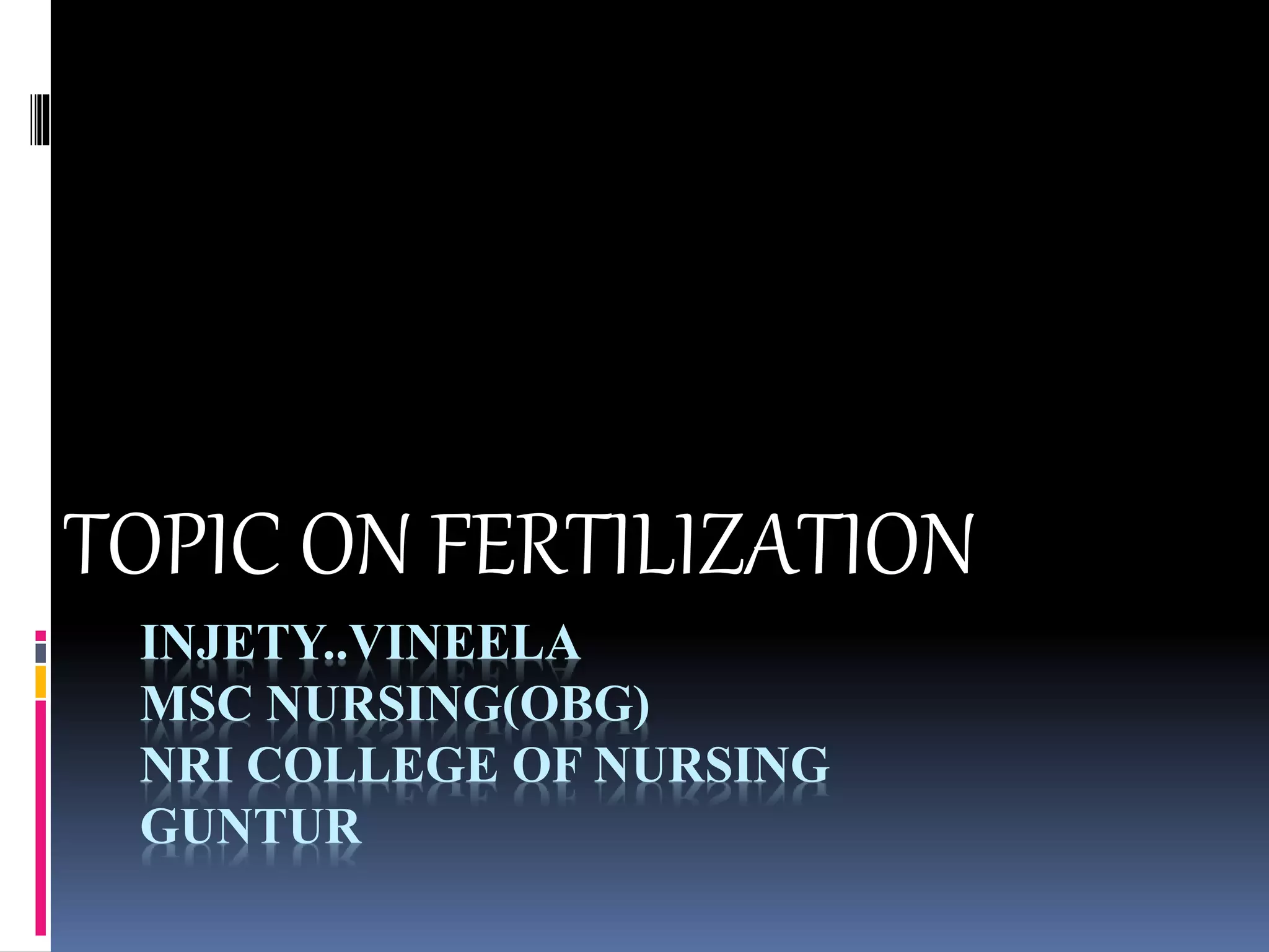
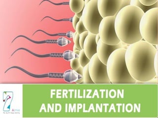
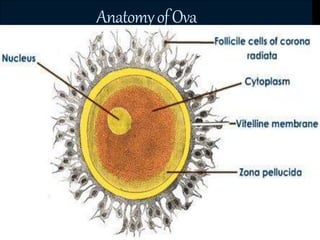
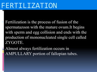
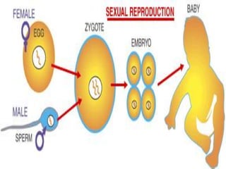
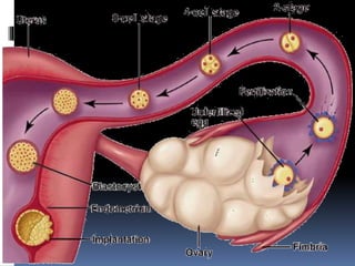
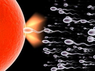
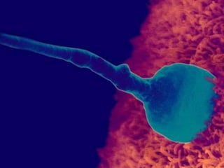
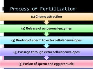
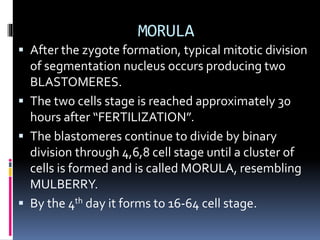
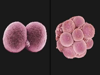
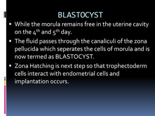
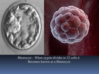
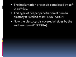
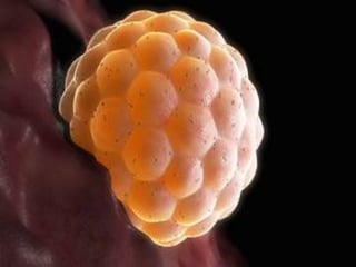
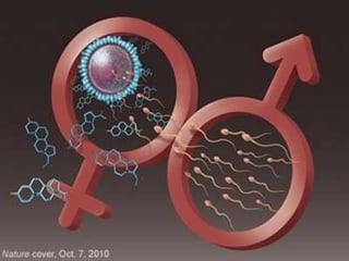
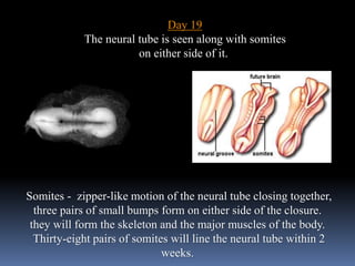
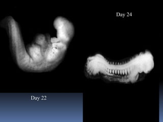
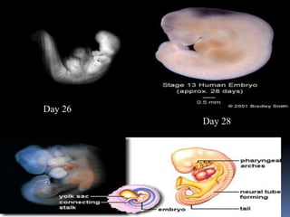
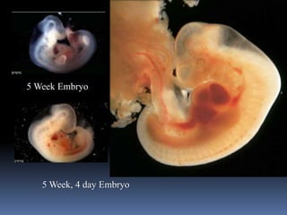
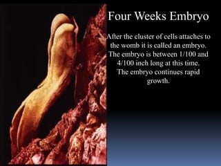
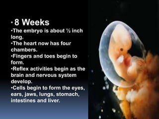
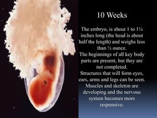
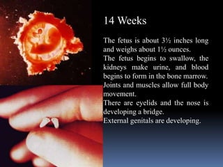
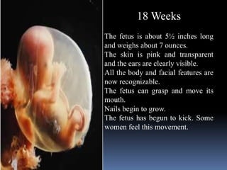
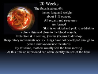
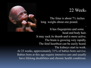
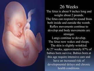
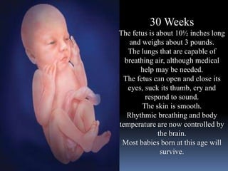
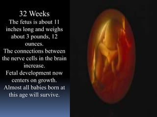
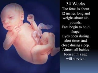
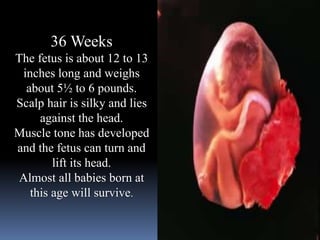
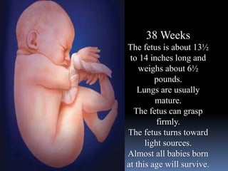
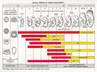
INJETY..VINEELA
MSC NURSING(OBG)
NRI COLLEGE OF NURSING
GUNTUR
TOPIC ON FERTILIZATION
Anatomy of Sperm
AnatomyofOva
FERTILIZATION
 Fertilization is the process of fusion of the
spermatazoon with the mature ovum.It begins
with sperm and egg collision and ends with the
production of mononucleated single cell called
ZYGOTE.
 Almost always fertilization occurs in
AMPULLARY portion of fallopian tubes.
APPROXIMATION OF GAMETES
 The ovum immediately following ovulation is
picked up by the tubal fimbriae which partly
envelope the ovary, specially at the time of
ovulation .
 The pick up action might be muscular or by a kind
of suction by ciliary action or by a postivie
chemotaxis exerted by the tubal secretion.
 The ovum is rapidly transported to the
ampullary part.
 Fertilisable life span of oocyte ranges from 12
to 24 hours where as that of sperm is 48 to 72
hours.
 Out of hundred of millions of sperms
deposited in the vagina at single ejaculation.
Only thousands capacitated spermataozoa
enter the uterine tube while only 300-500
reach the ovum.
 The tubal transport is facilitated by muscular
contraction and aspiration action of the
uterine tube.
CONTACT AND FUSION OF THE
GAMETES
 Complete dissolution of the cells of the corona
radiata occurs by the chemical action of the
HYALURONIDASE liberated from the Acrosomal
cap of the hundreds of sperm present at the site.
 Penetration of Zona pellucida is facilitated by
Hyaluronidase from the acrosomal cap.
 Soon after the Sperm fusion ,penetration of other
sperm is prevented by ZONA REACTION and
COLEMA BLOCK.
Process of Fertilization
(5) Fusion of sperm and egg pronuclei
(4) Passage through extra cellular envelopes
(3) Binding of sperm to extra cellular envelopes
(2) Release of acrosomal enzymes
(1) Chemo attraction
 Sex of the child is determined by the pattern of
sex chromosome supplied by the
SPERMATOZOON.
 If the SPERMATOZOON contains ‘X’
Chromosome a female embryo (46 XX).
 If it contains ‘Y’ Chromosome a Male embryo (46
XY) is formed.
MORULA
 After the zygote formation, typical mitotic division
of segmentation nucleus occurs producing two
BLASTOMERES.
 The two cells stage is reached approximately 30
hours after “FERTILIZATION”.
 The blastomeres continue to divide by binary
division through 4,6,8 cell stage until a cluster of
cells is formed and is called MORULA, resembling
MULBERRY.
 By the 4th day it forms to 16-64 cell stage.
BLASTOCYST
 While the morula remains free in the uterine cavity
on the 4th and 5th day.
 The fluid passes through the canaliculi of the zona
pellucida which seperates the cells of morula and is
now termed as BLASTOCYST.
 Zona Hatching is next step so that trophectoderm
cells interact with endometrial cells and
implantation occurs.
Blastocyst – When zygote divides to 32 cells it
Becomes known as a Blastocyst
IMPLANTATION/NIDATION/NESTING
 Implantation occurs in the endometrium of the
anterior or posterior wall of the body near the
fundus on the 6th day which corresponds,to the 20th
day of regular menstural cycle.
 IMPLANTATION occurs through four stages.
 Apposition
 Adhesion
 Penetration
 Invasion
 The implantation process is completed by 10th
or 11th day.
 This type of deeper penetration of human
blastocyst is called as IMPLANTATION.
 Now the blastocyst is covered all sides by the
endometrium (DECIDUA).
Day 15
The
primitive
streak can
be seen on
the left side
Day 17
The primitive streak can still
be seen, and the opposite end
of the embryo is starting to
fold up.
The Blastocyst begins to
collapse
Day 19
The neural tube is seen along with somites
on either side of it.
Somites - zipper-like motion of the neural tube closing together,
three pairs of small bumps form on either side of the closure.
they will form the skeleton and the major muscles of the body.
Thirty-eight pairs of somites will line the neural tube within 2
weeks.
Day 22
Day 24
Day 26
Day 28
5 Week Embryo
5 Week, 4 day Embryo
Four Weeks Embryo
After the cluster of cells attaches to
the womb it is called an embryo.
The embryo is between 1/100 and
4/100 inch long at this time.
The embryo continues rapid
growth.
* 8 Weeks
•The embryo is about ½ inch
long.
•The heart now has four
chambers.
•Fingers and toes begin to
form.
•Reflex activities begin as the
brain and nervous system
develop.
•Cells begin to form the eyes,
ears, jaws, lungs, stomach,
intestines and liver.
10 Weeks
The embryo, is about 1 to 1¼
inches long (the head is about
half the length) and weighs less
than ½ ounce.
The beginnings of all key body
parts are present, but they are
not completed.
Structures that will form eyes,
ears, arms and legs can be seen.
Muscles and skeleton are
developing and the nervous
system becomes more
responsive.
12 Weeks
The fetus is about 2½ inches
long and weighs about ½ ounce.
Fingers and toes are distinct
and have nails.
Hair begins to develop, but
won't be seen until later in the
pregnancy.
The fetus begins small, random
movements, too slight to be felt.
The fetal heartbeat can be detected with a heart monitor.
All major external body features have appeared.
Muscles continue to develop.
14 Weeks
The fetus is about 3½ inches long
and weighs about 1½ ounces.
The fetus begins to swallow, the
kidneys make urine, and blood
begins to form in the bone marrow.
Joints and muscles allow full body
movement.
There are eyelids and the nose is
developing a bridge.
External genitals are developing.
18 Weeks
The fetus is about 5½ inches long
and weighs about 7 ounces.
The skin is pink and transparent
and the ears are clearly visible.
All the body and facial features are
now recognizable.
The fetus can grasp and move its
mouth.
Nails begin to grow.
The fetus has begun to kick. Some
women feel this movement.
20 Weeks
The fetus is about 6¼
inches long and weighs
about 11½ ounces.
All organs and structures
are formed
Skin is wrinkled and pink to reddish in
color - thin and close to the blood vessels.
Protective skin coating, (vernix) begins to develop.
Respiratory movements occur - lungs have not developed enough to
permit survival outside the uterus.
By this time, mothers usually feel the fetus moving.
At this time an ultrasound can often identify the sex of the fetus.
22 Weeks
The fetus is about 7½ inches
long weighs about one pound.
.
It has fingerprints and some
head and body hair.
It may suck its thumb and is more active.
The brain is growing very rapidly.
The fetal heartbeat can be easily heard.
The kidneys start to work.
At 23 weeks, approximately 31% of babies born survive.
Babies born at this age require intensive care and usually
have lifelong disabilities and chronic health conditions.
24 Weeks
The fetus is about 8¼ inches long and
weighs about 1¼ pounds.
Bones of the ears harden making sound
conduction possible. The fetus hears
mother’s sounds such as breathing,
heartbeat and voice.
The first layers of fat are beginning to
form.
This is the beginning of substantial
weight gain for the fetus.
Lungs continue developing
At 25 weeks, approximately 68% of
babies born survive. Babies born at this
age require intensive care and usually
have life-long disabilities and chronic
health conditions.
26 Weeks
The fetus is about 9 inches long and
weighs about 2 pounds.
The fetus can respond to sound from
both inside and outside the womb.
Reflex movements continue to
develop and body movements are
stronger.
Lungs continue to develop.
The fetus now wakes and sleeps.
The skin is slightly wrinkled.
At 27 weeks, approximately 87% of
babies born survive. Babies born at
this age require intensive care and
have an increased risk of
developmental delays and chronic
health conditions.
28 Weeks
The fetus is about 10 inches
long and weighs about 2
pounds, 3 ounces.
Mouth and lips show more
sensitivity.
The eyes are partially open
and can perceive light.
More than 90% of babies born
at this age will survive. Some
survivors have developmental
delays and chronic health
conditions.
30 Weeks
The fetus is about 10½ inches long
and weighs about 3 pounds.
The lungs that are capable of
breathing air, although medical
help may be needed.
The fetus can open and close its
eyes, suck its thumb, cry and
respond to sound.
The skin is smooth.
Rhythmic breathing and body
temperature are now controlled by
the brain.
Most babies born at this age will
survive.
32 Weeks
The fetus is about 11
inches long and weighs
about 3 pounds, 12
ounces.
The connections between
the nerve cells in the brain
increase.
Fetal development now
centers on growth.
Almost all babies born at
this age will survive.
34 Weeks
The fetus is about
12 inches long and
weighs about 4½
pounds.
Ears begin to hold
shape.
Eyes open during
alert times and
close during sleep.
Almost all babies
born at this age
will survive.
36 Weeks
The fetus is about 12 to 13
inches long and weighs
about 5½ to 6 pounds.
Scalp hair is silky and lies
against the head.
Muscle tone has developed
and the fetus can turn and
lift its head.
Almost all babies born at
this age will survive.
38 Weeks
The fetus is about 13½
to 14 inches long and
weighs about 6½
pounds.
Lungs are usually
mature.
The fetus can grasp
firmly.
The fetus turns toward
light sources.
Almost all babies born
at this age will survive.
40 Weeks
The fetus is about 18
to 20 inches long
and may weigh
about 7½ pounds.
At the time of birth,
a baby has more
than 70 reflex
behaviors, which are
automatic behaviors
necessary for
survival.
The baby is full-term
and ready to be born.
Photo and Text
Courtesy of
Georgia Department of Human Resources,
Division of Public Health
Fetal Development
 Objectives:
 Understand the growth of a
human fetus.
 Follow the development
from conception to birth
 Introduction to the birth
process
Photo and Text
Courtesy of
Georgia Department of Human Resources,
Division of Public Health

Fertilization,implantation and fetal development

  • 1.
    INJETY..VINEELA MSC NURSING(OBG) NRI COLLEGEOF NURSING GUNTUR TOPIC ON FERTILIZATION
  • 3.
  • 4.
  • 5.
    FERTILIZATION  Fertilization isthe process of fusion of the spermatazoon with the mature ovum.It begins with sperm and egg collision and ends with the production of mononucleated single cell called ZYGOTE.  Almost always fertilization occurs in AMPULLARY portion of fallopian tubes.
  • 7.
    APPROXIMATION OF GAMETES The ovum immediately following ovulation is picked up by the tubal fimbriae which partly envelope the ovary, specially at the time of ovulation .  The pick up action might be muscular or by a kind of suction by ciliary action or by a postivie chemotaxis exerted by the tubal secretion.
  • 9.
     The ovumis rapidly transported to the ampullary part.  Fertilisable life span of oocyte ranges from 12 to 24 hours where as that of sperm is 48 to 72 hours.  Out of hundred of millions of sperms deposited in the vagina at single ejaculation. Only thousands capacitated spermataozoa enter the uterine tube while only 300-500 reach the ovum.  The tubal transport is facilitated by muscular contraction and aspiration action of the uterine tube.
  • 11.
    CONTACT AND FUSIONOF THE GAMETES  Complete dissolution of the cells of the corona radiata occurs by the chemical action of the HYALURONIDASE liberated from the Acrosomal cap of the hundreds of sperm present at the site.  Penetration of Zona pellucida is facilitated by Hyaluronidase from the acrosomal cap.  Soon after the Sperm fusion ,penetration of other sperm is prevented by ZONA REACTION and COLEMA BLOCK.
  • 13.
    Process of Fertilization (5)Fusion of sperm and egg pronuclei (4) Passage through extra cellular envelopes (3) Binding of sperm to extra cellular envelopes (2) Release of acrosomal enzymes (1) Chemo attraction
  • 15.
     Sex ofthe child is determined by the pattern of sex chromosome supplied by the SPERMATOZOON.  If the SPERMATOZOON contains ‘X’ Chromosome a female embryo (46 XX).  If it contains ‘Y’ Chromosome a Male embryo (46 XY) is formed.
  • 16.
    MORULA  After thezygote formation, typical mitotic division of segmentation nucleus occurs producing two BLASTOMERES.  The two cells stage is reached approximately 30 hours after “FERTILIZATION”.  The blastomeres continue to divide by binary division through 4,6,8 cell stage until a cluster of cells is formed and is called MORULA, resembling MULBERRY.  By the 4th day it forms to 16-64 cell stage.
  • 18.
    BLASTOCYST  While themorula remains free in the uterine cavity on the 4th and 5th day.  The fluid passes through the canaliculi of the zona pellucida which seperates the cells of morula and is now termed as BLASTOCYST.  Zona Hatching is next step so that trophectoderm cells interact with endometrial cells and implantation occurs.
  • 19.
    Blastocyst – Whenzygote divides to 32 cells it Becomes known as a Blastocyst
  • 20.
    IMPLANTATION/NIDATION/NESTING  Implantation occursin the endometrium of the anterior or posterior wall of the body near the fundus on the 6th day which corresponds,to the 20th day of regular menstural cycle.  IMPLANTATION occurs through four stages.  Apposition  Adhesion  Penetration  Invasion
  • 21.
     The implantationprocess is completed by 10th or 11th day.  This type of deeper penetration of human blastocyst is called as IMPLANTATION.  Now the blastocyst is covered all sides by the endometrium (DECIDUA).
  • 24.
    Day 15 The primitive streak can beseen on the left side Day 17 The primitive streak can still be seen, and the opposite end of the embryo is starting to fold up. The Blastocyst begins to collapse
  • 25.
    Day 19 The neuraltube is seen along with somites on either side of it. Somites - zipper-like motion of the neural tube closing together, three pairs of small bumps form on either side of the closure. they will form the skeleton and the major muscles of the body. Thirty-eight pairs of somites will line the neural tube within 2 weeks.
  • 26.
  • 27.
  • 28.
    5 Week Embryo 5Week, 4 day Embryo
  • 29.
    Four Weeks Embryo Afterthe cluster of cells attaches to the womb it is called an embryo. The embryo is between 1/100 and 4/100 inch long at this time. The embryo continues rapid growth.
  • 30.
    * 8 Weeks •Theembryo is about ½ inch long. •The heart now has four chambers. •Fingers and toes begin to form. •Reflex activities begin as the brain and nervous system develop. •Cells begin to form the eyes, ears, jaws, lungs, stomach, intestines and liver.
  • 31.
    10 Weeks The embryo,is about 1 to 1¼ inches long (the head is about half the length) and weighs less than ½ ounce. The beginnings of all key body parts are present, but they are not completed. Structures that will form eyes, ears, arms and legs can be seen. Muscles and skeleton are developing and the nervous system becomes more responsive.
  • 32.
    12 Weeks The fetusis about 2½ inches long and weighs about ½ ounce. Fingers and toes are distinct and have nails. Hair begins to develop, but won't be seen until later in the pregnancy. The fetus begins small, random movements, too slight to be felt. The fetal heartbeat can be detected with a heart monitor. All major external body features have appeared. Muscles continue to develop.
  • 33.
    14 Weeks The fetusis about 3½ inches long and weighs about 1½ ounces. The fetus begins to swallow, the kidneys make urine, and blood begins to form in the bone marrow. Joints and muscles allow full body movement. There are eyelids and the nose is developing a bridge. External genitals are developing.
  • 34.
    18 Weeks The fetusis about 5½ inches long and weighs about 7 ounces. The skin is pink and transparent and the ears are clearly visible. All the body and facial features are now recognizable. The fetus can grasp and move its mouth. Nails begin to grow. The fetus has begun to kick. Some women feel this movement.
  • 35.
    20 Weeks The fetusis about 6¼ inches long and weighs about 11½ ounces. All organs and structures are formed Skin is wrinkled and pink to reddish in color - thin and close to the blood vessels. Protective skin coating, (vernix) begins to develop. Respiratory movements occur - lungs have not developed enough to permit survival outside the uterus. By this time, mothers usually feel the fetus moving. At this time an ultrasound can often identify the sex of the fetus.
  • 36.
    22 Weeks The fetusis about 7½ inches long weighs about one pound. . It has fingerprints and some head and body hair. It may suck its thumb and is more active. The brain is growing very rapidly. The fetal heartbeat can be easily heard. The kidneys start to work. At 23 weeks, approximately 31% of babies born survive. Babies born at this age require intensive care and usually have lifelong disabilities and chronic health conditions.
  • 37.
    24 Weeks The fetusis about 8¼ inches long and weighs about 1¼ pounds. Bones of the ears harden making sound conduction possible. The fetus hears mother’s sounds such as breathing, heartbeat and voice. The first layers of fat are beginning to form. This is the beginning of substantial weight gain for the fetus. Lungs continue developing At 25 weeks, approximately 68% of babies born survive. Babies born at this age require intensive care and usually have life-long disabilities and chronic health conditions.
  • 38.
    26 Weeks The fetusis about 9 inches long and weighs about 2 pounds. The fetus can respond to sound from both inside and outside the womb. Reflex movements continue to develop and body movements are stronger. Lungs continue to develop. The fetus now wakes and sleeps. The skin is slightly wrinkled. At 27 weeks, approximately 87% of babies born survive. Babies born at this age require intensive care and have an increased risk of developmental delays and chronic health conditions.
  • 39.
    28 Weeks The fetusis about 10 inches long and weighs about 2 pounds, 3 ounces. Mouth and lips show more sensitivity. The eyes are partially open and can perceive light. More than 90% of babies born at this age will survive. Some survivors have developmental delays and chronic health conditions.
  • 40.
    30 Weeks The fetusis about 10½ inches long and weighs about 3 pounds. The lungs that are capable of breathing air, although medical help may be needed. The fetus can open and close its eyes, suck its thumb, cry and respond to sound. The skin is smooth. Rhythmic breathing and body temperature are now controlled by the brain. Most babies born at this age will survive.
  • 41.
    32 Weeks The fetusis about 11 inches long and weighs about 3 pounds, 12 ounces. The connections between the nerve cells in the brain increase. Fetal development now centers on growth. Almost all babies born at this age will survive.
  • 42.
    34 Weeks The fetusis about 12 inches long and weighs about 4½ pounds. Ears begin to hold shape. Eyes open during alert times and close during sleep. Almost all babies born at this age will survive.
  • 43.
    36 Weeks The fetusis about 12 to 13 inches long and weighs about 5½ to 6 pounds. Scalp hair is silky and lies against the head. Muscle tone has developed and the fetus can turn and lift its head. Almost all babies born at this age will survive.
  • 44.
    38 Weeks The fetusis about 13½ to 14 inches long and weighs about 6½ pounds. Lungs are usually mature. The fetus can grasp firmly. The fetus turns toward light sources. Almost all babies born at this age will survive.
  • 45.
    40 Weeks The fetusis about 18 to 20 inches long and may weigh about 7½ pounds. At the time of birth, a baby has more than 70 reflex behaviors, which are automatic behaviors necessary for survival. The baby is full-term and ready to be born. Photo and Text Courtesy of Georgia Department of Human Resources, Division of Public Health
  • 47.
    Fetal Development  Objectives: Understand the growth of a human fetus.  Follow the development from conception to birth  Introduction to the birth process Photo and Text Courtesy of Georgia Department of Human Resources, Division of Public Health