SlideShare a Scribd company logo
Electrocardiography
Analysis
Maggie Cornish
 Electrical conduction system of the heart
 ECG
 Phases: polarization, depolarization,
repolarization
 Electrode Placement
 Interpreting ECG strips & waves
 Cardiac Rhythms
 Rhythm Quiz
Agenda
 Most important aspect heart’s anatomy & physiology
to master in ECG analysis
 Composed of myocardial tissue
 ECG supplies information how well
conduction system of the heart is
functioning
 Neurons serve conduction function
for the rest of the body
 Myocardial tissue has 2 advantages over nerve tissue
Electrical Conduction System
 Sinoatrial node, “pacemaker” of the heart
 Small concentration of myocardial tissue
 Initiates impulses that depolarize the rest of the
heart
 Located in upper portion of the right atrium
 Electrical impulse that triggers each heartbeat
 Spreads through the atria
 Muscle tissue to contract in a coordinated
manner
 Inherent rate of 60-100 beats per minute
SA Node
 Atrioventricular node
 Large bundle of myocardial tissue
 Internodal tracts connect SA node to AV node
 2 pathways: beat & alpha
 Delays impulses from SA node to allowing atria to
complete their contraction and empty completely
 Atrial kick
 Prevents extra beats from being conducted
 Distal portion of AV node is the bundle of His that
splits into two branches: left & right bundle branches
AV Node
 Spread across respective ventricles and become
purkinje fibers
 Ventricles contract
 Electrical signal having passed through both atria and
ventricles making them contract = one heart beat
AV Node
 Records the electrical activity of the heart
 Detected by electrodes
 Electrical activity of myocardial cells result in complex
electrical reaction occurring in each fiber of myocardium
 Millions of fibers of the myocardium react electrically to an
impulse creating a wave of energy detected by the ECG
 ECG pattern: characteristic wave forms produced by
electrical activity
 Painless
 Three phases of electrochemical activity: polarization,
depolarization, and repolarization
ECG
 Cells ready to contract
 SA node ready to initiate impulse
 Extracellular ions: Na and Ca strong positive electrical
charge
 Intracellular ion: K weak positive electrical charge
 Difference of strength of electrical charges create a
positive charge on outside of the cell membrane &
negative charge on the inside
Polarization
 “Action phase”
 Shift in ion concentrations
 Na ions rush into the cell, followed by slower Ca ions
 K ions pushed out of the cell creating polarity of cell
to become reversed
 Negative charge along outside of the cell membrane
& positive charge inside the cell membrane
 Shortens fibers producing contractions
 Contraction increases pressure, pushing blood out of
heart chambers “systole”
Depolarization
 “Recovery phase”
 Sodium Potassium pump
 Pump out Na and Ca and pump in K reestablishing
electrical charge along cell membrane
 Muscle fibers of myocardium are relaxing returning to
preconstraction state
 Relaxation decreasing pressure, heart chambers fill
“diastole”
Repolarization
 1) cleanse skin removing: oils, sweat, dirt and hair
 2) place electrode sticky side down and begin placing on
one corner, avoiding touching sticky side
 3) smooth electrode to skin in circular motion and connect
monitor wire
 5 lead ECG:
-white: RA second intercostal space (right side)
-brown: chest (middle)
-green: RL (right side)
-red: LL (left side)
-black: LA second intercostal space (left side)
Electrode Placement
Electrode Placement
Smoke over fire, snow over grass, brown in the middle
Interpreting ECG Strips
Each small box= 0.04 seconds, 1mm
Each large box= 0.20 seconds, 5mm
25mm/second papers
 Atrial depolarization
 Time for electrical impulse from SA node to spread
through the atrial muscle
 Amplitude: 2-2.5 mm in height
 Duration: 0.06-0.11 seconds
 P-R interval:
-time takes an impulse to travel from the atria through
the AV node, bundle of his, and bundle branches to the
Purkinje fibres
-beginning of p wave to beginning of QRS complex
-duration: 0.12-0.20 seconds
P wave & P-R interval
P-R Interval
 Ventricular depolarization
 3 waves: Q,R,S
 Q wave: beginning, sometimes not present
 R wave: positive deflection
 S wave: negative deflection
 Duration: no longer than 10 seconds
QRS Complex
 T wave: repolarization of ventricles
 S-T segment: end of ventricular depolarization and
beginning of ventricular repolarization
 QT interval:
-time necessary for ventricle depolarization &
repolarization
-beginning of QRS complex to end of T wave
T wave, Q-T interval, S-T segment
Sinus Rhythm
• Normal
• Electrical stimuli initiated in SA node, conducted through AV
node, bundle of His, bundle branches, and Purkinje fibers
• Depolarization and repolarization of atria and ventricles
Sinus Bradycardia
• HR: <60 bpm
• Rhythm unchanged
• Normal sinus slowed down
• Waves further apart
Sinus Tachycardia
• HR: >100 bpm
• Sinus rhythm greater than 100 bpm
• Waves closer together
Atrial Fibrillation
• No P wave since SA node isn’t functioning
• Atria generates up to 600 stimuli/min
• Instead seeing P wave, number of small waves of different sizes
“fibrillatory waves” (F waves)
• Stimuli not strong enough to depolarize the AV node, rhythm is
irregular since number of stimuli being generated in atria
• Fast rhythm: 110-140 bpm
• QRS normal since abnormality above the ventricles
Atrial Flutter
• Similar to atrial fibrillation
• Abnormality of conduction of atria
• Flutter waves bombard Av node but more organized and regular
fashion
• “saw-tooth” pattern
Ventricular Fibrillation
• Life threatening
• Chaotic depolarization of ventricles
• No clear P waves or QRS complexes
Myocardial Infarction
• Heart attack
• 1st hyperacute T wave: taller and pointed
• 2nd ST segment elevation: when heart muscle is being injured by lack of
blood flow and oxygen
NAME THAT
RHYTHM
QUIZ TIME!!
Atrial Fibrillation
Sinus Bradycardia
Ventricular fibrillation
Normal Sinus
Atrial flutter
Sinus Tachycardia
 Ambulance Technician Study. (2006). ECG basics. Retrieved from
http://www.ambulancetechnicianstudy.co.uk/ecgbasics.html#.UKu1b4aA8uc
 Catalano, J.T. (2002). Guide to ECG analysis (2nd ed.).Philadelphia, PA: Lippincott
Williams & Wilkins
 Lewis, S.L., Heitkemper, M.M., Dirksen, S.R, O’Brien, P.G., & Bucher, L. (2010).
Medical-surgical nursing in canada (2nd ed.) Toronto, ON: Elsevier Canada
 Perry, P.A. & Potter, A.G. (2009). Canadian fundamentals of nursing (4th ed.)
Toronto, ON: Elsevier Canada
 University of Nottingham School of Nursing. (2012). Beginners guide to normal
heart function, sinus rhythm & common arrhythmias. Retrieved from
http://www.nottingham.ac.uk/nursing/practice/resources/cardiology/index.php
References

More Related Content

What's hot

Pre anaesthetic assessment and preoperative fasting guidelines
Pre anaesthetic assessment and preoperative fasting guidelinesPre anaesthetic assessment and preoperative fasting guidelines
Pre anaesthetic assessment and preoperative fasting guidelines
Anor Abidin
 
Cardiac risk stratification
Cardiac risk stratificationCardiac risk stratification
Cardiac risk stratification
Dr.S.N.Bhagirath ..
 
Basics of ECG
Basics of ECGBasics of ECG
Basics of ECG
Dr. Sreedhar Rao
 
Basics of ECG.ppt dr.k.subramanyam
Basics of ECG.ppt dr.k.subramanyamBasics of ECG.ppt dr.k.subramanyam
Basics of ECG.ppt dr.k.subramanyam
Adarsh
 
Monitoring in ICU
Monitoring in ICUMonitoring in ICU
12 lead electrocardiogram
12 lead electrocardiogram12 lead electrocardiogram
12 lead electrocardiogram
Ahmad Amirdash
 
ECG
ECGECG
Golden rules of anesthesia
Golden rules of anesthesiaGolden rules of anesthesia
Golden rules of anesthesia
Elevating Minds
 
Defibrillater
DefibrillaterDefibrillater
Defibrillater
mans4ani
 
Ecg
EcgEcg
Cardiac contractility
Cardiac contractilityCardiac contractility
Cardiac contractility
Kiran Goushika
 
ELECTROCARDIOGRAM BASIC
ELECTROCARDIOGRAM BASICELECTROCARDIOGRAM BASIC
ELECTROCARDIOGRAM BASIC
Dr Nilesh Kate
 
Transcutaneous pacing
Transcutaneous pacingTranscutaneous pacing
Transcutaneous pacing
Sasanth Krishnan
 
Defibrillator (ppt)
Defibrillator (ppt)Defibrillator (ppt)
Defibrillator (ppt)
Nitesh Kumar
 
Pulseoximeter and Plethysmography by Pandian M
Pulseoximeter and Plethysmography by Pandian MPulseoximeter and Plethysmography by Pandian M
Pulseoximeter and Plethysmography by Pandian M
Pandian M
 
Breathing System
Breathing SystemBreathing System
Breathing System
Khalid
 
BASICS OF ECG
BASICS OF ECGBASICS OF ECG
BASICS OF ECG
AmithaC2
 
Uses of Ultrasound in Anesthesiology
Uses of Ultrasound in AnesthesiologyUses of Ultrasound in Anesthesiology
Uses of Ultrasound in Anesthesiology
Saneesh P J
 
The coronary circulation
The coronary circulationThe coronary circulation
The coronary circulation
Idris Siddiqui
 
ECG, step by step approach (Updated)
ECG, step by step approach (Updated)ECG, step by step approach (Updated)
ECG, step by step approach (Updated)
Kerolus Shehata
 

What's hot (20)

Pre anaesthetic assessment and preoperative fasting guidelines
Pre anaesthetic assessment and preoperative fasting guidelinesPre anaesthetic assessment and preoperative fasting guidelines
Pre anaesthetic assessment and preoperative fasting guidelines
 
Cardiac risk stratification
Cardiac risk stratificationCardiac risk stratification
Cardiac risk stratification
 
Basics of ECG
Basics of ECGBasics of ECG
Basics of ECG
 
Basics of ECG.ppt dr.k.subramanyam
Basics of ECG.ppt dr.k.subramanyamBasics of ECG.ppt dr.k.subramanyam
Basics of ECG.ppt dr.k.subramanyam
 
Monitoring in ICU
Monitoring in ICUMonitoring in ICU
Monitoring in ICU
 
12 lead electrocardiogram
12 lead electrocardiogram12 lead electrocardiogram
12 lead electrocardiogram
 
ECG
ECGECG
ECG
 
Golden rules of anesthesia
Golden rules of anesthesiaGolden rules of anesthesia
Golden rules of anesthesia
 
Defibrillater
DefibrillaterDefibrillater
Defibrillater
 
Ecg
EcgEcg
Ecg
 
Cardiac contractility
Cardiac contractilityCardiac contractility
Cardiac contractility
 
ELECTROCARDIOGRAM BASIC
ELECTROCARDIOGRAM BASICELECTROCARDIOGRAM BASIC
ELECTROCARDIOGRAM BASIC
 
Transcutaneous pacing
Transcutaneous pacingTranscutaneous pacing
Transcutaneous pacing
 
Defibrillator (ppt)
Defibrillator (ppt)Defibrillator (ppt)
Defibrillator (ppt)
 
Pulseoximeter and Plethysmography by Pandian M
Pulseoximeter and Plethysmography by Pandian MPulseoximeter and Plethysmography by Pandian M
Pulseoximeter and Plethysmography by Pandian M
 
Breathing System
Breathing SystemBreathing System
Breathing System
 
BASICS OF ECG
BASICS OF ECGBASICS OF ECG
BASICS OF ECG
 
Uses of Ultrasound in Anesthesiology
Uses of Ultrasound in AnesthesiologyUses of Ultrasound in Anesthesiology
Uses of Ultrasound in Anesthesiology
 
The coronary circulation
The coronary circulationThe coronary circulation
The coronary circulation
 
ECG, step by step approach (Updated)
ECG, step by step approach (Updated)ECG, step by step approach (Updated)
ECG, step by step approach (Updated)
 

Viewers also liked

Structure and function of heart by Shashank Bandre Class 10 CBSE
Structure and function of heart by Shashank Bandre Class 10 CBSE Structure and function of heart by Shashank Bandre Class 10 CBSE
Structure and function of heart by Shashank Bandre Class 10 CBSE
Shashank Bandre
 
electrocardiography
electrocardiographyelectrocardiography
electrocardiography
Reeba Baby thomas
 
Circulation and exercise
Circulation and exerciseCirculation and exercise
Circulation and exercise
Gabriel Zaharia
 
Introduction to Electrocardiography
Introduction to ElectrocardiographyIntroduction to Electrocardiography
Introduction to Electrocardiography
Ranjeet Dalvi
 
Respiratory system and gaseous exchange
Respiratory system and gaseous exchangeRespiratory system and gaseous exchange
Respiratory system and gaseous exchange
https://sites.google.com/site/lifesciencesonthemove/
 
Exercise physiology 7
Exercise physiology 7 Exercise physiology 7
Exercise physiology 7
Sheldon Nelson
 
L3 conduction system of the heart
L3 conduction system of the heartL3 conduction system of the heart
L3 conduction system of the heart
Reach Na
 
The heart conduction system
The heart   conduction systemThe heart   conduction system
The heart conduction system
Lloyd Dean
 
Gas exchange
Gas exchangeGas exchange
Gas exchange
RichardBader
 
Conduction of the heart
Conduction of the heartConduction of the heart
Conduction of the heart
isakwisa willey
 
NURSING PROCEDURE OBTAIN 12 LEAD ECG
NURSING PROCEDURE OBTAIN 12 LEAD ECGNURSING PROCEDURE OBTAIN 12 LEAD ECG
NURSING PROCEDURE OBTAIN 12 LEAD ECG
Jeya Rajathurai
 
Structure and function of the Heart
Structure and function of the HeartStructure and function of the Heart
Structure and function of the Heart
Kpras Prasanna
 
Human Heart and its function
Human Heart and its functionHuman Heart and its function
Conduction system of heart
Conduction system of heartConduction system of heart
Conduction system of heart
Jyotindra Singh
 
Blood supply of heart (1)
Blood supply of heart (1)Blood supply of heart (1)
Blood supply of heart (1)
puneet mahajan
 
Blood clotting
Blood clottingBlood clotting
Blood clotting
Saurav Das
 
Conduction System of the Heart
Conduction System of the HeartConduction System of the Heart
Conduction System of the Heart
DR. CARLOS Azañero
 
Heart Structure and Function
Heart Structure and FunctionHeart Structure and Function
Heart Structure and Function
Faiq Bin Zahid
 
Shortcut to ECG
Shortcut to ECGShortcut to ECG
Shortcut to ECG
The Medical Post
 
6 response of the cardiovascular system to exercise
6 response of the cardiovascular system to exercise6 response of the cardiovascular system to exercise
6 response of the cardiovascular system to exercise
Siham Gritly
 

Viewers also liked (20)

Structure and function of heart by Shashank Bandre Class 10 CBSE
Structure and function of heart by Shashank Bandre Class 10 CBSE Structure and function of heart by Shashank Bandre Class 10 CBSE
Structure and function of heart by Shashank Bandre Class 10 CBSE
 
electrocardiography
electrocardiographyelectrocardiography
electrocardiography
 
Circulation and exercise
Circulation and exerciseCirculation and exercise
Circulation and exercise
 
Introduction to Electrocardiography
Introduction to ElectrocardiographyIntroduction to Electrocardiography
Introduction to Electrocardiography
 
Respiratory system and gaseous exchange
Respiratory system and gaseous exchangeRespiratory system and gaseous exchange
Respiratory system and gaseous exchange
 
Exercise physiology 7
Exercise physiology 7 Exercise physiology 7
Exercise physiology 7
 
L3 conduction system of the heart
L3 conduction system of the heartL3 conduction system of the heart
L3 conduction system of the heart
 
The heart conduction system
The heart   conduction systemThe heart   conduction system
The heart conduction system
 
Gas exchange
Gas exchangeGas exchange
Gas exchange
 
Conduction of the heart
Conduction of the heartConduction of the heart
Conduction of the heart
 
NURSING PROCEDURE OBTAIN 12 LEAD ECG
NURSING PROCEDURE OBTAIN 12 LEAD ECGNURSING PROCEDURE OBTAIN 12 LEAD ECG
NURSING PROCEDURE OBTAIN 12 LEAD ECG
 
Structure and function of the Heart
Structure and function of the HeartStructure and function of the Heart
Structure and function of the Heart
 
Human Heart and its function
Human Heart and its functionHuman Heart and its function
Human Heart and its function
 
Conduction system of heart
Conduction system of heartConduction system of heart
Conduction system of heart
 
Blood supply of heart (1)
Blood supply of heart (1)Blood supply of heart (1)
Blood supply of heart (1)
 
Blood clotting
Blood clottingBlood clotting
Blood clotting
 
Conduction System of the Heart
Conduction System of the HeartConduction System of the Heart
Conduction System of the Heart
 
Heart Structure and Function
Heart Structure and FunctionHeart Structure and Function
Heart Structure and Function
 
Shortcut to ECG
Shortcut to ECGShortcut to ECG
Shortcut to ECG
 
6 response of the cardiovascular system to exercise
6 response of the cardiovascular system to exercise6 response of the cardiovascular system to exercise
6 response of the cardiovascular system to exercise
 

Similar to Electrocardiography

ECG complete lecture notes along with interpretation
ECG complete lecture notes along with interpretationECG complete lecture notes along with interpretation
ECG complete lecture notes along with interpretation
DrSUVANATH
 
1electrophysiology-110623133004-phpapp02.pdf
1electrophysiology-110623133004-phpapp02.pdf1electrophysiology-110623133004-phpapp02.pdf
1electrophysiology-110623133004-phpapp02.pdf
AriAlBataranila
 
ECG complete lecture presentation, ECG waveform and leads placement
ECG complete lecture presentation, ECG waveform and leads placementECG complete lecture presentation, ECG waveform and leads placement
ECG complete lecture presentation, ECG waveform and leads placement
DrSUVANATH
 
Ecg basics & cardicac physiology
Ecg basics & cardicac physiologyEcg basics & cardicac physiology
Ecg basics & cardicac physiology
Subhanjan Das
 
Electrocardiography (ecg)
Electrocardiography (ecg)Electrocardiography (ecg)
Electrocardiography (ecg)
Madah Khan
 
Ecg
EcgEcg
ECG.ppt
ECG.pptECG.ppt
ECG.ppt
sumedhbhagat5
 
12 Lead Basics
12 Lead Basics12 Lead Basics
12 Lead Basics
TCDodson
 
Ecg part i
Ecg part iEcg part i
Ecg part i
khameesbandar
 
Antiarrhythmic drugs class Bidya.pdf
Antiarrhythmic drugs class Bidya.pdfAntiarrhythmic drugs class Bidya.pdf
Antiarrhythmic drugs class Bidya.pdf
SaishDalvi
 
Heart
HeartHeart
Heart
shabeel pn
 
Heart
HeartHeart
Basic ECG notes
Basic ECG notesBasic ECG notes
Basic ECG notes
Kerolus Shehata
 
Term paper on ecg and cardiac arrhythmias
Term paper on ecg  and cardiac arrhythmiasTerm paper on ecg  and cardiac arrhythmias
Term paper on ecg and cardiac arrhythmias
ROMENABEGUM
 
Term paper on ECG and cardiac arrhythmias
Term paper on ECG  and cardiac arrhythmiasTerm paper on ECG  and cardiac arrhythmias
Term paper on ECG and cardiac arrhythmias
Romena Begum
 
Electrocardiogram(ecg)
Electrocardiogram(ecg)Electrocardiogram(ecg)
Electrocardiogram(ecg)
New Leaf Rehab
 
Cardiac Electrophysiology
Cardiac ElectrophysiologyCardiac Electrophysiology
Cardiac Electrophysiology
TeleClinEd
 
BASIC CADIOVASCULAR PHYSIOLOGY.ppt
BASIC CADIOVASCULAR PHYSIOLOGY.pptBASIC CADIOVASCULAR PHYSIOLOGY.ppt
BASIC CADIOVASCULAR PHYSIOLOGY.ppt
LawalMajolagbe
 
05 ecg 2011
05 ecg 201105 ecg 2011
05 ecg 2011
mrtangextrahelp
 
Heart physiology, blood physiology
Heart physiology, blood physiologyHeart physiology, blood physiology
Heart physiology, blood physiology
MaruMengeshaWorku18B
 

Similar to Electrocardiography (20)
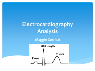
ECG complete lecture notes along with interpretation
ECG complete lecture notes along with interpretationECG complete lecture notes along with interpretation
ECG complete lecture notes along with interpretation
 
1electrophysiology-110623133004-phpapp02.pdf
1electrophysiology-110623133004-phpapp02.pdf1electrophysiology-110623133004-phpapp02.pdf
1electrophysiology-110623133004-phpapp02.pdf
 
ECG complete lecture presentation, ECG waveform and leads placement
ECG complete lecture presentation, ECG waveform and leads placementECG complete lecture presentation, ECG waveform and leads placement
ECG complete lecture presentation, ECG waveform and leads placement
 
Ecg basics & cardicac physiology
Ecg basics & cardicac physiologyEcg basics & cardicac physiology
Ecg basics & cardicac physiology
 
Electrocardiography (ecg)
Electrocardiography (ecg)Electrocardiography (ecg)
Electrocardiography (ecg)
 
Ecg
EcgEcg
Ecg
 
ECG.ppt
ECG.pptECG.ppt
ECG.ppt
 
12 Lead Basics
12 Lead Basics12 Lead Basics
12 Lead Basics
 
Ecg part i
Ecg part iEcg part i
Ecg part i
 
Antiarrhythmic drugs class Bidya.pdf
Antiarrhythmic drugs class Bidya.pdfAntiarrhythmic drugs class Bidya.pdf
Antiarrhythmic drugs class Bidya.pdf
 
Heart
HeartHeart
Heart
 
Heart
HeartHeart
Heart
 
Basic ECG notes
Basic ECG notesBasic ECG notes
Basic ECG notes
 
Term paper on ecg and cardiac arrhythmias
Term paper on ecg  and cardiac arrhythmiasTerm paper on ecg  and cardiac arrhythmias
Term paper on ecg and cardiac arrhythmias
 
Term paper on ECG and cardiac arrhythmias
Term paper on ECG  and cardiac arrhythmiasTerm paper on ECG  and cardiac arrhythmias
Term paper on ECG and cardiac arrhythmias
 
Electrocardiogram(ecg)
Electrocardiogram(ecg)Electrocardiogram(ecg)
Electrocardiogram(ecg)
 
Cardiac Electrophysiology
Cardiac ElectrophysiologyCardiac Electrophysiology
Cardiac Electrophysiology
 
BASIC CADIOVASCULAR PHYSIOLOGY.ppt
BASIC CADIOVASCULAR PHYSIOLOGY.pptBASIC CADIOVASCULAR PHYSIOLOGY.ppt
BASIC CADIOVASCULAR PHYSIOLOGY.ppt
 
05 ecg 2011
05 ecg 201105 ecg 2011
05 ecg 2011
 
Heart physiology, blood physiology
Heart physiology, blood physiologyHeart physiology, blood physiology
Heart physiology, blood physiology
 

Electrocardiography

  • 2.  Electrical conduction system of the heart  ECG  Phases: polarization, depolarization, repolarization  Electrode Placement  Interpreting ECG strips & waves  Cardiac Rhythms  Rhythm Quiz Agenda
  • 3.  Most important aspect heart’s anatomy & physiology to master in ECG analysis  Composed of myocardial tissue  ECG supplies information how well conduction system of the heart is functioning  Neurons serve conduction function for the rest of the body  Myocardial tissue has 2 advantages over nerve tissue Electrical Conduction System
  • 4.  Sinoatrial node, “pacemaker” of the heart  Small concentration of myocardial tissue  Initiates impulses that depolarize the rest of the heart  Located in upper portion of the right atrium  Electrical impulse that triggers each heartbeat  Spreads through the atria  Muscle tissue to contract in a coordinated manner  Inherent rate of 60-100 beats per minute SA Node
  • 5.  Atrioventricular node  Large bundle of myocardial tissue  Internodal tracts connect SA node to AV node  2 pathways: beat & alpha  Delays impulses from SA node to allowing atria to complete their contraction and empty completely  Atrial kick  Prevents extra beats from being conducted  Distal portion of AV node is the bundle of His that splits into two branches: left & right bundle branches AV Node
  • 6.  Spread across respective ventricles and become purkinje fibers  Ventricles contract  Electrical signal having passed through both atria and ventricles making them contract = one heart beat AV Node
  • 7.  Records the electrical activity of the heart  Detected by electrodes  Electrical activity of myocardial cells result in complex electrical reaction occurring in each fiber of myocardium  Millions of fibers of the myocardium react electrically to an impulse creating a wave of energy detected by the ECG  ECG pattern: characteristic wave forms produced by electrical activity  Painless  Three phases of electrochemical activity: polarization, depolarization, and repolarization ECG
  • 8.  Cells ready to contract  SA node ready to initiate impulse  Extracellular ions: Na and Ca strong positive electrical charge  Intracellular ion: K weak positive electrical charge  Difference of strength of electrical charges create a positive charge on outside of the cell membrane & negative charge on the inside Polarization
  • 9.  “Action phase”  Shift in ion concentrations  Na ions rush into the cell, followed by slower Ca ions  K ions pushed out of the cell creating polarity of cell to become reversed  Negative charge along outside of the cell membrane & positive charge inside the cell membrane  Shortens fibers producing contractions  Contraction increases pressure, pushing blood out of heart chambers “systole” Depolarization
  • 10.  “Recovery phase”  Sodium Potassium pump  Pump out Na and Ca and pump in K reestablishing electrical charge along cell membrane  Muscle fibers of myocardium are relaxing returning to preconstraction state  Relaxation decreasing pressure, heart chambers fill “diastole” Repolarization
  • 11.  1) cleanse skin removing: oils, sweat, dirt and hair  2) place electrode sticky side down and begin placing on one corner, avoiding touching sticky side  3) smooth electrode to skin in circular motion and connect monitor wire  5 lead ECG: -white: RA second intercostal space (right side) -brown: chest (middle) -green: RL (right side) -red: LL (left side) -black: LA second intercostal space (left side) Electrode Placement
  • 12. Electrode Placement Smoke over fire, snow over grass, brown in the middle
  • 13. Interpreting ECG Strips Each small box= 0.04 seconds, 1mm Each large box= 0.20 seconds, 5mm 25mm/second papers
  • 14.  Atrial depolarization  Time for electrical impulse from SA node to spread through the atrial muscle  Amplitude: 2-2.5 mm in height  Duration: 0.06-0.11 seconds  P-R interval: -time takes an impulse to travel from the atria through the AV node, bundle of his, and bundle branches to the Purkinje fibres -beginning of p wave to beginning of QRS complex -duration: 0.12-0.20 seconds P wave & P-R interval
  • 16.  Ventricular depolarization  3 waves: Q,R,S  Q wave: beginning, sometimes not present  R wave: positive deflection  S wave: negative deflection  Duration: no longer than 10 seconds QRS Complex
  • 17.  T wave: repolarization of ventricles  S-T segment: end of ventricular depolarization and beginning of ventricular repolarization  QT interval: -time necessary for ventricle depolarization & repolarization -beginning of QRS complex to end of T wave T wave, Q-T interval, S-T segment
  • 18. Sinus Rhythm • Normal • Electrical stimuli initiated in SA node, conducted through AV node, bundle of His, bundle branches, and Purkinje fibers • Depolarization and repolarization of atria and ventricles
  • 19. Sinus Bradycardia • HR: <60 bpm • Rhythm unchanged • Normal sinus slowed down • Waves further apart
  • 20. Sinus Tachycardia • HR: >100 bpm • Sinus rhythm greater than 100 bpm • Waves closer together
  • 21. Atrial Fibrillation • No P wave since SA node isn’t functioning • Atria generates up to 600 stimuli/min • Instead seeing P wave, number of small waves of different sizes “fibrillatory waves” (F waves) • Stimuli not strong enough to depolarize the AV node, rhythm is irregular since number of stimuli being generated in atria • Fast rhythm: 110-140 bpm • QRS normal since abnormality above the ventricles
  • 22. Atrial Flutter • Similar to atrial fibrillation • Abnormality of conduction of atria • Flutter waves bombard Av node but more organized and regular fashion • “saw-tooth” pattern
  • 23. Ventricular Fibrillation • Life threatening • Chaotic depolarization of ventricles • No clear P waves or QRS complexes
  • 24. Myocardial Infarction • Heart attack • 1st hyperacute T wave: taller and pointed • 2nd ST segment elevation: when heart muscle is being injured by lack of blood flow and oxygen
  • 26.
  • 28.
  • 30.
  • 32.
  • 34.
  • 36.
  • 38.  Ambulance Technician Study. (2006). ECG basics. Retrieved from http://www.ambulancetechnicianstudy.co.uk/ecgbasics.html#.UKu1b4aA8uc  Catalano, J.T. (2002). Guide to ECG analysis (2nd ed.).Philadelphia, PA: Lippincott Williams & Wilkins  Lewis, S.L., Heitkemper, M.M., Dirksen, S.R, O’Brien, P.G., & Bucher, L. (2010). Medical-surgical nursing in canada (2nd ed.) Toronto, ON: Elsevier Canada  Perry, P.A. & Potter, A.G. (2009). Canadian fundamentals of nursing (4th ed.) Toronto, ON: Elsevier Canada  University of Nottingham School of Nursing. (2012). Beginners guide to normal heart function, sinus rhythm & common arrhythmias. Retrieved from http://www.nottingham.ac.uk/nursing/practice/resources/cardiology/index.php References

Editor's Notes

  1. Smoke over fire, snow over grass, brown in the middle