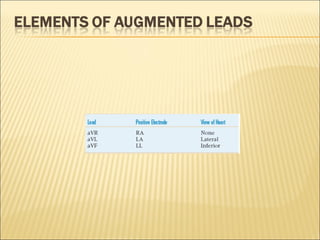
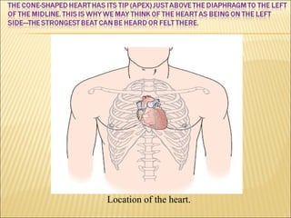
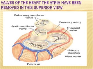
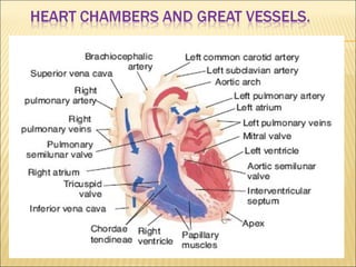
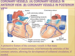
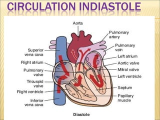
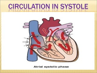
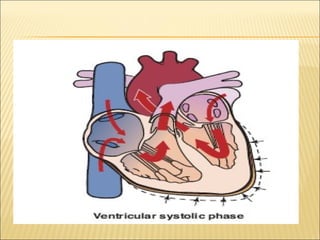
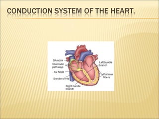
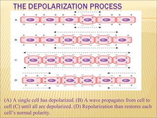
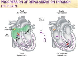
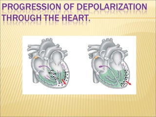
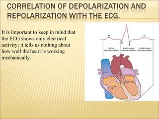
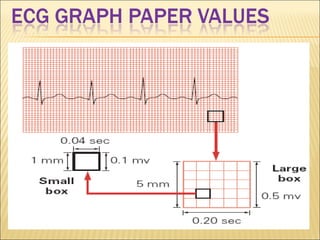
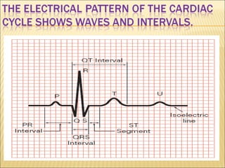
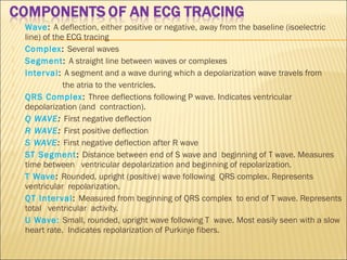
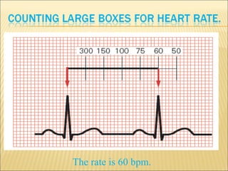
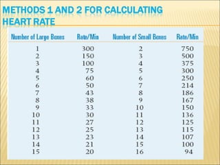
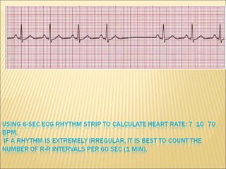
ECG BasicsECG Basics
Dr. Md.Toufiqur Rahman
MBBS, FCPS, MD, FACC, FESC, FRCP, FSCAI,
FAPSC, FAPSIC, FAHA
Associate Professor of Cardiology
National Institute of Cardiovascular Diseases
Sher-e-Bangla Nagar, Dhaka-1207
Consultant, Medinova,
Malbagh branch.
Location of the heart.
A protective feature of the coronary vessels is that many
interconnections, or anastomoses, exist between the arterioles of the
coronary arteries, allowing for development of collateral circulation, if
needed.
(A) A single cell has depolarized. (B) A wave propagates from cell to
cell (C) until all are depolarized. (D) Repolarization than restores each
cell’s normal polarity.
It is important to keep in mind that
the ECG shows only electrical
activity; it tells us nothing about
how well the heart is working
mechanically.
Lead MCL1 electrode placement.
 Wave: A deflection, either positive or negative, away from the baseline (isoelectric
line) of the ECG tracing
 Complex: Several waves
 Segment: A straight line between waves or complexes
 Interval: A segment and a wave during which a depolarization wave travels from
 the atria to the ventricles.
 QRS Complex: Three deflections following P wave. Indicates ventricular
depolarization (and contraction).
 Q WAVE: First negative deflection
 R WAVE: First positive deflection
 S WAVE: First negative deflection after R wave
 ST Segment: Distance between end of S wave and beginning of T wave. Measures
time between ventricular depolarization and beginning of repolarization.
 T Wave: Rounded, upright (positive) wave following QRS complex. Represents
ventricular repolarization.
 QT Interval: Measured from beginning of QRS complex to end of T wave. Represents
total ventricular activity.
 U Wave: Small, rounded, upright wave following T wave. Most easily seen with a slow
heart rate. Indicates repolarization of Purkinje fibers.
The rate is 60 bpm.
Rate: Normal (60–100 bpm) Rhythm: Regular P Waves: Normal
(upright and uniform PR Interval: Normal (0.12–0.20 sec) QRS:
Normal (0.06–0.10 sec)
Rate: Slow (60 bpm) Rhythm: Regular P Waves: Normal (upright and
uniform ) PR Interval: Normal (0.12–0.20 sec) QRS:Normal (0.06–
0.10 sec)
Sinus bradycardia is normal in athletes and during sleep. In acute MI,
it may be protective and beneficial or the slow rate may compromise
cardiac output. Certain medications, such as beta blockers, may also
cause sinus bradycardia.
 • An ECG is a series of waves and deflections recording the
heart’s electrical activity from a certain “view.”
 • Many views, each called a lead, monitor voltage changes
between electrodes placed in different positions on the
body.
 • Leads I, II, and III are bipolar leads, which consist of two
electrodes of opposite polarity (positive and negative). The
third (ground) electrode minimizes electrical activity from
other sources.
 • Leads aVR, aVL, and aVF are unipolar leads and consist
of a single positive electrode and a reference point (with
zero electrical potential) that lies in the center of the
heart’s electrical field.
 • Leads V1 through V6 are unipolar leads and consist of a
single positive electrode with a negative reference point,
found at the electrical center of the heart.
 • Voltage changes are amplified and visually displayed
on an oscilloscope and graph paper.
 • An ECG tracing looks different in each lead because
the recorded angle of electrical activity changes with
each lead.
 Several different angles allow a more accurate
perspective than a single one would.
 • The ECG machine can be adjusted to make any skin
electrode positive or negative. The polarity depends on
which lead the machine is recording.
 • A cable attached to the patient is divided into several
different-colored wires: three, four, or five for monitoring
purposes, or ten for a 12- lead ECG.
 • Incorrect placement of electrodes may turn a normal
ECG tracing into an abnormal one.
Thank you !Thank you !

ECG Elementary

  • 1.
    ECG BasicsECG Basics Dr.Md.Toufiqur Rahman MBBS, FCPS, MD, FACC, FESC, FRCP, FSCAI, FAPSC, FAPSIC, FAHA Associate Professor of Cardiology National Institute of Cardiovascular Diseases Sher-e-Bangla Nagar, Dhaka-1207 Consultant, Medinova, Malbagh branch.
  • 2.
  • 6.
    A protective featureof the coronary vessels is that many interconnections, or anastomoses, exist between the arterioles of the coronary arteries, allowing for development of collateral circulation, if needed.
  • 14.
    (A) A singlecell has depolarized. (B) A wave propagates from cell to cell (C) until all are depolarized. (D) Repolarization than restores each cell’s normal polarity.
  • 17.
    It is importantto keep in mind that the ECG shows only electrical activity; it tells us nothing about how well the heart is working mechanically.
  • 27.
  • 35.
     Wave: Adeflection, either positive or negative, away from the baseline (isoelectric line) of the ECG tracing  Complex: Several waves  Segment: A straight line between waves or complexes  Interval: A segment and a wave during which a depolarization wave travels from  the atria to the ventricles.  QRS Complex: Three deflections following P wave. Indicates ventricular depolarization (and contraction).  Q WAVE: First negative deflection  R WAVE: First positive deflection  S WAVE: First negative deflection after R wave  ST Segment: Distance between end of S wave and beginning of T wave. Measures time between ventricular depolarization and beginning of repolarization.  T Wave: Rounded, upright (positive) wave following QRS complex. Represents ventricular repolarization.  QT Interval: Measured from beginning of QRS complex to end of T wave. Represents total ventricular activity.  U Wave: Small, rounded, upright wave following T wave. Most easily seen with a slow heart rate. Indicates repolarization of Purkinje fibers.
  • 36.
    The rate is60 bpm.
  • 41.
    Rate: Normal (60–100bpm) Rhythm: Regular P Waves: Normal (upright and uniform PR Interval: Normal (0.12–0.20 sec) QRS: Normal (0.06–0.10 sec)
  • 42.
    Rate: Slow (60bpm) Rhythm: Regular P Waves: Normal (upright and uniform ) PR Interval: Normal (0.12–0.20 sec) QRS:Normal (0.06– 0.10 sec) Sinus bradycardia is normal in athletes and during sleep. In acute MI, it may be protective and beneficial or the slow rate may compromise cardiac output. Certain medications, such as beta blockers, may also cause sinus bradycardia.
  • 43.
     • AnECG is a series of waves and deflections recording the heart’s electrical activity from a certain “view.”  • Many views, each called a lead, monitor voltage changes between electrodes placed in different positions on the body.  • Leads I, II, and III are bipolar leads, which consist of two electrodes of opposite polarity (positive and negative). The third (ground) electrode minimizes electrical activity from other sources.  • Leads aVR, aVL, and aVF are unipolar leads and consist of a single positive electrode and a reference point (with zero electrical potential) that lies in the center of the heart’s electrical field.  • Leads V1 through V6 are unipolar leads and consist of a single positive electrode with a negative reference point, found at the electrical center of the heart.
  • 44.
     • Voltagechanges are amplified and visually displayed on an oscilloscope and graph paper.  • An ECG tracing looks different in each lead because the recorded angle of electrical activity changes with each lead.  Several different angles allow a more accurate perspective than a single one would.  • The ECG machine can be adjusted to make any skin electrode positive or negative. The polarity depends on which lead the machine is recording.  • A cable attached to the patient is divided into several different-colored wires: three, four, or five for monitoring purposes, or ten for a 12- lead ECG.  • Incorrect placement of electrodes may turn a normal ECG tracing into an abnormal one.
  • 45.

Editor's Notes

  • #2 Good morning ladies and gentlemen, it is my pleasure to be able to present and discuss with you this morning in such a prestigious international meeting. My presentation on Hypertension shall include the following aspects. I
  • #18 The following summarizes a basic ECG: • An ECG is a series of waves and deflections recording the heart’s electrical activity from a certain “view.” • Many views, each called a lead, monitor voltage changes between electrodes placed in different positions on the body. • Leads I, II, and III are bipolar leads, which consist of two electrodes of opposite polarity (positive and negative). The third (ground) electrode minimizes electrical activity from other sources. • Leads aVR, aVL, and aVF are unipolar leads and consist of a single positive electrode and a reference point (with zero electrical potential) that lies in the center of the heart’s electrical field. • Leads V1 through V6 are unipolar leads and consist of a single positive electrode with a negative reference point, found at the electrical center of the heart. • Voltage changes are amplified and visually displayed on an oscilloscope and graph paper. • An ECG tracing looks different in each lead because the recorded angle of electrical activity changes with each lead. Several different angles allow a more accurate perspective than a single one would. • The ECG machine can be adjusted to make any skin electrode positive or negative. The polarity depends on which lead the machine is recording. • A cable attached to the patient is divided into several different-colored wires: three, four, or five for monitoring purposes, or ten for a 12- lead ECG. • Incorrect placement of electrodes may turn a normal ECG tracing into an abnormal one.
  • #43 Sinus bradycardia is normal in athletes and during sleep. In acute MI, it may be protective and beneficial or the slow rate may compromise cardiac output. Certain medications, such as beta blockers, may also cause sinus bradycardia.