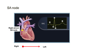
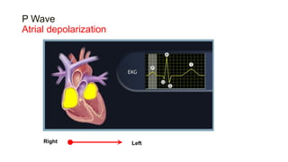
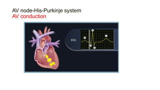
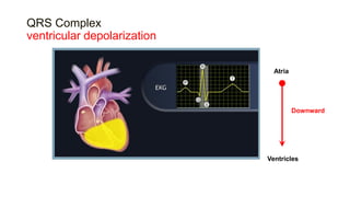
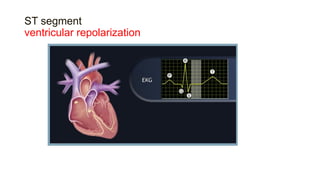
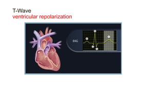
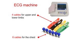
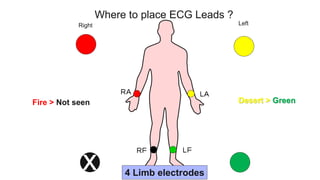
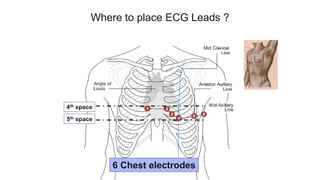
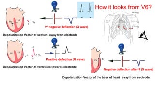
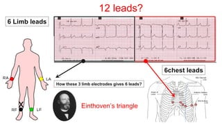
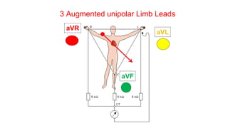
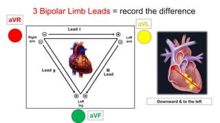
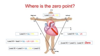
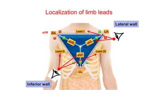
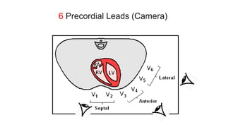
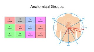
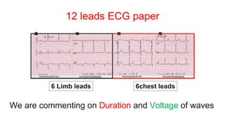
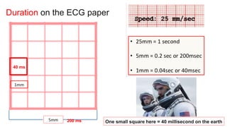
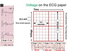
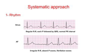
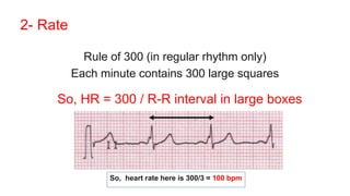
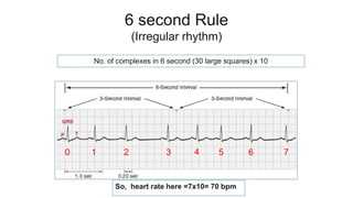
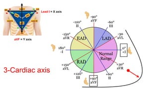
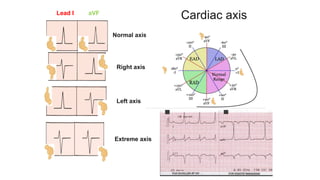
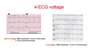
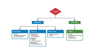
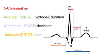
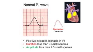
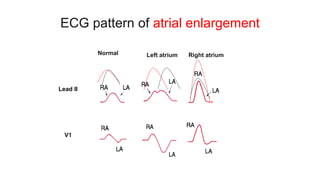
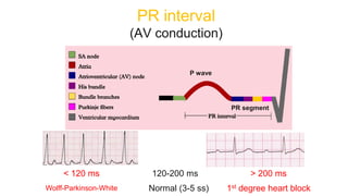
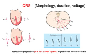
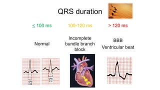
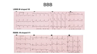
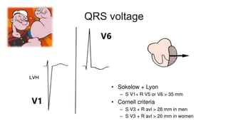
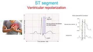
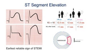
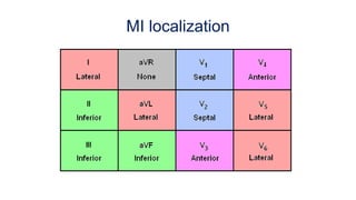
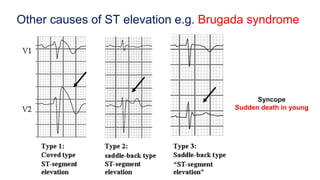
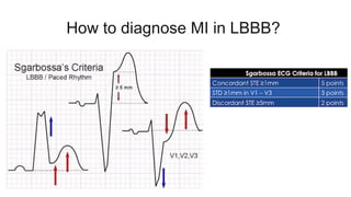
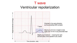
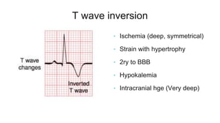
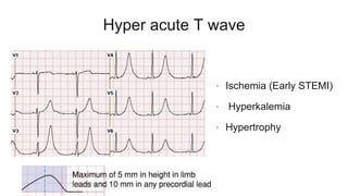
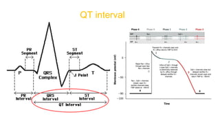
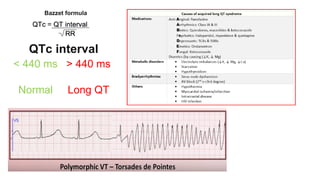
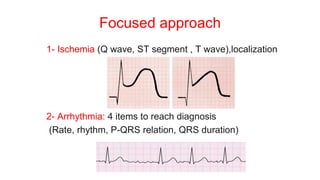
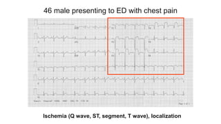
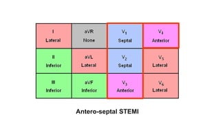
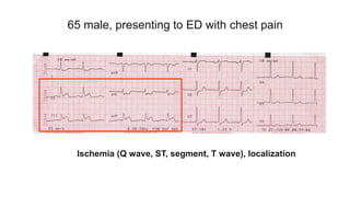
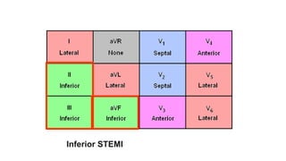
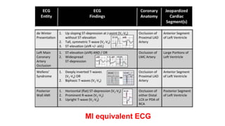
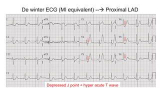
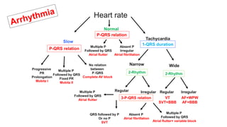
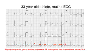
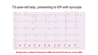
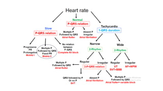
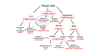
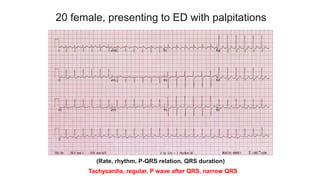
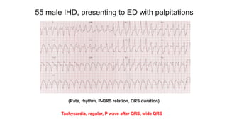
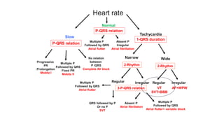
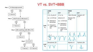
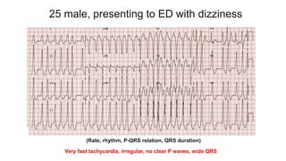
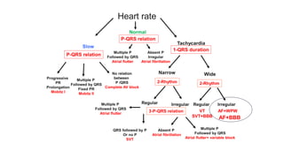
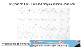
How to read ECG?
Ahmed Elborae, MSc, MD, PhD, MRCP UK (London)
Assistant Lecturer of Cardiology, Cairo University
A 45-year-old male patient, presenting to ER with chest pain
ECG
“Electrical activity”
Echocardiography
“Ultrasound imaging”
Angiography
“Coronary supply”
A single modality rarely gives the whole truth
 Simple
 Rapid
 Cheap
 Available
Our conduction system
SA node
Right atrium
SA node
Right Left
P Wave
Atrial depolarization
Right Left
AV node-His-Purkinje system
AV conduction
QRS Complex
ventricular depolarization
Atria
Ventricles
Downward
ST segment
ventricular repolarization
T-Wave
ventricular repolarization
How to record our heart electricity?
ECG machine
4 cables for upper and
lower limbs
6 cables for the chest
Right Left
4 Limb electrodes
x
Where to place ECG Leads ?
Fire > Not seen Desert > Green
Where to place ECG Leads ?
6 Chest electrodes
4th space
5th space
V1,V2
V3,V4
V5,V6
Capturing the same object from different angles…
Depolarization Vector of ventricles towards electrode
Depolarization Vector of septum away from electrode
Positive deflection (R wave)
1st negative deflection (Q wave)
3
Depolarization Vector of the base of heart away from electrode
Negative deflection after R (S wave)
How it looks from V6?
Depolarization Vector of ventricles away from electrode
Depolarization Vector of septum towards electrode
Negative deflection after R (S wave)
1st Positive deflection (R wave)
How it looks from V1?
12 leads?
6 Limb leads
6chest leads
x
How these 3 limb electrodes gives 6 leads?
Einthoven’s triangle
3 Augmented unipolar Limb Leads
aVR
aVL
aVF
How QRS looks from unipolar limb leads?
3 Bipolar Limb Leads = record the difference
Lead
Lead
Lead
Downward & to the left
aVR
aVL
aVF
How QRS looks from Bipolar limb leads?
Lead I = LA - RA
Lead II = LL - RA Lead III = LL - LA
Where is the zero point?
(Lead III + Lead I) - Lead II = Zero
Lead III + lead I = LL – LA + LA – RA
Lead III + Lead I = LL – RA = Lead II
Localization of limb leads
aVR
Inferior wall
Lateral wall
6 Precordial Leads (Camera)
Anatomical Groups
12 leads ECG paper
6 Limb leads 6chest leads
We are commenting on Duration and Voltage of waves
Duration on the ECG paper
40 ms
200 ms One small square here = 40 millisecond on the earth
Voltage on the ECG paper
One small square
10
small
boxes
=Two
large
boxes
2 large 1 mv
Usual Half volt. Double volt.
1 large 1 mv 4 large 1 mv
2 large =1 mv
How to read ECG?
Systematic approach
Systematic approach
1- Rhythm
Sinus
AF
Regular R-R, each P followed by QRS, normal PR interval
Irregular R-R, absent P waves, fibrillation waves
2- Rate
Rule of 300 (in regular rhythm only)
Each minute contains 300 large squares
So, HR = 300 / R-R interval in large boxes
So, heart rate here is 300/3 = 100 bpm
6 second Rule
(Irregular rhythm)
No. of complexes in 6 second (30 large squares) x 10
0 1 2 3 4 5 6 7
So, heart rate here =7x10= 70 bpm
3-Cardiac axis
Lead I = X axis
aVF = Y axis
Lead I aVF
Normal axis
Right axis
Left axis
Extreme axis
Cardiac axis
4-ECG voltage
Normal voltage: QRS amplitude > 5 mm in limb leads
,> 10 mm precordial leads
Low voltage: QRS amplitude < 5 mm in all limb leads
5-Comment on
-Waves( P,QRS,T) voltage& duration
-Segments (PR,ST) deviation
-Intervals (PR,QT) time
Normal P- wave
• Positive in lead II, biphasic in V1
• Duration less than 3 small squares
• Amplitude less than 2.5 small squares
Right atrium
Left atrium
ECG pattern of atrial enlargement
Left atrium Right atrium
Normal
V1
Lead II
Lead II
PR interval
(AV conduction)
PR segment
P wave
< 120 ms 120-200 ms > 200 ms
Wolff-Parkinson-White Normal (3-5 ss) 1st degree heart block
QRS (Morphology, duration, voltage)
Poor R wave progression (R in V3 < 3 small squares) might denotes anterior ischemia
Pathological Q wave of MI
Pathological = More than 1x1 small squares
Pathological Q = Black hole
QRS duration
< 100 ms 100-120 ms > 120 ms
Normal
Incomplete
bundle branch
block
BBB
Ventricular beat
BBB
LBBB:M shaped V6
RBBB: M shaped V1
QRS voltage
ST segment
Ventricular repolarization
ST Segment Elevation
Earliest reliable sign of STEMI
MI localization
Other causes of ST elevation e.g. Brugada syndrome
Syncope
Sudden death in young
ST Segment Depression
Myocardial ischemia or NSTEMI (ST≥ 0.5 mm)
Depression are not localizing
How to diagnose MI in LBBB?
T wave
Ventricular repolarization
T wave inversion
• Ischemia (deep, symmetrical)
• Strain with hypertrophy
• 2ry to BBB
• Hypokalemia
• Intracranial hge (Very deep)
Hyper acute T wave
• Ischemia (Early STEMI)
• Hyperkalemia
• Hypertrophy
QT interval
QTc interval
< 440 ms > 440 ms
Normal Long QT Long QT
Bazzet formula
How to read ECG?
Rapid focused approach
Focused approach
1- Ischemia (Q wave, ST segment , T wave),localization
2- Arrhythmia: 4 items to reach diagnosis
(Rate, rhythm, P-QRS relation, QRS duration)
46 male presenting to ED with chest pain
Ischemia (Q wave, ST, segment, T wave), localization
Antero-septal STEMI
65 male, presenting to ED with chest pain
Ischemia (Q wave, ST, segment, T wave), localization
Inferior STEMI
MI equivalent ECG
De winter ECG (MI equivalent) -- Proximal LAD
Depressed J point + hyper acute T wave
Wellen ECG (MI equivalent) -- Proximal LAD
Type 2 > Biphasic T wave
Type 1 > deep symmetrical T wave inversion
(Left main or multi-vessel occlusion equivalent)
Diffuse ST depression+ aVR ST elevation
Posterior wall MI
Tall R in v2,V3+ ST depression
Mirror image
Posterior leads
40-year-old , acute dyspnea 1 week post-partum C/S
Pulmonary embolism
Tachycardia, S1Q3T3, tall R in aVR, RV strain (Inverted T in precordial leads)
Heart rate
Slow
Tachycardia
Normal
P-QRS relation
Progressive
PR
Prolongation
Mobitz I
Multiple P
Followed by QRS
Fixed PR
Mobitz II
No relation
between
P /QRS
Complete AV block
Multiple P
Followed by QRS
Atrial flutter
1-QRS duration
Narrow Wide
2-Rhythm 2-Rhythm
Regular Regular
Irregular Irregular
3-P-QRS relation VT
SVT+BBB
AF+WPW
AF+BBB
Multiple P
Followed by QRS
Atrial flutter
QRS followed by P
Or no P
SVT
Multiple P
Followed by QRS
Atrial flutter+ variable block
Absent P
Atrial fibrillation
P-QRS relation
Absent P
Irregular
Atrial fibrillation
33-year-old athlete, routine ECG
Slightly bradycardic, group beating, progressive PR prolongation then dropped beat, narrow QRS
x x
73-year-old lady, presenting to ER with syncope
Bradycardia, multiple P followed by QRS with fixed PR interval, narrow QRS
x x x x x x
75-year-old gentleman, presenting to ER with delirium
Bradycardia, multiple P followed by QRS with variable PR interval (Complete AV dissociation)
77 female presenting to ED with palpitations
(Rate, rhythm, P-QRS relation, QRS duration)
Normal rate, irregular, absent P wave, narrow QRS
60 female, presenting to ED with palpitations
(Rate, rhythm, P-QRS relation, QRS duration)
Normal rate, regular, Multiple P waves followed by QRS, narrow QRS
20 female, presenting to ED with palpitations
(Rate, rhythm, P-QRS relation, QRS duration)
Tachycardia, regular, P wave after QRS, narrow QRS
55 male IHD, presenting to ED with palpitations
(Rate, rhythm, P-QRS relation, QRS duration)
Tachycardia, regular, P wave after QRS, wide QRS
VT vs. SVT+BBB
25 male, presenting to ED with dizziness
Very fast tachycardia, irregular, no clear P waves, wide QRS
(Rate, rhythm, P-QRS relation, QRS duration)
AF+BBB
Hyperkalemia (Sine wave)
65-year-old ESKD, missed dialysis session, confused
Hyperkalemia stretch ECG (PR, QRS, T wave)
https://litfl.com/ecg-library/
For more practice…
THANK YOU !
Einthoven

ECG made easy