The document provides information about electrocardiograms (ECGs) including:
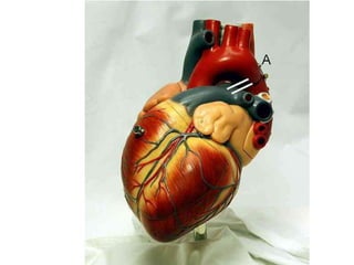
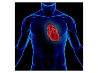
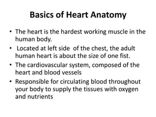
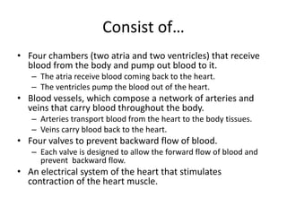
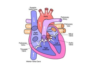
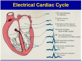
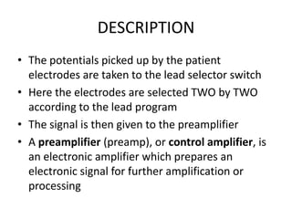
1) It describes the basic anatomy and electrical conduction system of the heart.
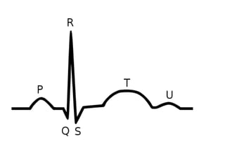
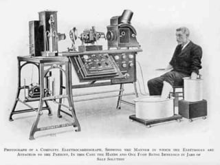
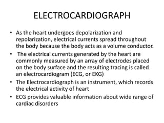
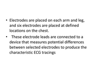
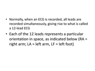
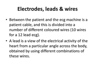
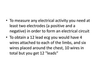
2) It explains what an ECG is and how it works by measuring the electrical signals produced by heart muscle depolarization and repolarization using electrodes placed on the body.
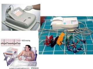
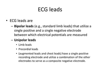
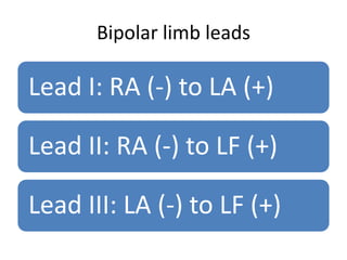
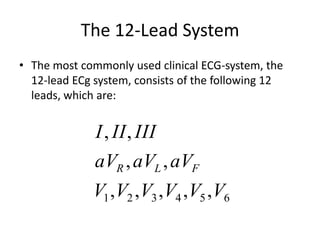
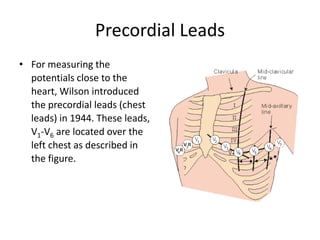
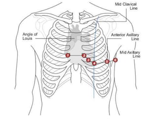
3) It details the 12-lead ECG system including the 10 wires attached to limbs and chest to measure electrical signals from different angles represented by 12 leads.


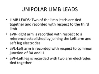
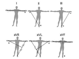
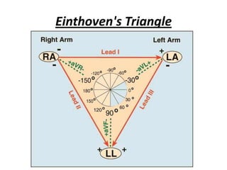
![Augmented unipolar limb leads
Lead aVR: RA (+) to [LA & LF] (-)
Lead aVL: LA (+) to [RA & LF] (-)
Lead aVF: LF (+) to [RA & LA] (-)](https://image.slidesharecdn.com/unitiecg-111226195137-phpapp01/85/ECG-Machine-25-320.jpg)


![Ambulatory blood pressure monitoring [abpm]](https://cdn.slidesharecdn.com/ss_thumbnails/ambulatorybloodpressuremonitoringabpm-140314094820-phpapp02-thumbnail.jpg?width=640&height=640&fit=bounds)


![ECG [electrocardiogram].pptx](https://cdn.slidesharecdn.com/ss_thumbnails/ecgelectrocardiogram-220416062706-thumbnail.jpg?width=640&height=640&fit=bounds)


![CTEV [ clubfoot] DR ARUN LAL ,DR MOHAMED ASHRAF travancore medical college k...](https://cdn.slidesharecdn.com/ss_thumbnails/ctevclubfootdrarunlaldrmohamedashraftravancoremedicalcollegekollamkeralaindia-260208063247-18fc466c-thumbnail.jpg?width=640&height=640&fit=bounds)


