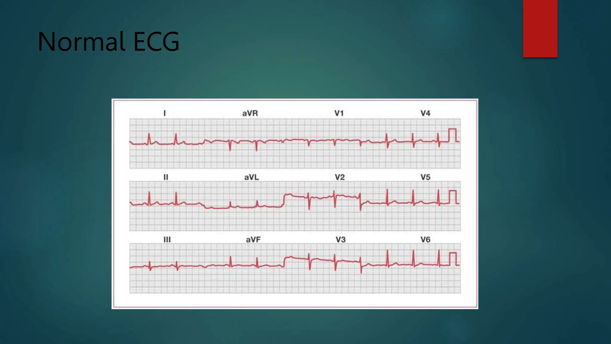
SlideShare a Scribd company logo
The ECG
ELECTROCARDIOGRAM
Cont:
An electrocardiogram — also called ECG Or EKG
INDICATION FOR USE
Noninvasive – Used to diagnose many common heart problems.
A health care provider might use an electrocardiogram to determine or detect the
following:
 Irregular heart rhythms (arrhythmias)
 If blocked or narrowed arteries in the heart (coronary artery disease) are causing
chest pain or a heart attack
 Whether you have had a previous heart attack
 How well certain heart disease treatments, such as a pacemaker, are working
Signs and symptoms
• Patients may need ECG if they have the following symptoms
• Chest pain
• Dizziness, lightheadedness or confusion
• Heart palpitations
• Rapid pulse
• Shortness of breath
• Weakness, fatigue or a decline in ability to exercise

Cont:
If symptoms come and go, they may not be detected during a standard ECG
recording.
A health care provider might recommend remote or continuous ECG monitoring .
Types
 Holter monitor. A Holter monitor is a small, wearable device that records a
continuous ECG, usually for 24 to 48 hours.
 Event monitor. This portable device is similar to a Holter monitor, but it records
only at certain times for a few minutes at a time.
 Can be worn longer than a Holter monitor, typically 30 days.
 Patient can push a button when they feel symptoms.
 Some devices automatically record when an irregular rhythm is detected.

Application
 During an ECG, up to 12 sensors (electrodes) are attached to the chest and limbs
 The electrodes are sticky patches with wires that connect to a monitor.
 They record the electrical signals that make the heart beat.
 A computer records the information and displays it as waves on a monitor or on
paper.
 Patient can breathe during the test, but will need to lie still.
 Keep the patient warm and ready to lie still.
 Moving, talking or shivering may interfere with the test results.
 A standard ECG takes a few minutes.
RESULTS OF ECG
ECG results can give a health care provider details about the following:
Heart rate.
 Usually, heart rate can be measured by checking the pulse.
 An ECG may be helpful if the pulse is difficult to feel or too fast or too irregular to
count accurately.
 An ECG can help identify an unusually fast heart rate (tachycardia) or an
unusually slow heart rate (bradycardia).
Heart rhythm.
 An ECG can detect irregular heartbeats (arrhythmias).
 An arrhythmia may occur when any part of the heart's electrical system doesn't
work properly.
Cont:
 Heart attack.
 An ECG can show evidence of a previous heart attack or one that's currently
happening.
 The patterns on the ECG may help determine which part of the heart has been
damaged, as well as the extent of the damage.
 Blood and oxygen supply to the heart.
 An ECG done while having symptoms can help health care provider determine
whether reduced blood flow to the heart muscle is causing the chest pain.
 Heart structure changes.
 An ECG can provide clues about an enlarged heart, heart defects and other heart
problems.
Application
 Ask the patient to undress down to the waist and lie down
 Remove excess hair where necessary
 Attach limb leads (anywhere on the limb)
 Attach the chest leads
o V1 and V2: either side of the sternum on the fourth rib (count down from the sternal angle, the second rib
insertion)
o V4: on the apex of the heart (feel for it)
o V3: halfway between V2 and V4
o V5 and V6: horizontally laterally from V4 (not up towards the axilla)
 Ask the patient to relax
 Press record

Cont:
Limb leads
 Three bipolar leads and three unipolar leads are obtained from three electrodes attached to
the left arm, the right arm, and the left leg, respectively.
 (An electrode is also attached to the right leg, but this is an earth electrode.)
 The bipolar limb leads reflect the potential difference between two of the three limb
electrodes:
 lead I: right arm–left arm
 lead II: right arm–left leg
 lead III: left leg–left arm

Cont:
 The unipolar leads reflect the potential difference between one of the three limb electrodes
and an estimate of zero potential – derived from the remaining two limb electrodes.
 These leads are known as augmented leads.
 The augmented leads and their respective limb electrodes are:
 aVR lead: right arm
 aVL lead: left arm
 aVF lead: left leg

Chest leads
 Another six electrodes, placed in standard positions on the chest wall, give rise to a further
six unipolar leads – the chest leads (also known as precordial leads), V1–V6.
 The potential difference of a chest lead is recorded between the relevant chest electrode and
an estimate of zero potential – derived from the average potential recorded from the three limb
leads.

Electrical Activity
Cont:
 P wave
 The P wave is a small deflection wave that represents atrial depolarization.
 PR interval
 The PR interval is the time between the first deflection of the P wave and the first deflection of
the QRS complex.
 QRS wave complex
 The three waves of the QRS complex represent ventricular depolarization.
 Note: the most confusing aspects of ECG reading is the labeling of these waves.
 The rule is: if the wave immediately after the P wave is an upward deflection, it is an R wave; if
it is a downward deflection, it is a Q wave:
Cont:
Small Q waves correspond to depolarization of the interventricular septum.
Q waves can also relate to breathing and are generally small and thin.
They can also signal an old myocardial infarction (in which case they are big and wide)
 the R wave reflects depolarization of the main mass of the ventricles –hence it is the largest
wave
 the S wave signifies the final depolarization of the ventricles, at the base of the heart

Cont:
 ST segment
 The ST segment, which is also known as the ST interval, is the time between the end of the
QRS complex and the start of the T wave.
 It reflects the period of zero potential between ventricular depolarization and repolarization.
 T wave
 T waves represent ventricular repolarization (atrial repolarization is obscured by the large
QRS complex).

Normal ECG
Heart Rate
 Identify the QRS complex (this is generally the biggest wave); count the number of large
squares between one QRS wave and the next; divide 300 by this number to determine the rate
Cont:
Number of large squares between QRS complexes Heart rate (bpm)
5 60
4 75
3 100
2 150
Heart Rhythm
 P waves are the key to determining whether a patient is in sinus rhythm or not.
 If P waves are not clearly visible in the chest leads, look for them in the other leads.
 The presence of P waves immediately before every QRS complex indicates sinus rhythm.
 If there are no P waves, note whether the QRS complexes are wide or narrow, regular or
irregular.

No P waves and irregular narrow QRS
Complexes
 This is the hallmark of atrial fibrillation.
 Sometimes the baseline appears "noisy" and sometimes it appears entirely flat.
 However, if there are no P waves and the QRS complexes appear at randomly irregular
intervals, the diagnosis is atrial fibrillation.

Atrial Fibrillation
Sawtooth P waves
 A sawtooth waveform signifies atrial flutter .
 The number of atrial contractions to one ventricular contraction should be specified.

Atrial Flutter
ST Segment Changes
 The ST segment extends from the end of the S wave to the start of the T wave.
 It should be flat or slightly upsloping and level with the baseline.
 Elevation of more than two small squares in the chest leads or one small square in the limb
leads, combined with a characteristic history, indicates the possibility of MI
 ST depression is diagnostic of ischemia .
 It is worth noting that although ST elevation can localize the lesion (eg, anterior MI,
inferior MI), ST depression cannot.
 Concave upwards ST elevation in all 12 leads is diagnostic of pericarditis.

T waves
 In a normal ECG, T waves are upright in every lead except aVR.
 T-wave inversion can represent current ischemia or previous infarction .
 In combination with LVH and ST depression, it can represent "strain". This form of LVH
carries a poor prognosis.

T Wave Inversion
QT
 Long QT syndrome may also be drug-induced.
 Once this occurs, the responsible drug needs to be discontinued.
Question
 A Nurse is caring for a patient with Atrial Fibrillation, in addition to
antidysrhythmic, what other medication does the nurse administer?
 a. Heparin
 b. Atropin
 c. Amoxicillin
 d. Magnesium sulphate

More Related Content

Similar to ICU - The ECG - ELECTROCARDIOGRAM.pptx

Ecg for candidate 2021
Ecg for candidate 2021Ecg for candidate 2021
Ecg for candidate 2021
aljamhori teaching hospital
 
Electrocardiography
ElectrocardiographyElectrocardiography
Electrocardiography
Anand Patel
 
EKG 12 Leads
EKG 12 LeadsEKG 12 Leads
EKG 12 Leads
bajah423
 
ECG
ECGECG
ecg.pdf
ecg.pdfecg.pdf
ECG 2.pdf
ECG 2.pdfECG 2.pdf
ECG 2.pdf
LHusna
 
Electrocardiogram(ecg)
Electrocardiogram(ecg)Electrocardiogram(ecg)
Electrocardiogram(ecg)
New Leaf Rehab
 
ECG by Adil.pptx
ECG by Adil.pptxECG by Adil.pptx
ECG by Adil.pptx
Dradil9949669613
 
ECG
ECGECG
ecg-130715033518-phpapp02-1.pdf
ecg-130715033518-phpapp02-1.pdfecg-130715033518-phpapp02-1.pdf
ecg-130715033518-phpapp02-1.pdf
AtiqurRahman7153
 
Anesthesia related presentation very helpful
Anesthesia related presentation very helpfulAnesthesia related presentation very helpful
Anesthesia related presentation very helpful
MalikArifUllah
 
Ecg power point
Ecg power pointEcg power point
Ecg power point
Pradeep Indurkar
 
Electrocardiography Basics
Electrocardiography BasicsElectrocardiography Basics
topic ECG and heart diseases+treatments
topic ECG and heart diseases+treatmentstopic ECG and heart diseases+treatments
topic ECG and heart diseases+treatments
mariarani2
 
Basic ecg
Basic ecgBasic ecg
Basic ecg
dratin75
 
ECG & Heart block [doctors online]
ECG & Heart block [doctors online]ECG & Heart block [doctors online]
ECG & Heart block [doctors online]Chandan Banerjee
 
ecg 3.pptx
ecg 3.pptxecg 3.pptx
ecg 3.pptx
Anwar Siddiqui
 
ecg3-220822200141-06ac3a90 (1).pdf
ecg3-220822200141-06ac3a90 (1).pdfecg3-220822200141-06ac3a90 (1).pdf
ecg3-220822200141-06ac3a90 (1).pdf
WaelSaid16
 
ECG for NPs
ECG for NPsECG for NPs
ECG for NPsdimaurer
 

Similar to ICU - The ECG - ELECTROCARDIOGRAM.pptx (20)

Ecg for candidate 2021
Ecg for candidate 2021Ecg for candidate 2021
Ecg for candidate 2021
 
MJ cardio
MJ cardioMJ cardio
MJ cardio
 
Electrocardiography
ElectrocardiographyElectrocardiography
Electrocardiography
 
EKG 12 Leads
EKG 12 LeadsEKG 12 Leads
EKG 12 Leads
 
ECG
ECGECG
ECG
 
ecg.pdf
ecg.pdfecg.pdf
ecg.pdf
 
ECG 2.pdf
ECG 2.pdfECG 2.pdf
ECG 2.pdf
 
Electrocardiogram(ecg)
Electrocardiogram(ecg)Electrocardiogram(ecg)
Electrocardiogram(ecg)
 
ECG by Adil.pptx
ECG by Adil.pptxECG by Adil.pptx
ECG by Adil.pptx
 
ECG
ECGECG
ECG
 
ecg-130715033518-phpapp02-1.pdf
ecg-130715033518-phpapp02-1.pdfecg-130715033518-phpapp02-1.pdf
ecg-130715033518-phpapp02-1.pdf
 
Anesthesia related presentation very helpful
Anesthesia related presentation very helpfulAnesthesia related presentation very helpful
Anesthesia related presentation very helpful
 
Ecg power point
Ecg power pointEcg power point
Ecg power point
 
Electrocardiography Basics
Electrocardiography BasicsElectrocardiography Basics
Electrocardiography Basics
 
topic ECG and heart diseases+treatments
topic ECG and heart diseases+treatmentstopic ECG and heart diseases+treatments
topic ECG and heart diseases+treatments
 
Basic ecg
Basic ecgBasic ecg
Basic ecg
 
ECG & Heart block [doctors online]
ECG & Heart block [doctors online]ECG & Heart block [doctors online]
ECG & Heart block [doctors online]
 
ecg 3.pptx
ecg 3.pptxecg 3.pptx
ecg 3.pptx
 
ecg3-220822200141-06ac3a90 (1).pdf
ecg3-220822200141-06ac3a90 (1).pdfecg3-220822200141-06ac3a90 (1).pdf
ecg3-220822200141-06ac3a90 (1).pdf
 
ECG for NPs
ECG for NPsECG for NPs
ECG for NPs
 

Recently uploaded

263778731218 Abortion Clinic /Pills In Harare ,
263778731218 Abortion Clinic /Pills In Harare ,263778731218 Abortion Clinic /Pills In Harare ,
263778731218 Abortion Clinic /Pills In Harare ,
sisternakatoto
 
Top-Vitamin-Supplement-Brands-in-India.pptx
Top-Vitamin-Supplement-Brands-in-India.pptxTop-Vitamin-Supplement-Brands-in-India.pptx
Top-Vitamin-Supplement-Brands-in-India.pptx
SwisschemDerma
 
basicmodesofventilation2022-220313203758.pdf
basicmodesofventilation2022-220313203758.pdfbasicmodesofventilation2022-220313203758.pdf
basicmodesofventilation2022-220313203758.pdf
aljamhori teaching hospital
 
Colonic and anorectal physiology with surgical implications
Colonic and anorectal physiology with surgical implicationsColonic and anorectal physiology with surgical implications
Colonic and anorectal physiology with surgical implications
Dr Maria Tamanna
 
Role of Mukta Pishti in the Management of Hyperthyroidism
Role of Mukta Pishti in the Management of HyperthyroidismRole of Mukta Pishti in the Management of Hyperthyroidism
Role of Mukta Pishti in the Management of Hyperthyroidism
Dr. Jyothirmai Paindla
 
Superficial & Deep Fascia of the NECK.pptx
Superficial & Deep Fascia of the NECK.pptxSuperficial & Deep Fascia of the NECK.pptx
Superficial & Deep Fascia of the NECK.pptx
Dr. Rabia Inam Gandapore
 
SURGICAL ANATOMY OF THE RETROPERITONEUM, ADRENALS, KIDNEYS AND URETERS.pptx
SURGICAL ANATOMY OF THE RETROPERITONEUM, ADRENALS, KIDNEYS AND URETERS.pptxSURGICAL ANATOMY OF THE RETROPERITONEUM, ADRENALS, KIDNEYS AND URETERS.pptx
SURGICAL ANATOMY OF THE RETROPERITONEUM, ADRENALS, KIDNEYS AND URETERS.pptx
Bright Chipili
 
Cervical & Brachial Plexus By Dr. RIG.pptx
Cervical & Brachial Plexus By Dr. RIG.pptxCervical & Brachial Plexus By Dr. RIG.pptx
Cervical & Brachial Plexus By Dr. RIG.pptx
Dr. Rabia Inam Gandapore
 
Physiology of Chemical Sensation of smell.pdf
Physiology of Chemical Sensation of smell.pdfPhysiology of Chemical Sensation of smell.pdf
Physiology of Chemical Sensation of smell.pdf
MedicoseAcademics
 
Best Ayurvedic medicine for Gas and Indigestion
Best Ayurvedic medicine for Gas and IndigestionBest Ayurvedic medicine for Gas and Indigestion
Best Ayurvedic medicine for Gas and Indigestion
SwastikAyurveda
 
Light House Retreats: Plant Medicine Retreat Europe
Light House Retreats: Plant Medicine Retreat EuropeLight House Retreats: Plant Medicine Retreat Europe
Light House Retreats: Plant Medicine Retreat Europe
Lighthouse Retreat
 
Flu Vaccine Alert in Bangalore Karnataka
Flu Vaccine Alert in Bangalore KarnatakaFlu Vaccine Alert in Bangalore Karnataka
Flu Vaccine Alert in Bangalore Karnataka
addon Scans
 
Ophthalmology Clinical Tests for OSCE exam
Ophthalmology Clinical Tests for OSCE examOphthalmology Clinical Tests for OSCE exam
Ophthalmology Clinical Tests for OSCE exam
KafrELShiekh University
 
Top 10 Best Ayurvedic Kidney Stone Syrups in India
Top 10 Best Ayurvedic Kidney Stone Syrups in IndiaTop 10 Best Ayurvedic Kidney Stone Syrups in India
Top 10 Best Ayurvedic Kidney Stone Syrups in India
SwastikAyurveda
 
Non-respiratory Functions of the Lungs.pdf
Non-respiratory Functions of the Lungs.pdfNon-respiratory Functions of the Lungs.pdf
Non-respiratory Functions of the Lungs.pdf
MedicoseAcademics
 
A Classical Text Review on Basavarajeeyam
A Classical Text Review on BasavarajeeyamA Classical Text Review on Basavarajeeyam
A Classical Text Review on Basavarajeeyam
Dr. Jyothirmai Paindla
 
Tom Selleck Health: A Comprehensive Look at the Iconic Actor’s Wellness Journey
Tom Selleck Health: A Comprehensive Look at the Iconic Actor’s Wellness JourneyTom Selleck Health: A Comprehensive Look at the Iconic Actor’s Wellness Journey
Tom Selleck Health: A Comprehensive Look at the Iconic Actor’s Wellness Journey
greendigital
 
Effective-Soaps-for-Fungal-Skin-Infections.pptx
Effective-Soaps-for-Fungal-Skin-Infections.pptxEffective-Soaps-for-Fungal-Skin-Infections.pptx
Effective-Soaps-for-Fungal-Skin-Infections.pptx
SwisschemDerma
 
KDIGO 2024 guidelines for diabetologists
KDIGO 2024 guidelines for diabetologistsKDIGO 2024 guidelines for diabetologists
KDIGO 2024 guidelines for diabetologists
د.محمود نجيب
 
Pharynx and Clinical Correlations BY Dr.Rabia Inam Gandapore.pptx
Pharynx and Clinical Correlations BY Dr.Rabia Inam Gandapore.pptxPharynx and Clinical Correlations BY Dr.Rabia Inam Gandapore.pptx
Pharynx and Clinical Correlations BY Dr.Rabia Inam Gandapore.pptx
Dr. Rabia Inam Gandapore
 

Recently uploaded (20)

263778731218 Abortion Clinic /Pills In Harare ,
263778731218 Abortion Clinic /Pills In Harare ,263778731218 Abortion Clinic /Pills In Harare ,
263778731218 Abortion Clinic /Pills In Harare ,
 
Top-Vitamin-Supplement-Brands-in-India.pptx
Top-Vitamin-Supplement-Brands-in-India.pptxTop-Vitamin-Supplement-Brands-in-India.pptx
Top-Vitamin-Supplement-Brands-in-India.pptx
 
basicmodesofventilation2022-220313203758.pdf
basicmodesofventilation2022-220313203758.pdfbasicmodesofventilation2022-220313203758.pdf
basicmodesofventilation2022-220313203758.pdf
 
Colonic and anorectal physiology with surgical implications
Colonic and anorectal physiology with surgical implicationsColonic and anorectal physiology with surgical implications
Colonic and anorectal physiology with surgical implications
 
Role of Mukta Pishti in the Management of Hyperthyroidism
Role of Mukta Pishti in the Management of HyperthyroidismRole of Mukta Pishti in the Management of Hyperthyroidism
Role of Mukta Pishti in the Management of Hyperthyroidism
 
Superficial & Deep Fascia of the NECK.pptx
Superficial & Deep Fascia of the NECK.pptxSuperficial & Deep Fascia of the NECK.pptx
Superficial & Deep Fascia of the NECK.pptx
 
SURGICAL ANATOMY OF THE RETROPERITONEUM, ADRENALS, KIDNEYS AND URETERS.pptx
SURGICAL ANATOMY OF THE RETROPERITONEUM, ADRENALS, KIDNEYS AND URETERS.pptxSURGICAL ANATOMY OF THE RETROPERITONEUM, ADRENALS, KIDNEYS AND URETERS.pptx
SURGICAL ANATOMY OF THE RETROPERITONEUM, ADRENALS, KIDNEYS AND URETERS.pptx
 
Cervical & Brachial Plexus By Dr. RIG.pptx
Cervical & Brachial Plexus By Dr. RIG.pptxCervical & Brachial Plexus By Dr. RIG.pptx
Cervical & Brachial Plexus By Dr. RIG.pptx
 
Physiology of Chemical Sensation of smell.pdf
Physiology of Chemical Sensation of smell.pdfPhysiology of Chemical Sensation of smell.pdf
Physiology of Chemical Sensation of smell.pdf
 
Best Ayurvedic medicine for Gas and Indigestion
Best Ayurvedic medicine for Gas and IndigestionBest Ayurvedic medicine for Gas and Indigestion
Best Ayurvedic medicine for Gas and Indigestion
 
Light House Retreats: Plant Medicine Retreat Europe
Light House Retreats: Plant Medicine Retreat EuropeLight House Retreats: Plant Medicine Retreat Europe
Light House Retreats: Plant Medicine Retreat Europe
 
Flu Vaccine Alert in Bangalore Karnataka
Flu Vaccine Alert in Bangalore KarnatakaFlu Vaccine Alert in Bangalore Karnataka
Flu Vaccine Alert in Bangalore Karnataka
 
Ophthalmology Clinical Tests for OSCE exam
Ophthalmology Clinical Tests for OSCE examOphthalmology Clinical Tests for OSCE exam
Ophthalmology Clinical Tests for OSCE exam
 
Top 10 Best Ayurvedic Kidney Stone Syrups in India
Top 10 Best Ayurvedic Kidney Stone Syrups in IndiaTop 10 Best Ayurvedic Kidney Stone Syrups in India
Top 10 Best Ayurvedic Kidney Stone Syrups in India
 
Non-respiratory Functions of the Lungs.pdf
Non-respiratory Functions of the Lungs.pdfNon-respiratory Functions of the Lungs.pdf
Non-respiratory Functions of the Lungs.pdf
 
A Classical Text Review on Basavarajeeyam
A Classical Text Review on BasavarajeeyamA Classical Text Review on Basavarajeeyam
A Classical Text Review on Basavarajeeyam
 
Tom Selleck Health: A Comprehensive Look at the Iconic Actor’s Wellness Journey
Tom Selleck Health: A Comprehensive Look at the Iconic Actor’s Wellness JourneyTom Selleck Health: A Comprehensive Look at the Iconic Actor’s Wellness Journey
Tom Selleck Health: A Comprehensive Look at the Iconic Actor’s Wellness Journey
 
Effective-Soaps-for-Fungal-Skin-Infections.pptx
Effective-Soaps-for-Fungal-Skin-Infections.pptxEffective-Soaps-for-Fungal-Skin-Infections.pptx
Effective-Soaps-for-Fungal-Skin-Infections.pptx
 
KDIGO 2024 guidelines for diabetologists
KDIGO 2024 guidelines for diabetologistsKDIGO 2024 guidelines for diabetologists
KDIGO 2024 guidelines for diabetologists
 
Pharynx and Clinical Correlations BY Dr.Rabia Inam Gandapore.pptx
Pharynx and Clinical Correlations BY Dr.Rabia Inam Gandapore.pptxPharynx and Clinical Correlations BY Dr.Rabia Inam Gandapore.pptx
Pharynx and Clinical Correlations BY Dr.Rabia Inam Gandapore.pptx
 

ICU - The ECG - ELECTROCARDIOGRAM.pptx

  • 2. Cont: An electrocardiogram — also called ECG Or EKG INDICATION FOR USE Noninvasive – Used to diagnose many common heart problems. A health care provider might use an electrocardiogram to determine or detect the following:  Irregular heart rhythms (arrhythmias)  If blocked or narrowed arteries in the heart (coronary artery disease) are causing chest pain or a heart attack  Whether you have had a previous heart attack  How well certain heart disease treatments, such as a pacemaker, are working
  • 3. Signs and symptoms • Patients may need ECG if they have the following symptoms • Chest pain • Dizziness, lightheadedness or confusion • Heart palpitations • Rapid pulse • Shortness of breath • Weakness, fatigue or a decline in ability to exercise 
  • 4. Cont: If symptoms come and go, they may not be detected during a standard ECG recording. A health care provider might recommend remote or continuous ECG monitoring .
  • 5. Types  Holter monitor. A Holter monitor is a small, wearable device that records a continuous ECG, usually for 24 to 48 hours.  Event monitor. This portable device is similar to a Holter monitor, but it records only at certain times for a few minutes at a time.  Can be worn longer than a Holter monitor, typically 30 days.  Patient can push a button when they feel symptoms.  Some devices automatically record when an irregular rhythm is detected. 
  • 6.
  • 7. Application  During an ECG, up to 12 sensors (electrodes) are attached to the chest and limbs  The electrodes are sticky patches with wires that connect to a monitor.  They record the electrical signals that make the heart beat.  A computer records the information and displays it as waves on a monitor or on paper.  Patient can breathe during the test, but will need to lie still.  Keep the patient warm and ready to lie still.  Moving, talking or shivering may interfere with the test results.  A standard ECG takes a few minutes.
  • 8. RESULTS OF ECG ECG results can give a health care provider details about the following: Heart rate.  Usually, heart rate can be measured by checking the pulse.  An ECG may be helpful if the pulse is difficult to feel or too fast or too irregular to count accurately.  An ECG can help identify an unusually fast heart rate (tachycardia) or an unusually slow heart rate (bradycardia). Heart rhythm.  An ECG can detect irregular heartbeats (arrhythmias).  An arrhythmia may occur when any part of the heart's electrical system doesn't work properly.
  • 9. Cont:  Heart attack.  An ECG can show evidence of a previous heart attack or one that's currently happening.  The patterns on the ECG may help determine which part of the heart has been damaged, as well as the extent of the damage.  Blood and oxygen supply to the heart.  An ECG done while having symptoms can help health care provider determine whether reduced blood flow to the heart muscle is causing the chest pain.  Heart structure changes.  An ECG can provide clues about an enlarged heart, heart defects and other heart problems.
  • 10. Application  Ask the patient to undress down to the waist and lie down  Remove excess hair where necessary  Attach limb leads (anywhere on the limb)  Attach the chest leads o V1 and V2: either side of the sternum on the fourth rib (count down from the sternal angle, the second rib insertion) o V4: on the apex of the heart (feel for it) o V3: halfway between V2 and V4 o V5 and V6: horizontally laterally from V4 (not up towards the axilla)  Ask the patient to relax  Press record 
  • 11. Cont:
  • 12. Limb leads  Three bipolar leads and three unipolar leads are obtained from three electrodes attached to the left arm, the right arm, and the left leg, respectively.  (An electrode is also attached to the right leg, but this is an earth electrode.)  The bipolar limb leads reflect the potential difference between two of the three limb electrodes:  lead I: right arm–left arm  lead II: right arm–left leg  lead III: left leg–left arm 
  • 13. Cont:  The unipolar leads reflect the potential difference between one of the three limb electrodes and an estimate of zero potential – derived from the remaining two limb electrodes.  These leads are known as augmented leads.  The augmented leads and their respective limb electrodes are:  aVR lead: right arm  aVL lead: left arm  aVF lead: left leg 
  • 14. Chest leads  Another six electrodes, placed in standard positions on the chest wall, give rise to a further six unipolar leads – the chest leads (also known as precordial leads), V1–V6.  The potential difference of a chest lead is recorded between the relevant chest electrode and an estimate of zero potential – derived from the average potential recorded from the three limb leads. 
  • 16. Cont:  P wave  The P wave is a small deflection wave that represents atrial depolarization.  PR interval  The PR interval is the time between the first deflection of the P wave and the first deflection of the QRS complex.  QRS wave complex  The three waves of the QRS complex represent ventricular depolarization.  Note: the most confusing aspects of ECG reading is the labeling of these waves.  The rule is: if the wave immediately after the P wave is an upward deflection, it is an R wave; if it is a downward deflection, it is a Q wave:
  • 17. Cont: Small Q waves correspond to depolarization of the interventricular septum. Q waves can also relate to breathing and are generally small and thin. They can also signal an old myocardial infarction (in which case they are big and wide)  the R wave reflects depolarization of the main mass of the ventricles –hence it is the largest wave  the S wave signifies the final depolarization of the ventricles, at the base of the heart 
  • 18. Cont:  ST segment  The ST segment, which is also known as the ST interval, is the time between the end of the QRS complex and the start of the T wave.  It reflects the period of zero potential between ventricular depolarization and repolarization.  T wave  T waves represent ventricular repolarization (atrial repolarization is obscured by the large QRS complex). 
  • 20. Heart Rate  Identify the QRS complex (this is generally the biggest wave); count the number of large squares between one QRS wave and the next; divide 300 by this number to determine the rate
  • 21. Cont: Number of large squares between QRS complexes Heart rate (bpm) 5 60 4 75 3 100 2 150
  • 22. Heart Rhythm  P waves are the key to determining whether a patient is in sinus rhythm or not.  If P waves are not clearly visible in the chest leads, look for them in the other leads.  The presence of P waves immediately before every QRS complex indicates sinus rhythm.  If there are no P waves, note whether the QRS complexes are wide or narrow, regular or irregular. 
  • 23. No P waves and irregular narrow QRS Complexes  This is the hallmark of atrial fibrillation.  Sometimes the baseline appears "noisy" and sometimes it appears entirely flat.  However, if there are no P waves and the QRS complexes appear at randomly irregular intervals, the diagnosis is atrial fibrillation. 
  • 25. Sawtooth P waves  A sawtooth waveform signifies atrial flutter .  The number of atrial contractions to one ventricular contraction should be specified. 
  • 27. ST Segment Changes  The ST segment extends from the end of the S wave to the start of the T wave.  It should be flat or slightly upsloping and level with the baseline.  Elevation of more than two small squares in the chest leads or one small square in the limb leads, combined with a characteristic history, indicates the possibility of MI  ST depression is diagnostic of ischemia .  It is worth noting that although ST elevation can localize the lesion (eg, anterior MI, inferior MI), ST depression cannot.  Concave upwards ST elevation in all 12 leads is diagnostic of pericarditis. 
  • 28. T waves  In a normal ECG, T waves are upright in every lead except aVR.  T-wave inversion can represent current ischemia or previous infarction .  In combination with LVH and ST depression, it can represent "strain". This form of LVH carries a poor prognosis. 
  • 30. QT  Long QT syndrome may also be drug-induced.  Once this occurs, the responsible drug needs to be discontinued.
  • 31. Question  A Nurse is caring for a patient with Atrial Fibrillation, in addition to antidysrhythmic, what other medication does the nurse administer?  a. Heparin  b. Atropin  c. Amoxicillin  d. Magnesium sulphate