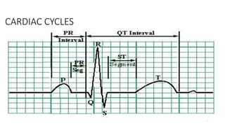
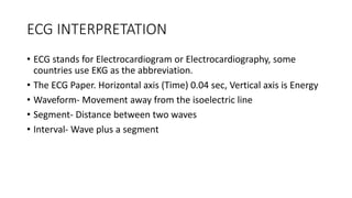
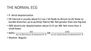
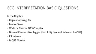
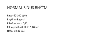
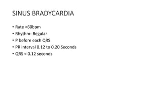
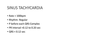
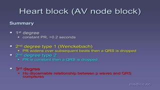
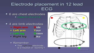
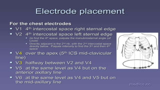
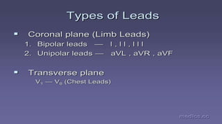
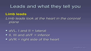
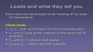
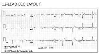
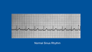
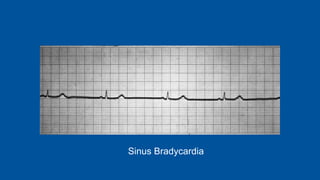
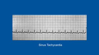
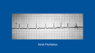
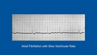
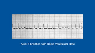
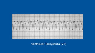
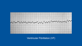
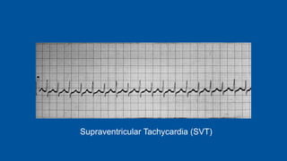
ECG & 12 LEAD ECG
MIRITI M.D
MT ELGON COUNTY HOSPITAL
COUNTY GOVERNMENT OF TRANS NZOIA
2011 American Heart Association: Advanced Cardiovascular Life Support
Provider Manual.
QUICK REVIEW OF THE HEART
• Purpose- Pumps blood
• Basic anatomy – 4 chambers, 2 sides, 4 Valves
• The Conductive System- SA NODE (60-100), Internodal pathway, AV
Node ( 40-60), Bundle of HIS, Bundle branches, Purkinje fibers.
Ventricular nodes (20-60)
• Cardiac cells- Automaticity, Excitability, Conductivity, Contractility.
3
THE CONDUCTING SYSTEM OF THE HEART
• SA Node
• Inter-nodal pathway
• AV Node
• Bundle of HIS
• Bundle Branches
• Purkinje Fibers
4
RELATIONSHIP
5
CARDIAC CYCLES
ECG INTERPRETATION
• ECG stands for Electrocardiogram or Electrocardiography, some
countries use EKG as the abbreviation.
• The ECG Paper. Horizontal axis (Time) 0.04 sec, Vertical axis is Energy
• Waveform- Movement away from the isoelectric line
• Segment- Distance between two waves
• Interval- Wave plus a segment
THE NORMAL ECG
• P- Atrial depolarization
• PR Interval is usually about 0.2 sec ( SA Node to Atrium to AV Node to
bundle branches up to purkinje fibers) NB: Not greater than one big box.
• QRS-Ventricular depolarization about 0.12 sec NB: Not more than 3
small boxes
• RATE=
300
𝑁𝑜 𝑜𝑓 𝐵𝐼𝐺 𝑏𝑜𝑥𝑒𝑠 𝑖𝑛 𝑏𝑒𝑡𝑤𝑒𝑒𝑛 𝑅−𝑅
OR
1500
𝑛𝑜 𝑜𝑓 𝑠𝑚𝑎𝑙𝑙 𝑏𝑜𝑥𝑒𝑠 𝑖𝑛 𝑏𝑒𝑡𝑤𝑒𝑒𝑛 𝑅−𝑅
• Rhythm- Regular
ECG INTERPRETATION BASIC QUESTIONS
Is the Rhythm
• Regular or Irregular
• Fast or Slow
• Wide or Narrow QRS Complex
• Normal P wave (Not bigger than 1 big box and followed by QRS)
• PR Interval
• Is QRS Normal
NORMAL SINUS RHYTM
Rate- 60-100 bpm
Rhythm- Regular
P before each QRS
PR interval = 0.12 to 0.20 sec
QRS= < 0.12 sec
SINUS BRADYCARDIA
• Rate <60bpm
• Rhythm- Regular
• P before each QRS
• PR interval 0.12 to 0.20 Seconds
• QRS < 0.12 seconds
SINUS TACHYCARDIA
• Rate > 100bpm
• Rhythm- Regular
• P before each QRS Complex
• PR Interval =0.12 to 0.20 sec
• QRS < 0.12 sec
I
IIIII
0
°
aVR
aVF
aVL
v4
v3
v2v1
v5 v6
Precordial Limb Leads
21
HOOKING UP THE
12-LEAD ECG
• Proper skin prep
• Placement of the limb electrodes
• Placement of the chest electrodes
22
ELECTRODE PLACEMENT
• LIMB LEADS
• CHEST LEAD
28
12-LEAD ECG LAYOUT
BASIC ECG STRIPS
FOR ACLS PROTOCOLS
Normal Sinus Rhythm
Sinus Bradycardia
Sinus Tachycardia
Atrial Fibrillation
Atrial Fibrillation with Slow Ventricular Rate
Atrial Fibrillation with Rapid Ventricular Rate
First Degree A-V Block
Second Degree A-V Block – Mobitz Type I –
Wenckebach Block
Second Degree A-V Block – Mobitz Type II
Third Degree (Complete) A-V Block
Ventricular Tachycardia (VT)
Ventricular Fibrillation (VF)
Supraventricular Tachycardia (SVT)

Ecg &amp; 12 lead ecg