Downloaded 701 times


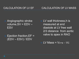


Left ventricular angiography is used to assess global and regional left ventricular function and anatomy. It involves inserting a catheter into the left ventricle and injecting contrast dye to visualize the ventricle on x-ray imaging. The procedure provides key information on mitral valve function, ventricular shape and wall motion abnormalities, and congenital defects like VSD. LV volumes and ejection fraction are calculated from the images to quantify function. Regional wall motion is graded and correlated to coronary artery territories. Characteristic appearances are seen in conditions like cardiomyopathy, mitral regurgitation, and septal defects. Potential complications include arrhythmias and endocardial injury.


