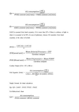
This document provides an overview of segmental analysis for congenital heart disease. It discusses the key segments that are analyzed which include thoraco-abdominal situs, pulmonary situs, atrial situs, ventricular situs and looping, connections between segments (venous, atrioventricular, ventriculoarterial), and abnormalities that can occur in each segment. The document emphasizes evaluating each segment in a systematic, sequential manner to identify abnormalities.
This document provides an overview of standard transthoracic echocardiography (TTE) and transesophageal echocardiography (TEE) views. It describes the imaging windows, planes, and positions for obtaining basic and modified TTE views such as parasternal, apical, and subcostal views. It also outlines TEE imaging levels and how to manipulate the probe to obtain standard midesophageal and transgastric views, including 4-chamber, 2-chamber, aortic valve, and left ventricular views. The document aims to guide practitioners in performing comprehensive TTE and TEE exams through appropriate patient positioning and transducer manipulation.
This document discusses the use of echocardiography in evaluating congenital heart diseases in adults. It outlines the indications for echocardiography and describes how to perform the examination and interpret findings. Key abnormalities that can be identified include atrial septal defects, ventricular septal defects, atrioventricular septal defects, anomalies of venous inflow, and abnormalities of ventricular morphology. Echocardiography is well-suited for diagnosing and monitoring these congenital heart conditions in adulthood.
Tissue Doppler Imaging (TDI) provides low velocity, high amplitude signals from the myocardium that can be used to assess systolic and diastolic function. TDI utilizes pulsed wave and color Doppler techniques to measure peak myocardial velocities. The E/E' ratio, where E is transmitral early diastolic velocity and E' is early diastolic mitral annular velocity, correlates well with left ventricular filling pressures and can help distinguish normal from elevated pressures. TDI parameters are useful for evaluating global and regional systolic function, diastolic function, ischemia, and viability as well as distinguishing between restrictive cardiomyopathy and constrictive pericarditis.
This document discusses the techniques used to detect, localize, and quantify intracardiac shunts in patients with congenital heart disease. An oximetry run is performed during cardiac catheterization to detect left-to-right shunts by measuring oxygen saturation levels in different chambers of the heart and identifying step-ups. The ratio of pulmonary to systemic blood flow (Qp:Qs) is also calculated using the Fick principle to quantify the size of the shunt. A Qp:Qs ratio >1.5 indicates a clinically significant left-to-right shunt.
The document discusses the embryology and anomalies of the systemic venous system. It begins by describing the normal development of the veins in a 4 week old embryo, including the vitelline, umbilical, cardinal, subcardinal, supracardinal and azygos veins. It then summarizes several anomalies of the superior vena cava including bilateral superior vena cava with normal drainage, bilateral superior vena cava with an unroofed coronary sinus, absent right superior vena cava, and retroaortic innominate vein. Diagnostic features and clinical manifestations of these anomalies are provided.
A lecture on the echocardiographic evaluation of hypertrophic cardiomyopathy. Starts with an overview of the topic then a systematic approach to diagnosis and then a differential diagnosis followed by take-home messages and conclusion.
segment approach to congenital heart diseasesSumiya Arshad
The Van Praagh classification system uses a "S, D, S" notation to systematically analyze congenital heart defects based on 1) visceroatrial situs, 2) ventricular loop orientation, and 3) position of the great arteries. This facilitates communication between physicians by providing a standardized approach to describing abnormalities in cardiac chamber position, connections, and vessel arrangements.
IVUS may not be clinically warranted in all interventions, and should be seen as an adjunct to angiography. IVUS provides information about vessel morphology, plaque topography, and therapeutic outcomes that is often either equivocal or unavailable in angiographic images.
There are 3 situations in which IVUS has the most clinical utility:
Small vessel stenting: Studies have shown that post-stent restenosis rates are higher in small vessels. This is particularly true for vessels with diameters of 3.0mm or less, wherein small increases in stent diameter have been shown to significantly decrease the rate of restenosis. A study by Moussa et al showed that, as measured by IVUS, the incidence of restenosis has an inverse relationship to the post-procedure in-stent lumen CSA1.
In-Stent restenosis: In these cases, IVUS helps to determine whether the restenosis is due to inadequate stent deployment (underexpansion or incomplete apposition) due to intimal hyperplasia. IVUS will also help you select the proper device size for treatment of the stented area.
Difficult to assess lesions: At times, images of a lesion and the adjacent reference segment are often hazy. IVUS should be used to identify whether the angiographic appearance is due to dissection, thrombus, residual plaque, or is benign.
M-mode echocardiography uses ultrasound to evaluate cardiac structures through motion over time. It can be used to assess morphology, movement, velocity and timing of valves and walls. Specific measurements are made of amplitudes, slopes, and time intervals which provide information on cardiac function. M-mode views of the mitral valve, aortic valve and left ventricle allow evaluation of values, wall motion, and calculation of ejection fraction, fractional shortening and left ventricular mass. Abnormal findings provide clues to conditions like valve disease, hypertrophy and ischemia.
This document discusses techniques for localizing the site of origin of ventricular tachycardia based on electrocardiogram characteristics. It describes that right ventricular outflow tract tachycardias typically present with left bundle branch block morphology while left ventricular sites may present with either right or left bundle branch block depending on exit site. Specific leads are discussed that can provide clues about anterior vs posterior, septal vs free wall origin within the outflow tracts. Other areas like fascicles, papillary muscles and mitral/tricuspid annuli are also summarized.
Basic haemodynamic assessment with echo (iHeartScan)SCGH ED CME
The document outlines the basic steps for assessing haemodynamic states using echocardiography:
1. Estimate left ventricular preload by measuring end diastolic volume or dimension, characterizing it as hypovolaemic, normal, or dilated.
2. Estimate systolic function by measuring ejection fraction, fractional shortening, or visual assessment.
3. Estimate left atrial pressure by examining interatrial septal motion and size, characterizing pressure as normal or high.
4. Make a final assessment of the primary dysfunction as normal, hypovolaemic, primary diastolic failure, primary systolic failure, systolic and diastolic failure, vasodilation
Basics in echocardiography - an initiative in evaluation of valvular heart di...Praveen Nagula
Echocardiography is commonly used to evaluate valvular heart disease. It uses ultrasound to produce images of cardiac structures and Doppler imaging to assess blood flow and hemodynamics. Echocardiography can be used to measure chamber size, evaluate valve function, detect shunts, and estimate pressures noninvasively. Hemodynamic assessments by echocardiography have largely replaced cardiac catheterization. Doppler echocardiography utilizes principles like Bernoulli's equation to estimate cardiac pressures and calculate values like stroke volume, cardiac output, and valvular regurgitant volumes. It provides reliable evaluation of valvular heart disease.
M-mode echocardiography uses rapid sampling of a region to create sequential parallel data lines, producing continuous horizontal lines representing points of brightness. This allows visualization of motion patterns over distance and time. Measurements of structures like the mitral valve can assess morphology, movement, velocity, and timing of cardiac events. Findings include increased wall thickness, reduced valve excursion, and fluttering indicating conditions like hypertrophy, stenosis, and regurgitation.
The document summarizes key aspects of cardiac catheterization and hemodynamic data collection. It describes the normal cardiac cycle, pressure measurement systems, normal pressure waveforms, methods to measure cardiac output like thermodilution and Fick, how to evaluate valvular stenosis and regurgitation, determine vascular resistance and shunts. Specific details are provided on assessing aortic stenosis, mitral stenosis, right-sided valves and quantifying regurgitant fractions. Oxygen saturation analysis and Fick principles are outlined for shunt determinations.
This document provides an overview of the basics of echocardiography. It discusses how ultrasound is directed and focused, and how it interacts with different media like soft tissue and gas. The principles of reflection, refraction, attenuation and acoustic impedance are explained. Imaging planes and windows for echocardiography are outlined. Normal dimensions of cardiac structures are provided. Quantitative measures of left ventricular function like fractional shortening and volumes are defined. Three-dimensional echocardiography is introduced as an advancement that improves accuracy of volume measurements and provides comprehensive views of valves and congenital defects.
PRESSURE MEASUREMENT by Cardiac catheterisation_Dr Amol Patil.pptxAshishSharma907946
1) Cardiac catheterization allows measurement of pressures within the heart by inserting catheters connected to transducers.
2) Proper equipment selection and setup is important to minimize artifacts and obtain accurate pressure tracings.
3) Different catheter types are used depending on the specific chamber being measured.
This document discusses atrioventricular canal defects (AVSDs), including their embryogenesis and pathophysiology. It describes the anatomy and classification of partial and complete AVSDs. Partial AVSDs involve a primum atrial septal defect with two distinct but contiguous AV valves, while complete AVSDs have a single common AV valve. The embryogenesis of AVSDs involves faulty development of the endocardial cushions. The document provides detailed descriptions and images of the anatomy and features of partial and complete AVSDs. It discusses the clinical aspects of AVSDs including prevalence, association with Down syndrome, surgical repair outcomes, and lifelong surveillance needs.
The document discusses guidelines for assessing diastolic dysfunction according to the ASE/EACVI 2016 guidelines. It defines diastolic dysfunction and describes the stages from grade I to grade IV. For each grade, it discusses the pathophysiology and key echocardiographic findings including mitral inflow patterns, tissue Doppler measurements, pulmonary vein flow, and left atrial size. The guidelines simplify the assessment of diastolic function into four grades based on parameters of left ventricular relaxation, left atrial pressure, mitral E/A ratio, E/e' ratio, pulmonary vein flow, and left atrial size.
The document discusses the history, anatomy, angiographic views, variations, and clinical relevance of coronary arteries. It provides a detailed overview of the typical anatomy and branches of the left main, left anterior descending, left circumflex, and right coronary arteries. It also describes common anatomical variations and anomalies seen in coronary arteries and their clinical implications. Angiographic classification methods for different coronary artery segments are presented.
The document discusses techniques for detecting intracardiac shunts including oximetry runs, indicator dilution curves, and angiography. It provides criteria for identifying left-to-right shunts using oxygen saturation step-ups in the oximetry run. Examples are given for detecting an atrial septal defect and ventricular septal defect. The ratio of pulmonary to systemic blood flow (Qp/Qs) is discussed as a measure of shunt magnitude.
This document discusses various methods for quantifying intracardiac shunts in patients with congenital heart lesions. It describes invasive oximetry and indicator dilution techniques as well as noninvasive Doppler echocardiography methods. For echocardiography, it outlines techniques for quantifying left-to-right shunts using pulmonary and aortic flow measurements, as well as a simplified method using diameter ratios. It also discusses limitations and sources of error for these quantification methods.
This document discusses contrast echocardiography, including the mechanism by which microbubble contrast agents improve echocardiographic imaging. Ideal contrast agents are described as being safe, metabolically inert, long-lasting, strong sound reflectors that are small enough to pass through capillaries. Several FDA-approved second generation contrast agents are mentioned along with their shell materials and gases. Optimal echocardiographic settings for contrast imaging are outlined. Clinical applications of contrast echocardiography include assessing shunts, venous anomalies, and leaks. Examples of its use in specific cases are provided.
A cardiac shunt is a pattern of blood flow in the heart that deviates from the normal circuit of the circulatory system. It may be described as right-left, left-right or bidirectional, or as systemic-to-pulmonary or pulmonary-to-systemic.
This document provides an overview of the anatomy and assessment of the mitral valve using transesophageal echocardiography (TEE). It describes the components of the mitral valve complex including the annulus, leaflets, chordae tendineae, and papillary muscles. It outlines different TEE views used to evaluate the mitral valve and provides details on quantifying mitral stenosis and regurgitation. Causes of mitral valve dysfunction like rheumatic heart disease and ischemic mitral regurgitation are summarized. Assessment of mitral valve repair is also discussed, including complications like paravalvular leaks and systolic anterior motion.
Cardiac catheterization is useful for assessing left-to-right shunts through three main techniques: oximetry runs to detect oxygen saturation step-ups, indicator dye dilution to detect early recirculation of dye injected into the proximal chamber, and angiocardiography to directly visualize the anatomic site of the shunt. While oximetry is best to localize the shunt, dye dilution can detect smaller shunts and angiography confirms anatomy. Together these techniques allow diagnosis and quantification of left-to-right intracardiac shunts.
Hemodynamics in echo lab by Dr. Ranjeet S.PalkarRanjeet Palkar
ECHO LAB AND CARDIOVASCULAR HEAMODYNAMICS. A simple cost effective,non invasive approach which when used appropriately can be boon for physicians and cardiologists in diagnosis and prognostication.
This document summarizes various echocardiography techniques for estimating pulmonary hypertension. It describes methods such as using tricuspid regurgitation peak velocity to estimate systolic pulmonary artery pressure, pulmonary regurgitation peak velocity to estimate mean pulmonary artery pressure, and right ventricular outflow tract acceleration time to estimate mean pulmonary artery pressure. It also discusses potential pitfalls and tricks for obtaining accurate measurements with each technique.
This document provides an overview of cardiovascular physiology, including:
1) The basic components and functioning of the cardiovascular system, including circulation through the heart, vessels, and regulation.
2) Properties of the pulmonary and systemic circulations such as pressures and resistances.
3) Cardiac physiology including the cardiac cycle, regulation of contractility, and the Frank-Starling mechanism.
4) Vascular physiology including blood flow and pressures, compliance, and fluid balance in the capillaries.
This document discusses how echocardiography can be used to assess hemodynamics by measuring blood flow velocities. Doppler echocardiography is validated for measuring stroke volume, cardiac output, regurgitant volumes, and pressures in the heart by relating flow velocities to pressure gradients. Measurements of velocities across valves and in vessels can be used to estimate parameters like pulmonary artery pressures, left ventricular end diastolic pressure, and left atrial pressure through validated Doppler equations.
The document provides an overview of cardiovascular physiology, including:
1. It describes the basic components and regulation of the cardiovascular system, including the heart, vessels, and regulatory mechanisms.
2. It discusses the pulmonary and systemic circulations in terms of pressure, resistance, and flow.
3. It covers the structure and function of the heart as a pump, including cardiac cycle, regulation of contractility, and the Frank-Starling mechanism.
This document discusses various hemodynamic parameters and waveforms seen in different cardiac conditions:
- It compares pressures, waveforms and compliance in conditions like constrictive pericarditis, right ventricular failure, cardiac tamponade and pulmonary hypertension.
- Key waveforms and pressures are described for valvular lesions like mitral stenosis, aortic stenosis, mitral regurgitation and aortic regurgitation.
- Equations for calculating cardiac output, shunt fractions and valvular areas are provided.
- Diagnostic criteria for pseudosevere versus true severe aortic stenosis on stress echocardiography are outlined.
- A case example is presented of a patient evaluated for pulmonary
1. Pulmonary hypertension is defined as a mean pulmonary artery pressure greater than 25 mm Hg at rest as measured by right heart catheterization. Severity is classified as mild, moderate, or severe based on pressure readings.
2. Echocardiography can be used to estimate pulmonary artery pressures and assess right heart structures for signs of pulmonary hypertension. Measurements like tricuspid regurgitation velocity, pulmonary regurgitation pressure gradient, and inferior vena cava size correlate with pulmonary artery pressures.
3. Additional findings on echo that indicate pulmonary hypertension include right ventricular dilation, septal flattening, reduced right atrial emptying, and pulmonary artery acceleration time. Together, echo findings can establish the diagnosis
Guytonian approach to shock - mean systemic filling pressure centeredCosmin Balan
In a world of binary decision there remains little room for applied maths and physiology. Or maybe not...
Parkin's approach brings out a forgotten tool-the volume state. Although reductionistic as well as Guyton's entire view, it might be a better language for us, for clinicians and for all those lost in translation when they've stumbled across loose binary decisions such as SVV,PPV,SPV etc.
Mean systemic filling pressure has been resurrected.
Parkin, Maas, Pinsky and Geerts have come a long way from Versprille.
The document describes a non-invasive device that uses pulse wave analysis from a blood pressure cuff to measure arterial waveforms and provide information about cardiac function and arterial stiffness. It analyzes the ejection wave from the heart and how it is modified as it travels into the arterial system. Recordings are made by reinflating the cuff above systolic pressure. Various physiological states and interventions that affect the measured waves are discussed.
These slides represent how to manage patients on a mechanical ventilator? Easy understanding of using ventilators. indication of mechanical ventilator use. How to wean a patient from a mechanical ventilator? How to fine-tune the ventilator settings?
This document discusses hypertension (high blood pressure) including definitions, stages, measurement techniques, targets for treatment, and general management strategies. Some key points:
- Hypertension is defined as a blood pressure level where treatment benefits outweigh risks based on clinical trials. The overall adult prevalence is 30-45%.
- Stages of hypertension include normal, elevated, stage 1, and stage 2 based on systolic and diastolic blood pressure levels. Isolated systolic and diastolic hypertension are also discussed.
- Lifestyle modifications like following a DASH diet, reducing salt and weight, exercising, and not smoking can help lower blood pressure. Various drug classes are also used to treat hypertension.
- Treatment targets
This document discusses different types of arrhythmias, including tachyarrhythmias like atrial fibrillation, ventricular tachycardia, and supraventricular tachycardia. It describes the causes, complications, and treatment options for each type. The document also covers bradyarrhythmias such as different degrees of atrioventricular block and sick sinus syndrome. Normal ECG findings are reviewed as well as how to properly read an ECG.
Acute coronary syndrome for undergraduatesMashiul Alam
Acute coronary syndrome (ACS) refers to conditions caused by reduced blood flow to the heart. It includes unstable angina, non-ST elevation myocardial infarction, and ST elevation myocardial infarction. Symptoms include chest pain, shortness of breath, nausea, and fatigue. Diagnosis involves ECG changes such as ST elevation or T wave changes and elevated cardiac biomarkers. Treatment focuses on reperfusion with medications or procedures like PCI, along with anticoagulants, antiplatelets, and lifestyle modifications. Complications can be mechanical, arrhythmic, embolic or inflammatory.
Heart failure is a complex clinical syndrome characterized by impaired myocardial performance and progressive maladaptive neurohormonal adaptation. It can be systolic or diastolic in nature. Common causes include ischemia, cardiomyopathy, hypertension, and valvular disorders. Symptoms include dyspnea, orthopnea, fatigue, and ankle swelling. Diagnostic evaluation involves ECG, echocardiogram, blood tests, and cardiac catheterization. Management depends on whether the heart failure is acute or chronic, and involves treatments like oxygen, vasodilators, diuretics, beta blockers, ACE inhibitors, and device therapies like ICDs or CRT. Lifestyle changes like exercise and diet are also important.
Valvular heart disease for undergraduatesMashiul Alam
This document summarizes different types of valvular heart disease, including their causes, clinical features, signs, and management. It discusses mitral stenosis, mitral regurgitation, aortic stenosis, and aortic regurgitation. For each condition, it outlines the typical causes such as rheumatic heart disease, describes common symptoms like shortness of breath and fatigue, lists exam findings, and recommends treatment approaches including medical therapy, interventions like valve repair/replacement, and surgery.
Stent thrombosis is a rare but devastating complication occurring in less than 1% of patients within 30 days of stenting and 0.2-6% annually afterwards. It is associated with higher thrombus burden and less procedural success, resulting in higher rates of death, recurrent heart attack, and recurrent stent thrombosis. Risk factors include stent-related issues like early versus late thrombosis, procedure-related issues like incomplete apposition or expansion, vessel-related issues like long lesions or small vessel size, and patient-related issues like diabetes, impaired heart function, renal disease, or non-compliance with dual anti-platelet therapy. Management depends on thrombus burden grade, with direct angioplasty and stenting for small burdens and
Temporary cardiac pacing is used to treat acute bradyarrhythmias or tachyarrhythmias until the underlying condition resolves or permanent pacing can be initiated. It aims to re-establish normal hemodynamics compromised by abnormal heart rates. Transvenous pacing is the preferred method, involving insertion of endocardial leads through veins to the heart. Precise lead placement is important and is confirmed with imaging. Pacing parameters like threshold, rate and sensing are optimized. Complications include those related to vascular access and device malfunction requiring troubleshooting. Close monitoring is needed to ensure proper pacing and detect any issues.
The Micra pacemaker is the world's smallest, about the size of a large vitamin. It is implanted inside the heart and attached to the heart wall using small tines. Some advantages are that it does not require any external wires or generator under the skin, eliminating pocket-related complications. The device has a battery longevity of 12 years. It is indicated for patients with AV block or bradycardia and can be safely used with MRI scans under specific conditions. Potential risks include oversensing, acceleration of arrhythmias, infection, and device embolization.
This document discusses the assessment of intracardiac shunts by cardiac catheterization. It describes how an oximetry run is performed to detect a left-to-right shunt by measuring oxygen saturation at various locations in the heart and great vessels. A significant step-up in oxygen saturation between the right atrium and ventricle or pulmonary artery suggests a left-to-right shunt. The document also discusses calculating shunt ratios using indicator dilution techniques and identifying shunt locations with angiography and pressure measurements. Complications of the procedure are outlined.
This document summarizes the echocardiographic assessment of mitral stenosis (MS). It describes the anatomy of the mitral valve and causes of MS. Methods for assessing MS severity include measuring the pressure gradient, mitral valve area using planimetry and pressure half-time, and pulmonary artery pressure. Suitability for percutaneous transvenous mitral commissurotomy is evaluated. Concomitant valve lesions are also identified. Stress echocardiography may be used when symptoms are equivocal. Transesophageal echocardiography is recommended in some cases.
This document discusses the echocardiographic assessment of atrial septal defects (ASDs). It describes the main types of ASDs and notes that 80% are secundum defects. Echocardiography is used to identify and characterize ASDs, detect associated anomalies, diagnose complications, and guide treatment. Transthoracic echocardiography is the initial study, while transesophageal echocardiography provides better views of the atrial septum. Key measurements include ASD size, location, rim dimensions, and quantifying shunt severity with Qp/Qs. Echocardiography guides decisions about ASD device closure or surgery.
Anticoagulation in atrial fibrillationMashiul Alam
This document discusses anticoagulation for atrial fibrillation (AF). It covers the epidemiology and pathophysiology of AF, as well as the risks of stroke. It describes scoring systems like CHADS2 and CHA2DS2-VASc that are used to determine stroke risk and recommend antithrombotic therapy. Newer oral anticoagulants like apixaban, dabigatran and rivaroxaban are discussed and compared to warfarin. Guidelines for anticoagulation in various clinical scenarios involving AF are provided, such as with stable ischemic heart disease, intracoronary stents, acute coronary syndrome, and cardioversion.
Intracoronaryopticalcoherencetomography 130909083234-Mashiul Alam
1. Intracoronary imaging techniques like intravascular ultrasound (IVUS), virtual histology, optical coherence tomography (OCT), and angioscopy can be used to image the coronary arteries.
2. OCT provides very high resolution images of the coronary arteries and has advantages over IVUS for identifying features like thin fibrous caps, intralesional macrophages, and intracoronary thrombi.
3. OCT is a safe imaging technique and is useful for evaluating plaque characteristics, guiding percutaneous coronary interventions, and assessing stent coverage and restenosis.
Intravascular ultrasonography (IVUS) provides images of coronary arteries and other blood vessels. It plays a critical role in understanding coronary disease and guiding interventional cardiology procedures. IVUS uses a catheter-mounted ultrasound transducer to create images. It can assess plaque, guide stent placement, detect complications, and characterize lesion morphology. IVUS provides detailed information to evaluate patients and optimize interventional strategies.
Warfarin and newer oral anticoagulants e.g. debigatran, rivaroxaban, apixaban were presented in cardiology morning session in Bangabandhu Sheikh Mujib Medical University.
Breast cancer: Post menopausal endocrine therapyDr. Sumit KUMAR
Breast cancer in postmenopausal women with hormone receptor-positive (HR+) status is a common and complex condition that necessitates a multifaceted approach to management. HR+ breast cancer means that the cancer cells grow in response to hormones such as estrogen and progesterone. This subtype is prevalent among postmenopausal women and typically exhibits a more indolent course compared to other forms of breast cancer, which allows for a variety of treatment options.
Diagnosis and Staging
The diagnosis of HR+ breast cancer begins with clinical evaluation, imaging, and biopsy. Imaging modalities such as mammography, ultrasound, and MRI help in assessing the extent of the disease. Histopathological examination and immunohistochemical staining of the biopsy sample confirm the diagnosis and hormone receptor status by identifying the presence of estrogen receptors (ER) and progesterone receptors (PR) on the tumor cells.
Staging involves determining the size of the tumor (T), the involvement of regional lymph nodes (N), and the presence of distant metastasis (M). The American Joint Committee on Cancer (AJCC) staging system is commonly used. Accurate staging is critical as it guides treatment decisions.
Treatment Options
Endocrine Therapy
Endocrine therapy is the cornerstone of treatment for HR+ breast cancer in postmenopausal women. The primary goal is to reduce the levels of estrogen or block its effects on cancer cells. Commonly used agents include:
Selective Estrogen Receptor Modulators (SERMs): Tamoxifen is a SERM that binds to estrogen receptors, blocking estrogen from stimulating breast cancer cells. It is effective but may have side effects such as increased risk of endometrial cancer and thromboembolic events.
Aromatase Inhibitors (AIs): These drugs, including anastrozole, letrozole, and exemestane, lower estrogen levels by inhibiting the aromatase enzyme, which converts androgens to estrogen in peripheral tissues. AIs are generally preferred in postmenopausal women due to their efficacy and safety profile compared to tamoxifen.
Selective Estrogen Receptor Downregulators (SERDs): Fulvestrant is a SERD that degrades estrogen receptors and is used in cases where resistance to other endocrine therapies develops.
Combination Therapies
Combining endocrine therapy with other treatments enhances efficacy. Examples include:
Endocrine Therapy with CDK4/6 Inhibitors: Palbociclib, ribociclib, and abemaciclib are CDK4/6 inhibitors that, when combined with endocrine therapy, significantly improve progression-free survival in advanced HR+ breast cancer.
Endocrine Therapy with mTOR Inhibitors: Everolimus, an mTOR inhibitor, can be added to endocrine therapy for patients who have developed resistance to aromatase inhibitors.
Chemotherapy
Chemotherapy is generally reserved for patients with high-risk features, such as large tumor size, high-grade histology, or extensive lymph node involvement. Regimens often include anthracyclines and taxanes.
Promoting Wellbeing - Applied Social Psychology - Psychology SuperNotesPsychoTech Services
A proprietary approach developed by bringing together the best of learning theories from Psychology, design principles from the world of visualization, and pedagogical methods from over a decade of training experience, that enables you to: Learn better, faster!
Co-Chairs, Val J. Lowe, MD, and Cyrus A. Raji, MD, PhD, prepared useful Practice Aids pertaining to Alzheimer’s disease for this CME/AAPA activity titled “Alzheimer’s Disease Case Conference: Gearing Up for the Expanding Role of Neuroradiology in Diagnosis and Treatment.” For the full presentation, downloadable Practice Aids, and complete CME/AAPA information, and to apply for credit, please visit us at https://bit.ly/3PvVY25. CME/AAPA credit will be available until June 28, 2025.
How to Control Your Asthma Tips by gokuldas hospital.Gokuldas Hospital
Respiratory issues like asthma are the most sensitive issue that is affecting millions worldwide. It hampers the daily activities leaving the body tired and breathless.
The key to a good grip on asthma is proper knowledge and management strategies. Understanding the patient-specific symptoms and carving out an effective treatment likewise is the best way to keep asthma under control.
The skin is the largest organ and its health plays a vital role among the other sense organs. The skin concerns like acne breakout, psoriasis, or anything similar along the lines, finding a qualified and experienced dermatologist becomes paramount.
DECLARATION OF HELSINKI - History and principlesanaghabharat01
This SlideShare presentation provides a comprehensive overview of the Declaration of Helsinki, a foundational document outlining ethical guidelines for conducting medical research involving human subjects.
Travel Clinic Cardiff: Health Advice for International TravelersNX Healthcare
Travel Clinic Cardiff offers comprehensive travel health services, including vaccinations, travel advice, and preventive care for international travelers. Our expert team ensures you are well-prepared and protected for your journey, providing personalized consultations tailored to your destination. Conveniently located in Cardiff, we help you travel with confidence and peace of mind. Visit us: www.nxhealthcare.co.uk
- Video recording of this lecture in English language: https://youtu.be/Pt1nA32sdHQ
- Video recording of this lecture in Arabic language: https://youtu.be/uFdc9F0rlP0
- Link to download the book free: https://nephrotube.blogspot.com/p/nephrotube-nephrology-books.html
- Link to NephroTube website: www.NephroTube.com
- Link to NephroTube social media accounts: https://nephrotube.blogspot.com/p/join-nephrotube-on-social-media.html
Demystifying Fallopian Tube Blockage- Grading the Differences and Implication...
Shunt calculation
1. CALCULATION FOR RIGHT HEART CATHETERIZATION
Name: Age:
Height: Weight:
BSA:
…….𝐻𝑡 ( 𝑐𝑚)+ ………𝑊𝑡 ( 𝑘𝑔)−60
100
=
Hb%: HR:
Oxymetry run: Pressure:
Location Saturation (%)
SVC High
Mid
Low
RA High
Mid
Low
RV RVOT
Mid
MPA
LPA
RPA
IVC
LV
O2 consumption = 125 ml/min /m2 BSA
O2 content = …..satof O2 x ……Hb (g/dl) x 1.36 x 10 (ml/L)
Location Pressure
(mmHg)
Mean
Pressure
RA Sys
Dia
RV Sys
Dia
PCWP
LA Sys
Dia
LV Sys
Dia
Aorta Sys
Dia
2. 𝑄𝑝 =
𝑂2 𝑐𝑜𝑛𝑠𝑢𝑚𝑝𝑡𝑖𝑜𝑛 (
𝑚𝑙
𝑚𝑖𝑛
)
𝑃𝑉𝑂2 𝑐𝑜𝑛𝑡𝑒𝑛𝑡 (𝑚𝑙/min) − PAO2 content ( 𝑚𝑙/min)
𝑄𝑠 =
𝑂2 𝑐𝑜𝑛𝑠𝑢𝑚𝑝𝑡𝑖𝑜𝑛 (
𝑚𝑙
𝑚𝑖𝑛
)
𝑆𝐴𝑂2 𝑐𝑜𝑛𝑡𝑒𝑛𝑡 (𝑚𝑙/min) − MVO2 content ( 𝑚𝑙/min)
SAO2 is assumed from hand oxymetry if it is more than 95%. If there is evidence of right to
shunt it is assumed to be 98%. In case of pulmonary disease O2 saturation from hand
oxymetry is the value to be held.
𝑀𝑉𝑂2 =
3 𝑆𝑉𝐶 𝑂2 + 1 𝐼𝑉𝐶 𝑂2
4
𝑆𝑉𝑅( 𝑤𝑜𝑜𝑑 𝑢𝑛𝑖𝑡) =
𝑀𝑒𝑎𝑛 𝐴𝑟𝑡𝑒𝑟𝑖𝑎𝑙 𝑃𝑟𝑒𝑠𝑠𝑢𝑟𝑒− 𝐶𝑉𝑃
𝐶𝑎𝑟𝑑𝑖𝑎𝑐 𝑜𝑢𝑡𝑝𝑢𝑡
𝑃𝑉𝑅 ( 𝑊𝑜𝑜𝑑 𝑢𝑛𝑖𝑡) =
𝑀𝑒𝑎𝑛 𝑃𝐴 𝑝𝑟𝑒𝑠𝑠𝑢𝑟𝑒− 𝑀𝑒𝑎𝑛 𝑃𝐶𝑊𝑃
𝐶𝑎𝑟𝑑𝑖𝑎𝑐 𝑜𝑢𝑡𝑝𝑢𝑡
Cardiac Output (CO) = SV x HR or
Fick Equation (l/min) =
𝑂2 𝑐𝑜𝑛𝑠𝑢𝑚𝑝𝑡𝑖𝑜𝑛 (
𝑚𝑙
𝑚𝑖𝑛
)
1.36 𝑥….𝐻𝑏 (
𝑔
𝑑𝑙
) 𝑥 10 (𝑆𝐴𝑂2−𝑆𝑉𝑂2)
Simple formula for shunt calculation
Qp / QS = SAO2 – SVO2 / PVO2 – PAO2
For bidirectional shunt
Qeff =
𝑂2 𝑐𝑜𝑛𝑠𝑢𝑚𝑝𝑡𝑖𝑜𝑛
𝑃𝑉𝑂2 𝑐𝑜𝑛𝑡𝑒𝑛𝑡−𝑀𝑉𝑂2 𝑐𝑜𝑛𝑡𝑒𝑛𝑡