SlideShare a Scribd company logo
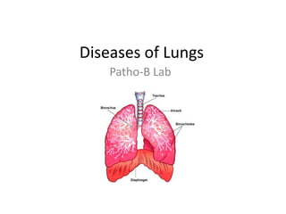
Diseases of Lungs Patho-B Lab
NEW SLIDES Atelectasis with congestion Bronchiectasis Emphysema Bronchial Asthma Adenocarcinoma Lung Pulmonary edema Bronchopneumoniae
OLD SLIDES Lobar Pneumoniae Viral Pneumoniae Lung Abscess Tumor Emboli- Lung Chronic Pulmonary Congestion TB Lungs
Atelectasis, known as Lung Collapse, is loss of lung volume caused by inadequate expansion of air spaces.
Types of Atelectasis Compression Atelectasis-  Usually associated with accumulation of fluids/blood/air with in the pleural cavity which mechanically collapse the adjacent lung.. Frequently occurs with pleural effusions caused most commonly by Conjustive Heart Failure. Reversible ResorptionAtelectasis- Occurs when an obstruction prevents the air from reaching distal airways. Most common cause of resorptionatelectasis is obstruction of bronchus by a mucopurulent plug. Reversible Contraction Atelectasis- Occurs when either local or generalized fibrotic changes in the lung/pleura hamper expansion and increase elastic recoil during expiration. Irreversible
Diagnosis:-  Radiological examination ----X-ray  Clinical Manifestation Dyspnea Productive Cough ***Significant adventitious sounds on Auscultation,dullness on percussion Treatment :- Treating the underlying cause. Bacteriological and Mycological examination of sputum( To treat the underlying cause if infectious in nature with relative intervention) Preventive measures for aspiration in childrens, post op surgical patients decreases the risk of Atelectasis Complication:- May worsen bronchial Asthma,Bronchiectasis.
LPO Hallmark Alveolar Collapse
Hallmark Alveolar Collapse LPO
**Congestion **Anthracosis **Alveolar collapse HPO Congestion  Here can give  A clue of the  Patient sufferering From  compressive Type of Atelectasis
Bronchiectasis is the permanent (abnormal) dilation of bronchi and bronchioles caused by destruction of the muscle and elastic supporting tissue, associated with chronic necrotizing infections. 2 Processes involved in Pathogenesis Obstruction Chronic persistent infection
Diagnosis:- Sputum culture – May reveal Pseudomonas aeruginosa, fungi such as Aspergillus and various Mycobacteria Radiological Examination – X-ray findings not significant unless very gross... CT scan is much more sensitive for Bronchiectasis Clinical Manifestation  Chronic productive cough(Foul smelling) Fever,Malaise, aand increased cough and sputum volume. Haemoptysis(can be slight of massive) Halitosis is a common feature Clubbing of fingers may develop  Management In patients with airflow obstruction, bronchodilators to enhance airway patency. Physiotherapy – to keep the dilated bronchi empty of secretions Antibiotic therapy(Ciprofloxacin, ceftizimide 12 hourly 25—750 mg IV infusion)
Normal Bronchus
Normal Bronchioles
LPO Dilated Bronchiole with secretion within
HPO Inflammatory changes in the wall of the bronchiole.. Necrotized smooth muscles and supporting tissue due to inflammation
LPO Bronchioles
Dilated Bronchi    LPO
Secretions within  The bronchi due to Accumulation of  Pus in dilated  bronchi LPO
Prominent Fibrotic change and  Inflammatory change In entire slide
Inflammatory and fibrotic  Changes found in the  Surrounding lung tissue
Bronchiectatic cavity  Filled with secretions , Lined by normal epithelium With some granulation tissue HPO
HPO Inflammatory  Changes in the Bronchial wall
** Inflammatory changes **Fibrotic changes **Anthracosis LPO
** Inflammatory changes **Fibrotic changes HPO
** Inflammatory changes **Fibrotic changes LPO
Characterized by abnormal permanent enlargement of the airspaces distal to the terminal bronchiole, accompanied by destruction of wall without obvious fibrosis Most common cause – Smoking (Nicotine) 2 Pathogenic Mechanisms are Excess cellular proteases with low antiprotease activity Excess of oxygen species
Types of Emphysema Centriacinar(Centrilobar) Emphysema Involves central of proximal acinisparring the distal alveoli. Panacinar(Panlobar) Emphysema Uniform enlargement of the acinifrom the level of respiratory bronchiole to terminal alveoli. Distal acinar(Panseptal) Emphysema Proximal part of acini is normal but distal part is involved Irregular Emphysema Irregular involved acini, almost invariably associated with scarring. Most common form of Emphysema
Diagnosis Alpha1-antiprotease assay CT is more sensitive that X-ray for Emphysema Clinical Manifestation Dyspnea is usually the first symptom(Progressive) Cough and Wheezing in patient with underlying Chronic bronchitis or Chronic asthmatic bronchitis. Weight loss Barrel Chest Management Smoking cessation Oxygen Therapy Pulmonary rehabilitation Mucolytic therapy
Thinning  And destruction of  Alveolar walls LPO
LPO Thinning  And destruction of  Alveolar walls
Thinning  And destruction of  Alveolar walls LPO
Characterized by chronic airway inflammation and increased airway hyper-responsiveness Funtionally characterized by the presence of airflow obstruction which is variable over the short periods of time or is reversible with treatment
Types of Asthma Atopic/Extrinsic Asthma – Most common type +ve Family History common +ve Allergy causing Attacks (Rhinitis, urticaria, eczema) Elevated Ig-E serum levels Non Atopic/ Intrinsic /Acquired Asthma–    Non immune in nature +ve Family history uncommon No associated Allergy Ig-E serum levels are normal Drug Induced Asthma Drug like Aspirin provoke asthma Patient with Aspirin sensitivity present with Recurrent Rhinitis,Bronchospasm,urticaria Occupational Asthma Stimulated by fumes(plastics,resins), organic and chemical dusts(wood,cotton) Attacks usually develop after repeated exposure to the inciting agents
Triad of Reversible airway obstruction Chronic inflammation with eosinophils Bronchial smooth muscle cell hypertrophy Hyperreactivity
Significant Morphology in Asthma Macroscopic Occlusion of the bronchi and bronchioles by thick, tenacious Mucous plugs. Microscopic Mucous plug contain whorls of shed epithelium(Curschmann spirals) Numerous eosinophils and Charcot leyden crystals(collections of crystals made of eosinophil proteins) are also present
Diagnosis Diagnosis is made on the basis of compatible clinical history combined with the demonstration of the airflow obstruction Pulmonary function tests Measurement of allergic status( Elevated sputum or peripheral blood eosinophil count ) Airway inflammation assessment Clinical Manifestation Typically symptoms includes recurrent episodes of wheezing, chest tightness, breathlessness  and cough Management Avoiding aggravating factors  Short acting inhaled beta2- Agonist (Bronchodilators) – For patients with mild intermittent asthma(Symptoms less than once a week for 3 months and fewer than 2 nocturnal episodes) Long acting beta2 agonist(salmeterol,formoterol) if patient remains poorly controlled Low/high dose steroid therapy depending on the severity of symptoms
**Airway Remodelling in Asthma** -Thickening of Basement membrane     of the bronchial epithelium -Edema and inflammatory    infiltrate in walls -Increase in the size   of the glands -Hypertrophy of the    bronchial muscle walls LPO
**Airway Remodelling in Asthma** -Thickening of Basement membrane     of the bronchial epithelium -Edema and inflammatory    infiltrates in wall -Increase in the size   of the glands -Hypertrophy of the    bronchial muscle walls LPO
HPO **Airway Remodelling in Asthma** -Thickening of Basement membrane     of the bronchial epithelium -Edema and inflammatory    infiltrates in wall -Increase in the size   of the glands -Hypertrophy of the    bronchial muscle walls
**Airway Remodelling in Asthma** -Thickening of Basement membrane     of the bronchial epithelium -Edema and inflammatory    infiltrates in wall -Increase in the size   of the glands -Hypertrophy of the    bronchial muscle walls HPO
Classification of Lung tumors for therapeutic purposes Small Cell Lung Carcinomas(SCLC) All small cell lung carcinomas have metastasized by the time they are diagnosed hence they cannot be cured by surgery, Chemotherapy and Radiation is the only treatment for SCLC Non small cell lung Carcinoma(NSCLC)- poor response to chemotherapy and are better treated by surgery. Squamous cell Adenocarcinoma ----- Most common of lung cancer in women and non smoker Large cell carcinoma
Most common of lung cancer in women and non smoker Precursor –Atypical adenomatous hyperplasia(AAH) Occur as central lesions but 	are more peripherally located Many arising in relation to peripheral lung scars      (scar carcinomas) Histological types Acinar(Gland forming) Papillary Solid
In general all Adenocarcinomasgrow slowly and form smaller masses than other subtypes but they tend to metastasize widely at an early stage. Diagnosis:- Histologic diagnosis Bronchoscopy Bronchial biopsy CT/Ultrasound Clinical Manifestation Cough is common early symptom  Haemoptysis Bronchial obstruction Recurrent pneumonia Lassitude and Weight loss- indicated metastatic spread Hoarseness Management Surgical resection for all Non small cell lung carcinoma-NSCLC
**Stromal invasion(Bronchial mucosa) **Desmoplasia (growth of fibrous  or connective tissue)
**Stromal invasion(Bronchial mucosa) **Desmoplasia (growth of fibrous  or connective tissue) ** Acinar type Gland forming Adenocarcinoma
**Stromal invasion **Desmoplasia (growth of fibrous  or connective tissue)
**Stromal invasion(Bronchial mucosa) **Desmoplasia (growth of fibrous  or connective tissue)
**Cytologicatypia **Pleomorphism
Caused due to accumulation of fluids in lungs Etiology Cardiogenic pulmonary edema Most common clinical problem seen frequently in left ventricular failure. Noncardiogenic pulmonary edema Renal failure Acute Respiratory distress syndrome(ARDS)
Signs:- JVP – increased, sweating, cool extremities, dullness and crepitation at base. Chest radiograph:- Cardiomegaly,pleural effusion. ECG- decreased left ventricular function Decreased PaO2 Can lead to Acute severe dyspnea. Management:-  Treating the underlying cause Usually the prognosis is poor(with survival 2-3 years) unless Heart-lung transplantation is performed.
4 stages of lobar/Broncho Pneumonia Congestion Affected lobe is heavy, red boggy,histologicallyvascular congestion can be seen with scattered neutrophils in the alveoli. Red hepatization Lung shows liver like consistency, the alveolar spaces are packed with neutrophils, red cells and fibrin. Gray hepatization Lung is dry,gray, and firm, because the red cells are lysed while the fibrinosuppurativeexudate persists within the alveoli Resolution Follows in umcomplicated cases, as exudates within the alveoli are enzymatically digested to produce granular, semifluid debris that is resorbed, ingested by macrophages, organized by fibroblast growing into it.
Pneumonia Diagnosis Radiological- X-ray(Pleural effusions) Microbiological- sputum direct smear Blood culture +ve for pneumococcal pneumonia Serology-For viral infections,mycoplasma,legionella Clinical Manifestation Typically presents as acute illness Systemic features – fever, shivering, vomiting,anorexia, headache. Pulmonary symptoms-cough(initially dry,painful and later accompanied by expectoration of mucopurulent sputum) Management Rest, cessation of smoking Antibiotic therapy Oxygen to all patients with tachypnea,hypoxemia,acidosis. Analgesic  for pleural pain
Pneumonia generally defined as any infection in the lung. Initial infection is in bronchi which gradually extends in adjacent alveoli. Patchy distribution of inflammation implies Bronchopneumonia and is usually involves more than one lobe.
Bronchiolitis
Vascular congestion
Congestion Red Hepatization Gray hepatization
Bronchiolitis
Congestion
Congestion Red Hepatization Gray hepatization
Red Hepatization Gray hepatization
LPO Patchy Inflammation of the  Interstitium and Septa Mononuclear infiltrates in alveolar wall
Inflammatory changes  only in the interstitium
LPO Edematous walls Mononuclear infiltrates in The alveolar walls
There is fibrosis in the alveoli  Along with purulent exudate  (at the pointer) HPO Congested alveolar wall
Purulent exudate in High power shows presence of neutrophils HPO
Exudates in alveoli
Exudates in alveoli
Bronchopneumonia Bronchopneumonia:-  Infection involving bronchi initially and then alveoli Viral pneumonia:- Interstitium involves Bacterial pneumonia:- Alveoli filled with exudates and fluid Viral pneumonia Bacterial pneumonia
Etiology:-Aspiration of Pyogenic organism most common cause of lung abscess  Liquefactive type of necrosis. In lung abscess there is localized collection of pus, or a cavity lined by chronic inflammatory tissue. It may also be produced by infection of previously healthy lung tissue with Staph. Aureus,Klebsiella pneumonia.
Diagnosis –  X-ray(segmental opacity with consolidation or collapse) Clinical Manifestation:- High Pyrexia Systemic upset Clubbing(10-14 days) Consolidation on chest examination Pleural rub common Management Oral Amoxicillin 6hourly effective in most of the patients- 2 weeks If Anaerobic – Metronidazole 400mg 8 hourly -2weeks
Complete destruction of lung parenchyma Due to the chronic inflammation
Neoplastic cells Within the vessel
Congestion is a passive process resulting from impaired venous return out of the tissue. It may occur systematically, as in cardiac failure or it may be local as in venous obstruction. Chronic Passive congestion is a long standing congestion in which the stasis of poorly oxygenated blood causes chronic hypoxia which in turn leads to the death of the parenchymal tissue. Productive cough associated with dyspnea is usually the first symptom It involves treating the underlying cause usually.
**Thickened & fibrotic septa ** Alveolar space contains  numerous Hemosiderin laden  Macrophages (heart failure cells)
TB-LUNGS Caseative type of necrosis caused by Mycobacterium. M. Tuberculosis Spreads through inhalation of aerosolised droplet nuclei from other infected patients. Characterized by granulomatous inflammation. Diagnosis:-  Usually confirmed by direct microscopy and culture of sputum samples.  Tuberculin test Fluid examination(pleural fluid) Clinical Manifestation Chronic cough often with haemoptysis Pyrexia  Unresolved pneumonia Exudativepleural effusion Weight loss Management Classical 4 drug TB therapy
Reference Robbins -8th Ed Davidson’s principles and practice of Medicine http://www.virtualmedicalcentre.com/humanatlas1/vmc_white.asp?anid=0004
Thanking to the entire Universe

More Related Content

What's hot

Bronchiectases
BronchiectasesBronchiectases
Bronchiectases
GAMANDEEP
 
Dyspnea
DyspneaDyspnea
Emphysema
EmphysemaEmphysema
Emphysema
Prasad CSBR
 
Atelectasis
AtelectasisAtelectasis
Atelectasis
education4227
 
Restrictive lungdiseases
Restrictive lungdiseasesRestrictive lungdiseases
Restrictive lungdiseases
Prasad CSBR
 
5.Bronchiectasis
5.Bronchiectasis5.Bronchiectasis
5.Bronchiectasisghalan
 
Bronchiectasis
BronchiectasisBronchiectasis
Bronchiectasis
Amr Eldakroury
 
Chronic obstructive pulmonary disorder (copd)
Chronic obstructive pulmonary disorder (copd)Chronic obstructive pulmonary disorder (copd)
Chronic obstructive pulmonary disorder (copd)
Jamilah AlQahtani
 
Pneumonia
PneumoniaPneumonia
Pneumonia
Eko Priyanto
 
Bronchiectasis
Bronchiectasis Bronchiectasis
Bronchiectasis
ANILKUMAR BR
 
Occupational lung diseases
Occupational lung diseasesOccupational lung diseases
Occupational lung diseases
Ashutosh Pakale
 
Pulmonary embolism ppt
Pulmonary embolism pptPulmonary embolism ppt
Pulmonary embolism ppt
resmigs
 
Pleural effusion
Pleural effusionPleural effusion
Pleural effusion
ANILKUMAR BR
 
Atelectasis
AtelectasisAtelectasis
Atelectasis
Abhay Rajpoot
 
Pneumonia
PneumoniaPneumonia
Pneumonia
Kamal Bharathi
 
Occupational lung diseases
Occupational lung diseasesOccupational lung diseases
Occupational lung diseases
Johny Wilbert
 
Obstructive Lung Diseases
Obstructive Lung DiseasesObstructive Lung Diseases
Obstructive Lung Diseases
autumnpianist
 
Pneumonia ppt
Pneumonia pptPneumonia ppt
Pneumonia ppt
Priya Sharma
 

What's hot (20)

Bronchiectases
BronchiectasesBronchiectases
Bronchiectases
 
Dyspnea
DyspneaDyspnea
Dyspnea
 
Emphysema
EmphysemaEmphysema
Emphysema
 
Atelectasis
AtelectasisAtelectasis
Atelectasis
 
Restrictive lungdiseases
Restrictive lungdiseasesRestrictive lungdiseases
Restrictive lungdiseases
 
Hemoptysis
HemoptysisHemoptysis
Hemoptysis
 
5.Bronchiectasis
5.Bronchiectasis5.Bronchiectasis
5.Bronchiectasis
 
Bronchiectasis
BronchiectasisBronchiectasis
Bronchiectasis
 
Chronic obstructive pulmonary disorder (copd)
Chronic obstructive pulmonary disorder (copd)Chronic obstructive pulmonary disorder (copd)
Chronic obstructive pulmonary disorder (copd)
 
Pneumonia
PneumoniaPneumonia
Pneumonia
 
Bronchiectasis
Bronchiectasis Bronchiectasis
Bronchiectasis
 
Occupational lung diseases
Occupational lung diseasesOccupational lung diseases
Occupational lung diseases
 
Pulmonary embolism ppt
Pulmonary embolism pptPulmonary embolism ppt
Pulmonary embolism ppt
 
Pleural effusion
Pleural effusionPleural effusion
Pleural effusion
 
Atelectasis
AtelectasisAtelectasis
Atelectasis
 
Pneumonia
PneumoniaPneumonia
Pneumonia
 
Occupational lung diseases
Occupational lung diseasesOccupational lung diseases
Occupational lung diseases
 
Obstructive Lung Diseases
Obstructive Lung DiseasesObstructive Lung Diseases
Obstructive Lung Diseases
 
Pneumonia ppt
Pneumonia pptPneumonia ppt
Pneumonia ppt
 
Copd
CopdCopd
Copd
 

Viewers also liked

Pathology Final Practical
Pathology Final PracticalPathology Final Practical
Pathology Final PracticalMD Specialclass
 
PPT -- 2010 UBO/UBU physical therapy-coding & billing
PPT --  2010 UBO/UBU physical therapy-coding & billingPPT --  2010 UBO/UBU physical therapy-coding & billing
PPT -- 2010 UBO/UBU physical therapy-coding & billingdr josh norine
 
Spring 2013: Kathy Mills Chang Notes
Spring 2013: Kathy Mills Chang NotesSpring 2013: Kathy Mills Chang Notes
Spring 2013: Kathy Mills Chang NotesIndiana State Chiros
 
pneumonia
 pneumonia pneumonia
pneumonia
Hasnah Noi
 
Healthy bodies - Healthy Lung
Healthy bodies - Healthy LungHealthy bodies - Healthy Lung
Healthy bodies - Healthy Lung
Janu Permana
 
Atheroma
AtheromaAtheroma
Atheroma
Edonna Jim
 
Acute inflammation /prosthodontic courses
Acute inflammation  /prosthodontic coursesAcute inflammation  /prosthodontic courses
Acute inflammation /prosthodontic courses
Indian dental academy
 
Students' Presentations 11-C
Students' Presentations 11-CStudents' Presentations 11-C
Students' Presentations 11-CRuben-Toranzo
 
Bronchial asthma
Bronchial asthma Bronchial asthma
Bronchial asthma
MohdKarim95
 
Lung pathology 2
Lung pathology 2Lung pathology 2
Pneumonia and asthma
Pneumonia and asthmaPneumonia and asthma
Pneumonia and asthma
MEEQAT HOSPITAL
 
Bronchial Asthma in Acute Exacerbation, Diabetes Mellitus-Type II, Hyperchole...
Bronchial Asthma in Acute Exacerbation, Diabetes Mellitus-Type II, Hyperchole...Bronchial Asthma in Acute Exacerbation, Diabetes Mellitus-Type II, Hyperchole...
Bronchial Asthma in Acute Exacerbation, Diabetes Mellitus-Type II, Hyperchole...
Jack Frost
 
Bronchial asthma
Bronchial asthmaBronchial asthma
Bronchial asthma
Kiran Reddy
 
Bronchial asthma by dr.bakul
Bronchial asthma by dr.bakulBronchial asthma by dr.bakul
Bronchial asthma by dr.bakul
Bakul Arora
 
Asthma
AsthmaAsthma
asthma management
asthma managementasthma management
asthma management
Dr. Bushra Hasan Khan
 
L2 bronchial asthma
L2 bronchial asthmaL2 bronchial asthma
L2 bronchial asthma
bilal natiq
 

Viewers also liked (20)

Asthma
Asthma Asthma
Asthma
 
Pathology Final Practical
Pathology Final PracticalPathology Final Practical
Pathology Final Practical
 
PPT -- 2010 UBO/UBU physical therapy-coding & billing
PPT --  2010 UBO/UBU physical therapy-coding & billingPPT --  2010 UBO/UBU physical therapy-coding & billing
PPT -- 2010 UBO/UBU physical therapy-coding & billing
 
Spring 2013: Kathy Mills Chang Notes
Spring 2013: Kathy Mills Chang NotesSpring 2013: Kathy Mills Chang Notes
Spring 2013: Kathy Mills Chang Notes
 
pneumonia
 pneumonia pneumonia
pneumonia
 
Healthy bodies - Healthy Lung
Healthy bodies - Healthy LungHealthy bodies - Healthy Lung
Healthy bodies - Healthy Lung
 
Atheroma
AtheromaAtheroma
Atheroma
 
Acute inflammation /prosthodontic courses
Acute inflammation  /prosthodontic coursesAcute inflammation  /prosthodontic courses
Acute inflammation /prosthodontic courses
 
Students' Presentations 11-C
Students' Presentations 11-CStudents' Presentations 11-C
Students' Presentations 11-C
 
Bronchial asthma
Bronchial asthma Bronchial asthma
Bronchial asthma
 
Lung pathology 2
Lung pathology 2Lung pathology 2
Lung pathology 2
 
Bronchial asthma (VK)
Bronchial asthma (VK) Bronchial asthma (VK)
Bronchial asthma (VK)
 
Pneumonia and asthma
Pneumonia and asthmaPneumonia and asthma
Pneumonia and asthma
 
Bronchial Asthma in Acute Exacerbation, Diabetes Mellitus-Type II, Hyperchole...
Bronchial Asthma in Acute Exacerbation, Diabetes Mellitus-Type II, Hyperchole...Bronchial Asthma in Acute Exacerbation, Diabetes Mellitus-Type II, Hyperchole...
Bronchial Asthma in Acute Exacerbation, Diabetes Mellitus-Type II, Hyperchole...
 
Bronchial asthma
Bronchial asthmaBronchial asthma
Bronchial asthma
 
Bronchial asthma by dr.bakul
Bronchial asthma by dr.bakulBronchial asthma by dr.bakul
Bronchial asthma by dr.bakul
 
Asthma
AsthmaAsthma
Asthma
 
Bronchial asthma
Bronchial asthmaBronchial asthma
Bronchial asthma
 
asthma management
asthma managementasthma management
asthma management
 
L2 bronchial asthma
L2 bronchial asthmaL2 bronchial asthma
L2 bronchial asthma
 

Similar to Diseases of lungs

Respiratory lectures
Respiratory lecturesRespiratory lectures
Respiratory lectures
DOCTOR WHO
 
Respiratory Diseases -Obstructive lung diseases.ppt
Respiratory Diseases -Obstructive lung diseases.pptRespiratory Diseases -Obstructive lung diseases.ppt
Respiratory Diseases -Obstructive lung diseases.ppt
ArpitaHalder8
 
Copd
CopdCopd
Copd
OM VERMA
 
Copd
CopdCopd
Respiratory pathology
Respiratory pathologyRespiratory pathology
cough and dyspnea
cough and dyspneacough and dyspnea
cough and dyspnea
Amit Goyal
 
Respiratory lecture
Respiratory lectureRespiratory lecture
Respiratory lecture
Chelsea Elise
 
FINAL PPT.pptx
FINAL PPT.pptxFINAL PPT.pptx
FINAL PPT.pptx
dypradio
 
Copd presentation dickson bns 3
Copd  presentation  dickson bns 3Copd  presentation  dickson bns 3
Copd presentation dickson bns 3
AKANKWATSA CV DICKSON
 
COPD - Chronic Obstructive Pulmonary Disease |medico X| Pathology
COPD - Chronic Obstructive Pulmonary Disease |medico X| PathologyCOPD - Chronic Obstructive Pulmonary Disease |medico X| Pathology
COPD - Chronic Obstructive Pulmonary Disease |medico X| Pathology
Dr. Devkumar Sahu
 
Resp physiology & respiratory failure in children
Resp physiology & respiratory failure in childrenResp physiology & respiratory failure in children
Resp physiology & respiratory failure in children
Sameekshya Pradhan
 
Chronic obstructive pulmonary diseases & Nursing care.
Chronic obstructive pulmonary diseases & Nursing care.Chronic obstructive pulmonary diseases & Nursing care.
Chronic obstructive pulmonary diseases & Nursing care.
V4Veeru25
 
COPD
COPDCOPD
Copd 2010
Copd 2010Copd 2010
Copd 2010
NorthTec
 
CHRONIC OBSTRUCTIVE PULMONARY DISEASE
CHRONIC OBSTRUCTIVE PULMONARY DISEASECHRONIC OBSTRUCTIVE PULMONARY DISEASE
CHRONIC OBSTRUCTIVE PULMONARY DISEASETomcy Thankachan
 
Pulmonary-Failure-.pptx
Pulmonary-Failure-.pptxPulmonary-Failure-.pptx
Pulmonary-Failure-.pptx
WengelRedkiss
 
Obstructive Lung Disease _ogos 22.pdf
Obstructive Lung Disease  _ogos 22.pdfObstructive Lung Disease  _ogos 22.pdf
Obstructive Lung Disease _ogos 22.pdf
May3007
 
Nursing Care of Emphysema patient .pptx
Nursing Care of Emphysema patient  .pptxNursing Care of Emphysema patient  .pptx
Nursing Care of Emphysema patient .pptx
Anandh Perera
 
Emphysema (COPD)
Emphysema (COPD)Emphysema (COPD)
Emphysema (COPD)
ROMAN BAJRANG
 
Approach to respiratory disorderv 3.pptx
Approach to respiratory disorderv 3.pptxApproach to respiratory disorderv 3.pptx
Approach to respiratory disorderv 3.pptx
tesa10
 

Similar to Diseases of lungs (20)

Respiratory lectures
Respiratory lecturesRespiratory lectures
Respiratory lectures
 
Respiratory Diseases -Obstructive lung diseases.ppt
Respiratory Diseases -Obstructive lung diseases.pptRespiratory Diseases -Obstructive lung diseases.ppt
Respiratory Diseases -Obstructive lung diseases.ppt
 
Copd
CopdCopd
Copd
 
Copd
CopdCopd
Copd
 
Respiratory pathology
Respiratory pathologyRespiratory pathology
Respiratory pathology
 
cough and dyspnea
cough and dyspneacough and dyspnea
cough and dyspnea
 
Respiratory lecture
Respiratory lectureRespiratory lecture
Respiratory lecture
 
FINAL PPT.pptx
FINAL PPT.pptxFINAL PPT.pptx
FINAL PPT.pptx
 
Copd presentation dickson bns 3
Copd  presentation  dickson bns 3Copd  presentation  dickson bns 3
Copd presentation dickson bns 3
 
COPD - Chronic Obstructive Pulmonary Disease |medico X| Pathology
COPD - Chronic Obstructive Pulmonary Disease |medico X| PathologyCOPD - Chronic Obstructive Pulmonary Disease |medico X| Pathology
COPD - Chronic Obstructive Pulmonary Disease |medico X| Pathology
 
Resp physiology & respiratory failure in children
Resp physiology & respiratory failure in childrenResp physiology & respiratory failure in children
Resp physiology & respiratory failure in children
 
Chronic obstructive pulmonary diseases & Nursing care.
Chronic obstructive pulmonary diseases & Nursing care.Chronic obstructive pulmonary diseases & Nursing care.
Chronic obstructive pulmonary diseases & Nursing care.
 
COPD
COPDCOPD
COPD
 
Copd 2010
Copd 2010Copd 2010
Copd 2010
 
CHRONIC OBSTRUCTIVE PULMONARY DISEASE
CHRONIC OBSTRUCTIVE PULMONARY DISEASECHRONIC OBSTRUCTIVE PULMONARY DISEASE
CHRONIC OBSTRUCTIVE PULMONARY DISEASE
 
Pulmonary-Failure-.pptx
Pulmonary-Failure-.pptxPulmonary-Failure-.pptx
Pulmonary-Failure-.pptx
 
Obstructive Lung Disease _ogos 22.pdf
Obstructive Lung Disease  _ogos 22.pdfObstructive Lung Disease  _ogos 22.pdf
Obstructive Lung Disease _ogos 22.pdf
 
Nursing Care of Emphysema patient .pptx
Nursing Care of Emphysema patient  .pptxNursing Care of Emphysema patient  .pptx
Nursing Care of Emphysema patient .pptx
 
Emphysema (COPD)
Emphysema (COPD)Emphysema (COPD)
Emphysema (COPD)
 
Approach to respiratory disorderv 3.pptx
Approach to respiratory disorderv 3.pptxApproach to respiratory disorderv 3.pptx
Approach to respiratory disorderv 3.pptx
 

More from MD Specialclass

Applied nutrition 3 rd presentation - diseases of liver, gall bladder, and ...
Applied nutrition   3 rd presentation - diseases of liver, gall bladder, and ...Applied nutrition   3 rd presentation - diseases of liver, gall bladder, and ...
Applied nutrition 3 rd presentation - diseases of liver, gall bladder, and ...MD Specialclass
 
Git diseases presentn no. 2 applied nutrition
Git diseases presentn no. 2 applied nutritionGit diseases presentn no. 2 applied nutrition
Git diseases presentn no. 2 applied nutritionMD Specialclass
 
Getting ahead of headaches
Getting ahead of headachesGetting ahead of headaches
Getting ahead of headachesMD Specialclass
 
EXCRETA, SEWAGE, REFUSE DISPOSAL
EXCRETA, SEWAGE, REFUSE DISPOSAL EXCRETA, SEWAGE, REFUSE DISPOSAL
EXCRETA, SEWAGE, REFUSE DISPOSAL MD Specialclass
 
EXCRETA, SEWAGE, REFUSE DISPOSAL
EXCRETA, SEWAGE, REFUSE DISPOSAL EXCRETA, SEWAGE, REFUSE DISPOSAL
EXCRETA, SEWAGE, REFUSE DISPOSAL MD Specialclass
 
Fever & id diet final
Fever & id diet finalFever & id diet final
Fever & id diet final
MD Specialclass
 
Hepatocellular carcinoma
Hepatocellular carcinomaHepatocellular carcinoma
Hepatocellular carcinomaMD Specialclass
 
Normal sleep and sleep disorders
Normal sleep and sleep disordersNormal sleep and sleep disorders
Normal sleep and sleep disordersMD Specialclass
 
Toxic and drug induced hepatitis
Toxic and drug induced hepatitisToxic and drug induced hepatitis
Toxic and drug induced hepatitisMD Specialclass
 
Complications of cirrhosis
Complications of cirrhosisComplications of cirrhosis
Complications of cirrhosisMD Specialclass
 

More from MD Specialclass (20)

Applied nutrition 3 rd presentation - diseases of liver, gall bladder, and ...
Applied nutrition   3 rd presentation - diseases of liver, gall bladder, and ...Applied nutrition   3 rd presentation - diseases of liver, gall bladder, and ...
Applied nutrition 3 rd presentation - diseases of liver, gall bladder, and ...
 
Git diseases presentn no. 2 applied nutrition
Git diseases presentn no. 2 applied nutritionGit diseases presentn no. 2 applied nutrition
Git diseases presentn no. 2 applied nutrition
 
Getting ahead of headaches
Getting ahead of headachesGetting ahead of headaches
Getting ahead of headaches
 
Acute brain attack 911
Acute brain attack  911Acute brain attack  911
Acute brain attack 911
 
Hema intro anemia
Hema intro anemiaHema intro anemia
Hema intro anemia
 
EXCRETA, SEWAGE, REFUSE DISPOSAL
EXCRETA, SEWAGE, REFUSE DISPOSAL EXCRETA, SEWAGE, REFUSE DISPOSAL
EXCRETA, SEWAGE, REFUSE DISPOSAL
 
EXCRETA, SEWAGE, REFUSE DISPOSAL
EXCRETA, SEWAGE, REFUSE DISPOSAL EXCRETA, SEWAGE, REFUSE DISPOSAL
EXCRETA, SEWAGE, REFUSE DISPOSAL
 
Fever & id diet final
Fever & id diet finalFever & id diet final
Fever & id diet final
 
Eating disorders
Eating disordersEating disorders
Eating disorders
 
Hepatocellular carcinoma
Hepatocellular carcinomaHepatocellular carcinoma
Hepatocellular carcinoma
 
Eating disorders
Eating disordersEating disorders
Eating disorders
 
Normal sleep and sleep disorders
Normal sleep and sleep disordersNormal sleep and sleep disorders
Normal sleep and sleep disorders
 
Tic disorder
Tic disorderTic disorder
Tic disorder
 
Thyroid
ThyroidThyroid
Thyroid
 
Dermatology dr.n.ramos
Dermatology   dr.n.ramosDermatology   dr.n.ramos
Dermatology dr.n.ramos
 
Tic disorder
Tic disorderTic disorder
Tic disorder
 
Toxic and drug induced hepatitis
Toxic and drug induced hepatitisToxic and drug induced hepatitis
Toxic and drug induced hepatitis
 
Pancreatic disease
Pancreatic diseasePancreatic disease
Pancreatic disease
 
Liver disease
Liver diseaseLiver disease
Liver disease
 
Complications of cirrhosis
Complications of cirrhosisComplications of cirrhosis
Complications of cirrhosis
 

Recently uploaded

Synthetic Fiber Construction in lab .pptx
Synthetic Fiber Construction in lab .pptxSynthetic Fiber Construction in lab .pptx
Synthetic Fiber Construction in lab .pptx
Pavel ( NSTU)
 
Supporting (UKRI) OA monographs at Salford.pptx
Supporting (UKRI) OA monographs at Salford.pptxSupporting (UKRI) OA monographs at Salford.pptx
Supporting (UKRI) OA monographs at Salford.pptx
Jisc
 
CACJapan - GROUP Presentation 1- Wk 4.pdf
CACJapan - GROUP Presentation 1- Wk 4.pdfCACJapan - GROUP Presentation 1- Wk 4.pdf
CACJapan - GROUP Presentation 1- Wk 4.pdf
camakaiclarkmusic
 
Operation Blue Star - Saka Neela Tara
Operation Blue Star   -  Saka Neela TaraOperation Blue Star   -  Saka Neela Tara
Operation Blue Star - Saka Neela Tara
Balvir Singh
 
Guidance_and_Counselling.pdf B.Ed. 4th Semester
Guidance_and_Counselling.pdf B.Ed. 4th SemesterGuidance_and_Counselling.pdf B.Ed. 4th Semester
Guidance_and_Counselling.pdf B.Ed. 4th Semester
Atul Kumar Singh
 
Chapter 3 - Islamic Banking Products and Services.pptx
Chapter 3 - Islamic Banking Products and Services.pptxChapter 3 - Islamic Banking Products and Services.pptx
Chapter 3 - Islamic Banking Products and Services.pptx
Mohd Adib Abd Muin, Senior Lecturer at Universiti Utara Malaysia
 
Francesca Gottschalk - How can education support child empowerment.pptx
Francesca Gottschalk - How can education support child empowerment.pptxFrancesca Gottschalk - How can education support child empowerment.pptx
Francesca Gottschalk - How can education support child empowerment.pptx
EduSkills OECD
 
Home assignment II on Spectroscopy 2024 Answers.pdf
Home assignment II on Spectroscopy 2024 Answers.pdfHome assignment II on Spectroscopy 2024 Answers.pdf
Home assignment II on Spectroscopy 2024 Answers.pdf
Tamralipta Mahavidyalaya
 
Azure Interview Questions and Answers PDF By ScholarHat
Azure Interview Questions and Answers PDF By ScholarHatAzure Interview Questions and Answers PDF By ScholarHat
Azure Interview Questions and Answers PDF By ScholarHat
Scholarhat
 
The approach at University of Liverpool.pptx
The approach at University of Liverpool.pptxThe approach at University of Liverpool.pptx
The approach at University of Liverpool.pptx
Jisc
 
Chapter -12, Antibiotics (One Page Notes).pdf
Chapter -12, Antibiotics (One Page Notes).pdfChapter -12, Antibiotics (One Page Notes).pdf
Chapter -12, Antibiotics (One Page Notes).pdf
Kartik Tiwari
 
The Accursed House by Émile Gaboriau.pptx
The Accursed House by Émile Gaboriau.pptxThe Accursed House by Émile Gaboriau.pptx
The Accursed House by Émile Gaboriau.pptx
DhatriParmar
 
S1-Introduction-Biopesticides in ICM.pptx
S1-Introduction-Biopesticides in ICM.pptxS1-Introduction-Biopesticides in ICM.pptx
S1-Introduction-Biopesticides in ICM.pptx
tarandeep35
 
Normal Labour/ Stages of Labour/ Mechanism of Labour
Normal Labour/ Stages of Labour/ Mechanism of LabourNormal Labour/ Stages of Labour/ Mechanism of Labour
Normal Labour/ Stages of Labour/ Mechanism of Labour
Wasim Ak
 
Language Across the Curriculm LAC B.Ed.
Language Across the  Curriculm LAC B.Ed.Language Across the  Curriculm LAC B.Ed.
Language Across the Curriculm LAC B.Ed.
Atul Kumar Singh
 
Thesis Statement for students diagnonsed withADHD.ppt
Thesis Statement for students diagnonsed withADHD.pptThesis Statement for students diagnonsed withADHD.ppt
Thesis Statement for students diagnonsed withADHD.ppt
EverAndrsGuerraGuerr
 
special B.ed 2nd year old paper_20240531.pdf
special B.ed 2nd year old paper_20240531.pdfspecial B.ed 2nd year old paper_20240531.pdf
special B.ed 2nd year old paper_20240531.pdf
Special education needs
 
The Challenger.pdf DNHS Official Publication
The Challenger.pdf DNHS Official PublicationThe Challenger.pdf DNHS Official Publication
The Challenger.pdf DNHS Official Publication
Delapenabediema
 
Overview on Edible Vaccine: Pros & Cons with Mechanism
Overview on Edible Vaccine: Pros & Cons with MechanismOverview on Edible Vaccine: Pros & Cons with Mechanism
Overview on Edible Vaccine: Pros & Cons with Mechanism
DeeptiGupta154
 
Digital Artifact 2 - Investigating Pavilion Designs
Digital Artifact 2 - Investigating Pavilion DesignsDigital Artifact 2 - Investigating Pavilion Designs
Digital Artifact 2 - Investigating Pavilion Designs
chanes7
 

Recently uploaded (20)

Synthetic Fiber Construction in lab .pptx
Synthetic Fiber Construction in lab .pptxSynthetic Fiber Construction in lab .pptx
Synthetic Fiber Construction in lab .pptx
 
Supporting (UKRI) OA monographs at Salford.pptx
Supporting (UKRI) OA monographs at Salford.pptxSupporting (UKRI) OA monographs at Salford.pptx
Supporting (UKRI) OA monographs at Salford.pptx
 
CACJapan - GROUP Presentation 1- Wk 4.pdf
CACJapan - GROUP Presentation 1- Wk 4.pdfCACJapan - GROUP Presentation 1- Wk 4.pdf
CACJapan - GROUP Presentation 1- Wk 4.pdf
 
Operation Blue Star - Saka Neela Tara
Operation Blue Star   -  Saka Neela TaraOperation Blue Star   -  Saka Neela Tara
Operation Blue Star - Saka Neela Tara
 
Guidance_and_Counselling.pdf B.Ed. 4th Semester
Guidance_and_Counselling.pdf B.Ed. 4th SemesterGuidance_and_Counselling.pdf B.Ed. 4th Semester
Guidance_and_Counselling.pdf B.Ed. 4th Semester
 
Chapter 3 - Islamic Banking Products and Services.pptx
Chapter 3 - Islamic Banking Products and Services.pptxChapter 3 - Islamic Banking Products and Services.pptx
Chapter 3 - Islamic Banking Products and Services.pptx
 
Francesca Gottschalk - How can education support child empowerment.pptx
Francesca Gottschalk - How can education support child empowerment.pptxFrancesca Gottschalk - How can education support child empowerment.pptx
Francesca Gottschalk - How can education support child empowerment.pptx
 
Home assignment II on Spectroscopy 2024 Answers.pdf
Home assignment II on Spectroscopy 2024 Answers.pdfHome assignment II on Spectroscopy 2024 Answers.pdf
Home assignment II on Spectroscopy 2024 Answers.pdf
 
Azure Interview Questions and Answers PDF By ScholarHat
Azure Interview Questions and Answers PDF By ScholarHatAzure Interview Questions and Answers PDF By ScholarHat
Azure Interview Questions and Answers PDF By ScholarHat
 
The approach at University of Liverpool.pptx
The approach at University of Liverpool.pptxThe approach at University of Liverpool.pptx
The approach at University of Liverpool.pptx
 
Chapter -12, Antibiotics (One Page Notes).pdf
Chapter -12, Antibiotics (One Page Notes).pdfChapter -12, Antibiotics (One Page Notes).pdf
Chapter -12, Antibiotics (One Page Notes).pdf
 
The Accursed House by Émile Gaboriau.pptx
The Accursed House by Émile Gaboriau.pptxThe Accursed House by Émile Gaboriau.pptx
The Accursed House by Émile Gaboriau.pptx
 
S1-Introduction-Biopesticides in ICM.pptx
S1-Introduction-Biopesticides in ICM.pptxS1-Introduction-Biopesticides in ICM.pptx
S1-Introduction-Biopesticides in ICM.pptx
 
Normal Labour/ Stages of Labour/ Mechanism of Labour
Normal Labour/ Stages of Labour/ Mechanism of LabourNormal Labour/ Stages of Labour/ Mechanism of Labour
Normal Labour/ Stages of Labour/ Mechanism of Labour
 
Language Across the Curriculm LAC B.Ed.
Language Across the  Curriculm LAC B.Ed.Language Across the  Curriculm LAC B.Ed.
Language Across the Curriculm LAC B.Ed.
 
Thesis Statement for students diagnonsed withADHD.ppt
Thesis Statement for students diagnonsed withADHD.pptThesis Statement for students diagnonsed withADHD.ppt
Thesis Statement for students diagnonsed withADHD.ppt
 
special B.ed 2nd year old paper_20240531.pdf
special B.ed 2nd year old paper_20240531.pdfspecial B.ed 2nd year old paper_20240531.pdf
special B.ed 2nd year old paper_20240531.pdf
 
The Challenger.pdf DNHS Official Publication
The Challenger.pdf DNHS Official PublicationThe Challenger.pdf DNHS Official Publication
The Challenger.pdf DNHS Official Publication
 
Overview on Edible Vaccine: Pros & Cons with Mechanism
Overview on Edible Vaccine: Pros & Cons with MechanismOverview on Edible Vaccine: Pros & Cons with Mechanism
Overview on Edible Vaccine: Pros & Cons with Mechanism
 
Digital Artifact 2 - Investigating Pavilion Designs
Digital Artifact 2 - Investigating Pavilion DesignsDigital Artifact 2 - Investigating Pavilion Designs
Digital Artifact 2 - Investigating Pavilion Designs
 

Diseases of lungs

  • 1. Diseases of Lungs Patho-B Lab
  • 2. NEW SLIDES Atelectasis with congestion Bronchiectasis Emphysema Bronchial Asthma Adenocarcinoma Lung Pulmonary edema Bronchopneumoniae
  • 3. OLD SLIDES Lobar Pneumoniae Viral Pneumoniae Lung Abscess Tumor Emboli- Lung Chronic Pulmonary Congestion TB Lungs
  • 4. Atelectasis, known as Lung Collapse, is loss of lung volume caused by inadequate expansion of air spaces.
  • 5. Types of Atelectasis Compression Atelectasis- Usually associated with accumulation of fluids/blood/air with in the pleural cavity which mechanically collapse the adjacent lung.. Frequently occurs with pleural effusions caused most commonly by Conjustive Heart Failure. Reversible ResorptionAtelectasis- Occurs when an obstruction prevents the air from reaching distal airways. Most common cause of resorptionatelectasis is obstruction of bronchus by a mucopurulent plug. Reversible Contraction Atelectasis- Occurs when either local or generalized fibrotic changes in the lung/pleura hamper expansion and increase elastic recoil during expiration. Irreversible
  • 6. Diagnosis:- Radiological examination ----X-ray Clinical Manifestation Dyspnea Productive Cough ***Significant adventitious sounds on Auscultation,dullness on percussion Treatment :- Treating the underlying cause. Bacteriological and Mycological examination of sputum( To treat the underlying cause if infectious in nature with relative intervention) Preventive measures for aspiration in childrens, post op surgical patients decreases the risk of Atelectasis Complication:- May worsen bronchial Asthma,Bronchiectasis.
  • 11. **Congestion **Anthracosis **Alveolar collapse HPO Congestion Here can give A clue of the Patient sufferering From compressive Type of Atelectasis
  • 12. Bronchiectasis is the permanent (abnormal) dilation of bronchi and bronchioles caused by destruction of the muscle and elastic supporting tissue, associated with chronic necrotizing infections. 2 Processes involved in Pathogenesis Obstruction Chronic persistent infection
  • 13. Diagnosis:- Sputum culture – May reveal Pseudomonas aeruginosa, fungi such as Aspergillus and various Mycobacteria Radiological Examination – X-ray findings not significant unless very gross... CT scan is much more sensitive for Bronchiectasis Clinical Manifestation Chronic productive cough(Foul smelling) Fever,Malaise, aand increased cough and sputum volume. Haemoptysis(can be slight of massive) Halitosis is a common feature Clubbing of fingers may develop Management In patients with airflow obstruction, bronchodilators to enhance airway patency. Physiotherapy – to keep the dilated bronchi empty of secretions Antibiotic therapy(Ciprofloxacin, ceftizimide 12 hourly 25—750 mg IV infusion)
  • 16. LPO Dilated Bronchiole with secretion within
  • 17. LPO Dilated Bronchiole with secretion within
  • 18. HPO Inflammatory changes in the wall of the bronchiole.. Necrotized smooth muscles and supporting tissue due to inflammation
  • 21. Secretions within The bronchi due to Accumulation of Pus in dilated bronchi LPO
  • 22. Prominent Fibrotic change and Inflammatory change In entire slide
  • 23. Inflammatory and fibrotic Changes found in the Surrounding lung tissue
  • 24. Bronchiectatic cavity Filled with secretions , Lined by normal epithelium With some granulation tissue HPO
  • 25. HPO Inflammatory Changes in the Bronchial wall
  • 26. ** Inflammatory changes **Fibrotic changes **Anthracosis LPO
  • 27. ** Inflammatory changes **Fibrotic changes HPO
  • 28. ** Inflammatory changes **Fibrotic changes LPO
  • 29. Characterized by abnormal permanent enlargement of the airspaces distal to the terminal bronchiole, accompanied by destruction of wall without obvious fibrosis Most common cause – Smoking (Nicotine) 2 Pathogenic Mechanisms are Excess cellular proteases with low antiprotease activity Excess of oxygen species
  • 30. Types of Emphysema Centriacinar(Centrilobar) Emphysema Involves central of proximal acinisparring the distal alveoli. Panacinar(Panlobar) Emphysema Uniform enlargement of the acinifrom the level of respiratory bronchiole to terminal alveoli. Distal acinar(Panseptal) Emphysema Proximal part of acini is normal but distal part is involved Irregular Emphysema Irregular involved acini, almost invariably associated with scarring. Most common form of Emphysema
  • 31. Diagnosis Alpha1-antiprotease assay CT is more sensitive that X-ray for Emphysema Clinical Manifestation Dyspnea is usually the first symptom(Progressive) Cough and Wheezing in patient with underlying Chronic bronchitis or Chronic asthmatic bronchitis. Weight loss Barrel Chest Management Smoking cessation Oxygen Therapy Pulmonary rehabilitation Mucolytic therapy
  • 32. Thinning And destruction of Alveolar walls LPO
  • 33. LPO Thinning And destruction of Alveolar walls
  • 34. Thinning And destruction of Alveolar walls LPO
  • 35. Thinning And destruction of Alveolar walls LPO
  • 36. Characterized by chronic airway inflammation and increased airway hyper-responsiveness Funtionally characterized by the presence of airflow obstruction which is variable over the short periods of time or is reversible with treatment
  • 37. Types of Asthma Atopic/Extrinsic Asthma – Most common type +ve Family History common +ve Allergy causing Attacks (Rhinitis, urticaria, eczema) Elevated Ig-E serum levels Non Atopic/ Intrinsic /Acquired Asthma– Non immune in nature +ve Family history uncommon No associated Allergy Ig-E serum levels are normal Drug Induced Asthma Drug like Aspirin provoke asthma Patient with Aspirin sensitivity present with Recurrent Rhinitis,Bronchospasm,urticaria Occupational Asthma Stimulated by fumes(plastics,resins), organic and chemical dusts(wood,cotton) Attacks usually develop after repeated exposure to the inciting agents
  • 38. Triad of Reversible airway obstruction Chronic inflammation with eosinophils Bronchial smooth muscle cell hypertrophy Hyperreactivity
  • 39. Significant Morphology in Asthma Macroscopic Occlusion of the bronchi and bronchioles by thick, tenacious Mucous plugs. Microscopic Mucous plug contain whorls of shed epithelium(Curschmann spirals) Numerous eosinophils and Charcot leyden crystals(collections of crystals made of eosinophil proteins) are also present
  • 40. Diagnosis Diagnosis is made on the basis of compatible clinical history combined with the demonstration of the airflow obstruction Pulmonary function tests Measurement of allergic status( Elevated sputum or peripheral blood eosinophil count ) Airway inflammation assessment Clinical Manifestation Typically symptoms includes recurrent episodes of wheezing, chest tightness, breathlessness and cough Management Avoiding aggravating factors Short acting inhaled beta2- Agonist (Bronchodilators) – For patients with mild intermittent asthma(Symptoms less than once a week for 3 months and fewer than 2 nocturnal episodes) Long acting beta2 agonist(salmeterol,formoterol) if patient remains poorly controlled Low/high dose steroid therapy depending on the severity of symptoms
  • 41. **Airway Remodelling in Asthma** -Thickening of Basement membrane of the bronchial epithelium -Edema and inflammatory infiltrate in walls -Increase in the size of the glands -Hypertrophy of the bronchial muscle walls LPO
  • 42. **Airway Remodelling in Asthma** -Thickening of Basement membrane of the bronchial epithelium -Edema and inflammatory infiltrate in walls -Increase in the size of the glands -Hypertrophy of the bronchial muscle walls LPO
  • 43. **Airway Remodelling in Asthma** -Thickening of Basement membrane of the bronchial epithelium -Edema and inflammatory infiltrate in walls -Increase in the size of the glands -Hypertrophy of the bronchial muscle walls LPO
  • 44. **Airway Remodelling in Asthma** -Thickening of Basement membrane of the bronchial epithelium -Edema and inflammatory infiltrate in walls -Increase in the size of the glands -Hypertrophy of the bronchial muscle walls LPO
  • 45. **Airway Remodelling in Asthma** -Thickening of Basement membrane of the bronchial epithelium -Edema and inflammatory infiltrate in walls -Increase in the size of the glands -Hypertrophy of the bronchial muscle walls LPO
  • 46. **Airway Remodelling in Asthma** -Thickening of Basement membrane of the bronchial epithelium -Edema and inflammatory infiltrates in wall -Increase in the size of the glands -Hypertrophy of the bronchial muscle walls LPO
  • 47. HPO **Airway Remodelling in Asthma** -Thickening of Basement membrane of the bronchial epithelium -Edema and inflammatory infiltrates in wall -Increase in the size of the glands -Hypertrophy of the bronchial muscle walls
  • 48. **Airway Remodelling in Asthma** -Thickening of Basement membrane of the bronchial epithelium -Edema and inflammatory infiltrates in wall -Increase in the size of the glands -Hypertrophy of the bronchial muscle walls HPO
  • 49. **Airway Remodelling in Asthma** -Thickening of Basement membrane of the bronchial epithelium -Edema and inflammatory infiltrates in wall -Increase in the size of the glands -Hypertrophy of the bronchial muscle walls HPO
  • 50. Classification of Lung tumors for therapeutic purposes Small Cell Lung Carcinomas(SCLC) All small cell lung carcinomas have metastasized by the time they are diagnosed hence they cannot be cured by surgery, Chemotherapy and Radiation is the only treatment for SCLC Non small cell lung Carcinoma(NSCLC)- poor response to chemotherapy and are better treated by surgery. Squamous cell Adenocarcinoma ----- Most common of lung cancer in women and non smoker Large cell carcinoma
  • 51. Most common of lung cancer in women and non smoker Precursor –Atypical adenomatous hyperplasia(AAH) Occur as central lesions but are more peripherally located Many arising in relation to peripheral lung scars (scar carcinomas) Histological types Acinar(Gland forming) Papillary Solid
  • 52. In general all Adenocarcinomasgrow slowly and form smaller masses than other subtypes but they tend to metastasize widely at an early stage. Diagnosis:- Histologic diagnosis Bronchoscopy Bronchial biopsy CT/Ultrasound Clinical Manifestation Cough is common early symptom Haemoptysis Bronchial obstruction Recurrent pneumonia Lassitude and Weight loss- indicated metastatic spread Hoarseness Management Surgical resection for all Non small cell lung carcinoma-NSCLC
  • 53. **Stromal invasion(Bronchial mucosa) **Desmoplasia (growth of fibrous or connective tissue)
  • 54. **Stromal invasion(Bronchial mucosa) **Desmoplasia (growth of fibrous or connective tissue) ** Acinar type Gland forming Adenocarcinoma
  • 55. **Stromal invasion **Desmoplasia (growth of fibrous or connective tissue)
  • 56. **Stromal invasion(Bronchial mucosa) **Desmoplasia (growth of fibrous or connective tissue)
  • 59. Caused due to accumulation of fluids in lungs Etiology Cardiogenic pulmonary edema Most common clinical problem seen frequently in left ventricular failure. Noncardiogenic pulmonary edema Renal failure Acute Respiratory distress syndrome(ARDS)
  • 60. Signs:- JVP – increased, sweating, cool extremities, dullness and crepitation at base. Chest radiograph:- Cardiomegaly,pleural effusion. ECG- decreased left ventricular function Decreased PaO2 Can lead to Acute severe dyspnea. Management:- Treating the underlying cause Usually the prognosis is poor(with survival 2-3 years) unless Heart-lung transplantation is performed.
  • 61.
  • 62.
  • 63.
  • 64.
  • 65. 4 stages of lobar/Broncho Pneumonia Congestion Affected lobe is heavy, red boggy,histologicallyvascular congestion can be seen with scattered neutrophils in the alveoli. Red hepatization Lung shows liver like consistency, the alveolar spaces are packed with neutrophils, red cells and fibrin. Gray hepatization Lung is dry,gray, and firm, because the red cells are lysed while the fibrinosuppurativeexudate persists within the alveoli Resolution Follows in umcomplicated cases, as exudates within the alveoli are enzymatically digested to produce granular, semifluid debris that is resorbed, ingested by macrophages, organized by fibroblast growing into it.
  • 66. Pneumonia Diagnosis Radiological- X-ray(Pleural effusions) Microbiological- sputum direct smear Blood culture +ve for pneumococcal pneumonia Serology-For viral infections,mycoplasma,legionella Clinical Manifestation Typically presents as acute illness Systemic features – fever, shivering, vomiting,anorexia, headache. Pulmonary symptoms-cough(initially dry,painful and later accompanied by expectoration of mucopurulent sputum) Management Rest, cessation of smoking Antibiotic therapy Oxygen to all patients with tachypnea,hypoxemia,acidosis. Analgesic for pleural pain
  • 67. Pneumonia generally defined as any infection in the lung. Initial infection is in bronchi which gradually extends in adjacent alveoli. Patchy distribution of inflammation implies Bronchopneumonia and is usually involves more than one lobe.
  • 70. Congestion Red Hepatization Gray hepatization
  • 71.
  • 74. Congestion Red Hepatization Gray hepatization
  • 75. Red Hepatization Gray hepatization
  • 76.
  • 77. LPO Patchy Inflammation of the Interstitium and Septa Mononuclear infiltrates in alveolar wall
  • 78. Inflammatory changes only in the interstitium
  • 79. LPO Edematous walls Mononuclear infiltrates in The alveolar walls
  • 80.
  • 81. There is fibrosis in the alveoli Along with purulent exudate (at the pointer) HPO Congested alveolar wall
  • 82. Purulent exudate in High power shows presence of neutrophils HPO
  • 85. Bronchopneumonia Bronchopneumonia:- Infection involving bronchi initially and then alveoli Viral pneumonia:- Interstitium involves Bacterial pneumonia:- Alveoli filled with exudates and fluid Viral pneumonia Bacterial pneumonia
  • 86. Etiology:-Aspiration of Pyogenic organism most common cause of lung abscess Liquefactive type of necrosis. In lung abscess there is localized collection of pus, or a cavity lined by chronic inflammatory tissue. It may also be produced by infection of previously healthy lung tissue with Staph. Aureus,Klebsiella pneumonia.
  • 87. Diagnosis – X-ray(segmental opacity with consolidation or collapse) Clinical Manifestation:- High Pyrexia Systemic upset Clubbing(10-14 days) Consolidation on chest examination Pleural rub common Management Oral Amoxicillin 6hourly effective in most of the patients- 2 weeks If Anaerobic – Metronidazole 400mg 8 hourly -2weeks
  • 88. Complete destruction of lung parenchyma Due to the chronic inflammation
  • 89. Complete destruction of lung parenchyma Due to the chronic inflammation
  • 92. Congestion is a passive process resulting from impaired venous return out of the tissue. It may occur systematically, as in cardiac failure or it may be local as in venous obstruction. Chronic Passive congestion is a long standing congestion in which the stasis of poorly oxygenated blood causes chronic hypoxia which in turn leads to the death of the parenchymal tissue. Productive cough associated with dyspnea is usually the first symptom It involves treating the underlying cause usually.
  • 93. **Thickened & fibrotic septa ** Alveolar space contains numerous Hemosiderin laden Macrophages (heart failure cells)
  • 94. **Thickened & fibrotic septa ** Alveolar space contains numerous Hemosiderin laden Macrophages (heart failure cells)
  • 95. TB-LUNGS Caseative type of necrosis caused by Mycobacterium. M. Tuberculosis Spreads through inhalation of aerosolised droplet nuclei from other infected patients. Characterized by granulomatous inflammation. Diagnosis:- Usually confirmed by direct microscopy and culture of sputum samples. Tuberculin test Fluid examination(pleural fluid) Clinical Manifestation Chronic cough often with haemoptysis Pyrexia Unresolved pneumonia Exudativepleural effusion Weight loss Management Classical 4 drug TB therapy
  • 96.
  • 97.
  • 98.
  • 99.
  • 100. Reference Robbins -8th Ed Davidson’s principles and practice of Medicine http://www.virtualmedicalcentre.com/humanatlas1/vmc_white.asp?anid=0004
  • 101. Thanking to the entire Universe