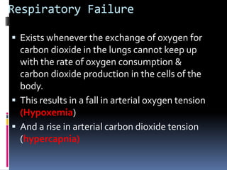
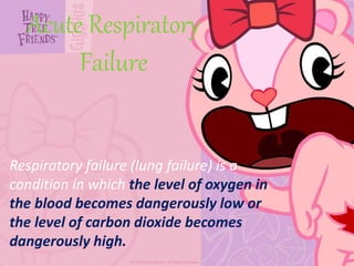
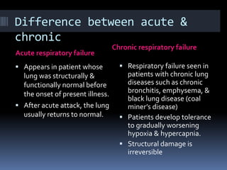
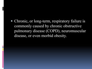
PULMONARY FAILURE
Respiratory Failure
 Exists whenever the exchange of oxygen for
carbon dioxide in the lungs cannot keep up
with the rate of oxygen consumption &
carbon dioxide production in the cells of the
body.
 This results in a fall in arterial oxygen tension
(Hypoxemia)
 And a rise in arterial carbon dioxide tension
(hypercapnia)
Acute Respiratory
Failure
Respiratory failure (lung failure) is a
condition in which the level of oxygen in
the blood becomes dangerously low or
the level of carbon dioxide becomes
dangerously high.
Difference between acute &
chronic
Acute respiratory failure
Chronic respiratory failure
 Appears in patient whose
lung was structurally &
functionally normal before
the onset of present illness.
 After acute attack, the lung
usually returns to normal.
 Respiratory failure seen in
patients with chronic lung
diseases such as chronic
bronchitis, emphysema, &
black lung disease (coal
miner’s disease)
 Patients develop tolerance
to gradually worsening
hypoxia & hypercapnia.
 Structural damage is
irreversible
 Chronic, or long-term, respiratory failure is
commonly caused by chronic obstructive
pulmonary disease (COPD), neuromuscular
disease, or even morbid obesity.
 Chronic Obstructive Pulmonary Disease
(COPD)
 Breathing is effortless and unconscious for most
people.
 Hundreds of times each day you breathe in
oxygen and exhale carbon dioxide while gas
exchange takes place in your lungs.
 Fresh, inhaled oxygen is absorbed into the blood
circulating through your lungs.
 Oxygen-rich blood then travels from the lungs to
the heart and is pumped into the arteries to be
distributed throughout your body.
 At the same time, carbon dioxide (a waste product
produced by your cells) travels to your heart through
your veins.
 It is then pumped into your lungs to be released as you
exhale.
 For people with chronic obstructive pulmonary disease
(COPD), breathing is not this easy.
 Damage to the lung’s airways and air sacs, typically
caused by smoking, environmental exposure or
genetics, makes it physically difficult to breathe in and
out.
 Inflammation of the airways blocks the movement of
air, and it becomes difficult to get oxygen into the
body.
 Breathlessness may occur during exercise, during very
little activity or even at rest depending on the severity
of COPD.
Causes of Acute Respiratory
Failure
 Respiratory failure results from inadequate
ventilation:
 Causes are:
 Upper airway obstruction
 Central nervous system depression
 Postoperative period ( major thoracic or upper abdominal
surgery)
 Pleural effusion, hemothorax, and pneumothorax
 Trauma caused by motor vehicle accidents
 Acute diseases like
 pneumonia,
 chemical pneumonitis,
 bronchial asthma,
 atelectasis,
 pulmonary embolism, &
 pulmonary edema
 Atelectasis
 is the collapse of part or all of a lung by blockage
of the air passages (bronchus or bronchioles), or
by very shallow breathing.
 Pulmonary Embolism
 blockage in an artery in the lungs caused by an
embolus (a free-floating blood clot) that travels
through the blood vessels (usually from a vein in a
leg or in the pelvic area) to the lungs.
 Pulmonary embolism causes damage to lung
tissue, disrupts the proper functioning of the
damaged lung, and can cause death
CNS Depression
 The respiratory center, which controls every
breath, lies in the lower part of the brain stem
(pons & medulla).
 Drug overdose, anesthesia, head injury, stroke,
brain tumors, encephalitis, meningitis, hypoxia, &
hypercapnia are all capable of depressing the
respiratory center.
 In these patients, respiration becomes slow
& shallow & respiratory arrest may occur in
severe cases.
 Any disease of the nerves, spinal cord, muscles,
or neuromuscular junction involved in
respiration will seriously affect ventilation.
 Examples:
 Polyneuritis
 Myasthenia gravis
 Damage to the cervical segment of spinal cord
 Large acute lesions of multiple sclerosis in the brain
stem
 poliomyelitis
 The impulse arising in the respiratory center
travel through nerves that extend from the brain
stem down the spinal cord to receptors in the
muscles of respiration.
Post operative period
 Effects of anesthetic drugs, analgesics, &
sedatives (pentobarbital & morphine)
 Pain in thoracic & abdominal area interferes
with deep breathing & coughing
 Muscle relaxants, some patients may have
difficulty in metabolizing or excreting these
drugs, so their effects last longer than usual,
making patients weak in postop period.
Pleural effusion,
hemothorax, & pneumothorax
 Interfere with ventilation by preventing
expansion of the lungs.
 Pleural Effusions
 is an accumulation of fluid between the layers of
the membrane that lines the lungs and chest
cavity.
Respiratory Failure
In practice, respiratory failure is defined as
a PaO2 value of less than 60 mm Hg while
breathing air or a PaCO2 of more than 50
mm Hg.
 Normal Values:
pH: 7.35-7.45
PO2: 80-100 mm Hg
PCO2: 35-45 mm Hg
% of O2 saturation: 95-100%
Signs and symptoms:
Anxious
 eyes are closed
 the accessory muscles of ventilation are fully used
Position, sitting forward with drooling secretions
 Hypoxia and hypercarbia produce characteristic
effects on the CNS and cardiovascular system
(CVS).
1 Hypoxia:
 CNS - Uncooperative, confused, drunken-like state
 CVS - Bradycardia, variable blood pressure,
cyanosis
2 Hypercarbia:
 CNS - Tremor and overt flap
 CVS - Raised pulse rate, peripheral vasodilation with
pink peripheries, blood pressure changes are
variable
Diagnostic Test
ABG analysis
CXR
ECG
Pulse oximetry
CBC
Serum electrolytes
Pulmonary artery
catheterization
Treatment and drugs:
Cautious oxygen therapy (nasal prongs or Venturi mask)
If Respiratory Acidosis persist, Mechanical ventilation
with an Edotracheal is attached or Tracheostomy
Antibiotics
Bronchodilators
Corticosteroids
 If cor pulmonale and cardiac output decreased
administer Inotropic agents,vasopresors, and diuretics
may ordered
Cor pulmonale
 disease of the heart:
 enlargement and failure of the right ventricle of
the heart, caused by disease of the lungs or
pulmonary blood vessels.
Management:
(a) Establish an airway
This applies particularly to the unconscious
patient, e.g. due to overdose, general
anaesthesia, CNS trauma and so on.
The patient is placed on the side with the
head down, and lower jaw pulled forward to
prevent the tongue falling back and obstructing
the upper airway.
 At this stage it may become obvious that the
obstruction is due primarily to foreign bodies or
vomit, so this must be cleared, if possible.
 Indications for artificial airways
 (1) Oropharyngeal: this is useful where it is
expected that the patient will soon recover
consciousness, e.g. post-operatively, or where there
is lack of expertise in endotracheal intubation. A
laryngeal mask may be used as an alternative in this
situation
 (2) Endotracheal tube (ETt): If unconsciousness is
expected to last for more than a matter of minutes,
as in drug overdose, then an ETT must be used both
to ensure and to protect the airway (e.g. from
aspiration of gastric contents). If ventilation is
depressed or inadequate due to trauma or disease,
than mechanical ventilation will be required.
(3) Cricothyrotomy and
tracheostomy obstruction above the cords due
to disease or infection may make intubation
impossible. Cricothyrotomy or tracheostomy is
then necessary to restore the airway.
(4) Bronchoscopy may also be required for
bronchial toilet, removing viscid mucous and
obtaining specimens for microscopy and culture
cricothyrotomy
 Tracheotomy by incision through the skin &
cricothyroid membrane esp. as an emergency
procedure for relief of an obstructed airway.
 (b) Administer oxygen to ensure adequate
tissue oxygenation
 It is of paramount importance to maintain
a PaO2 sufficient to give an arterial Hb
saturation of at least 85% (i.e. 8-9 kPa
(kilopascal) or 60-70 mmHg).
 Hyperoxia should be avoided, particularly
in the bronchitic who is a CO2 retainer and
dependent on hypoxic ventilatory drive.
(c) Maintain alveolar ventilation and treat
underlying cause
These two are inextricably linked. The causes of
ARF (acute respiratory failure) are many and
varied as are the requisite therapies. If treatment of
the underlying cause is not successful (i.e. steroids,
bronchodilators in asthma; physiotherapy,
antibiotics, mucolytics, bronchodilators in acute or
chronic bronchitis), then the carbon dioxide
tension will begin to rise, necessitating
intermittent positive pressure ventilation
(IPPV).
 There is little place for respiratory stimulants,
except perhaps narcotic antagonists in opiate
overdose. NB Infection is a cause of
exacerbation of ARF in bronchitics in less than
50% of cases. Other causes such as heart
failure, dysrthymias and pneumothorax must
be excluded and treated where necessary.
 In ARF due to chronic obstructive pulmonary
disease (COPD), muscle fatigue is a major
contributory factor to continuing hypoxia and
hypercarbia. Non-invasive methods of
ventilation (e.g. nasal mask) as well as
endotracheal intubation and IPPV may be
needed
NURSING MANAGEMENT:
> Orient the patient to the treatment unit to prevent anxiety
> To reverse hypoxemia, administer oxygen as ordered
> Maintain patent airway
> Monitor BP, RR and PR
> Place patient in semi-fowlers position
THANKS…

Pulmonary-Failure-.pptx

  • 1.
  • 2.
    Respiratory Failure  Existswhenever the exchange of oxygen for carbon dioxide in the lungs cannot keep up with the rate of oxygen consumption & carbon dioxide production in the cells of the body.  This results in a fall in arterial oxygen tension (Hypoxemia)  And a rise in arterial carbon dioxide tension (hypercapnia)
  • 3.
    Acute Respiratory Failure Respiratory failure(lung failure) is a condition in which the level of oxygen in the blood becomes dangerously low or the level of carbon dioxide becomes dangerously high.
  • 4.
    Difference between acute& chronic Acute respiratory failure Chronic respiratory failure  Appears in patient whose lung was structurally & functionally normal before the onset of present illness.  After acute attack, the lung usually returns to normal.  Respiratory failure seen in patients with chronic lung diseases such as chronic bronchitis, emphysema, & black lung disease (coal miner’s disease)  Patients develop tolerance to gradually worsening hypoxia & hypercapnia.  Structural damage is irreversible
  • 5.
     Chronic, orlong-term, respiratory failure is commonly caused by chronic obstructive pulmonary disease (COPD), neuromuscular disease, or even morbid obesity.
  • 6.
     Chronic ObstructivePulmonary Disease (COPD)  Breathing is effortless and unconscious for most people.  Hundreds of times each day you breathe in oxygen and exhale carbon dioxide while gas exchange takes place in your lungs.  Fresh, inhaled oxygen is absorbed into the blood circulating through your lungs.  Oxygen-rich blood then travels from the lungs to the heart and is pumped into the arteries to be distributed throughout your body.
  • 7.
     At thesame time, carbon dioxide (a waste product produced by your cells) travels to your heart through your veins.  It is then pumped into your lungs to be released as you exhale.  For people with chronic obstructive pulmonary disease (COPD), breathing is not this easy.  Damage to the lung’s airways and air sacs, typically caused by smoking, environmental exposure or genetics, makes it physically difficult to breathe in and out.  Inflammation of the airways blocks the movement of air, and it becomes difficult to get oxygen into the body.  Breathlessness may occur during exercise, during very little activity or even at rest depending on the severity of COPD.
  • 8.
    Causes of AcuteRespiratory Failure  Respiratory failure results from inadequate ventilation:  Causes are:  Upper airway obstruction  Central nervous system depression  Postoperative period ( major thoracic or upper abdominal surgery)  Pleural effusion, hemothorax, and pneumothorax  Trauma caused by motor vehicle accidents  Acute diseases like  pneumonia,  chemical pneumonitis,  bronchial asthma,  atelectasis,  pulmonary embolism, &  pulmonary edema
  • 9.
     Atelectasis  isthe collapse of part or all of a lung by blockage of the air passages (bronchus or bronchioles), or by very shallow breathing.
  • 10.
     Pulmonary Embolism blockage in an artery in the lungs caused by an embolus (a free-floating blood clot) that travels through the blood vessels (usually from a vein in a leg or in the pelvic area) to the lungs.  Pulmonary embolism causes damage to lung tissue, disrupts the proper functioning of the damaged lung, and can cause death
  • 11.
    CNS Depression  Therespiratory center, which controls every breath, lies in the lower part of the brain stem (pons & medulla).  Drug overdose, anesthesia, head injury, stroke, brain tumors, encephalitis, meningitis, hypoxia, & hypercapnia are all capable of depressing the respiratory center.  In these patients, respiration becomes slow & shallow & respiratory arrest may occur in severe cases.
  • 12.
     Any diseaseof the nerves, spinal cord, muscles, or neuromuscular junction involved in respiration will seriously affect ventilation.  Examples:  Polyneuritis  Myasthenia gravis  Damage to the cervical segment of spinal cord  Large acute lesions of multiple sclerosis in the brain stem  poliomyelitis  The impulse arising in the respiratory center travel through nerves that extend from the brain stem down the spinal cord to receptors in the muscles of respiration.
  • 13.
    Post operative period Effects of anesthetic drugs, analgesics, & sedatives (pentobarbital & morphine)  Pain in thoracic & abdominal area interferes with deep breathing & coughing  Muscle relaxants, some patients may have difficulty in metabolizing or excreting these drugs, so their effects last longer than usual, making patients weak in postop period.
  • 14.
    Pleural effusion, hemothorax, &pneumothorax  Interfere with ventilation by preventing expansion of the lungs.  Pleural Effusions  is an accumulation of fluid between the layers of the membrane that lines the lungs and chest cavity.
  • 15.
    Respiratory Failure In practice,respiratory failure is defined as a PaO2 value of less than 60 mm Hg while breathing air or a PaCO2 of more than 50 mm Hg.  Normal Values: pH: 7.35-7.45 PO2: 80-100 mm Hg PCO2: 35-45 mm Hg % of O2 saturation: 95-100%
  • 16.
    Signs and symptoms: Anxious eyes are closed  the accessory muscles of ventilation are fully used Position, sitting forward with drooling secretions  Hypoxia and hypercarbia produce characteristic effects on the CNS and cardiovascular system (CVS).
  • 17.
    1 Hypoxia:  CNS- Uncooperative, confused, drunken-like state  CVS - Bradycardia, variable blood pressure, cyanosis 2 Hypercarbia:  CNS - Tremor and overt flap  CVS - Raised pulse rate, peripheral vasodilation with pink peripheries, blood pressure changes are variable
  • 18.
    Diagnostic Test ABG analysis CXR ECG Pulseoximetry CBC Serum electrolytes Pulmonary artery catheterization
  • 19.
    Treatment and drugs: Cautiousoxygen therapy (nasal prongs or Venturi mask) If Respiratory Acidosis persist, Mechanical ventilation with an Edotracheal is attached or Tracheostomy Antibiotics Bronchodilators Corticosteroids  If cor pulmonale and cardiac output decreased administer Inotropic agents,vasopresors, and diuretics may ordered
  • 20.
    Cor pulmonale  diseaseof the heart:  enlargement and failure of the right ventricle of the heart, caused by disease of the lungs or pulmonary blood vessels.
  • 21.
    Management: (a) Establish anairway This applies particularly to the unconscious patient, e.g. due to overdose, general anaesthesia, CNS trauma and so on. The patient is placed on the side with the head down, and lower jaw pulled forward to prevent the tongue falling back and obstructing the upper airway.  At this stage it may become obvious that the obstruction is due primarily to foreign bodies or vomit, so this must be cleared, if possible.
  • 22.
     Indications forartificial airways  (1) Oropharyngeal: this is useful where it is expected that the patient will soon recover consciousness, e.g. post-operatively, or where there is lack of expertise in endotracheal intubation. A laryngeal mask may be used as an alternative in this situation  (2) Endotracheal tube (ETt): If unconsciousness is expected to last for more than a matter of minutes, as in drug overdose, then an ETT must be used both to ensure and to protect the airway (e.g. from aspiration of gastric contents). If ventilation is depressed or inadequate due to trauma or disease, than mechanical ventilation will be required.
  • 23.
    (3) Cricothyrotomy and tracheostomyobstruction above the cords due to disease or infection may make intubation impossible. Cricothyrotomy or tracheostomy is then necessary to restore the airway. (4) Bronchoscopy may also be required for bronchial toilet, removing viscid mucous and obtaining specimens for microscopy and culture
  • 24.
    cricothyrotomy  Tracheotomy byincision through the skin & cricothyroid membrane esp. as an emergency procedure for relief of an obstructed airway.
  • 25.
     (b) Administeroxygen to ensure adequate tissue oxygenation  It is of paramount importance to maintain a PaO2 sufficient to give an arterial Hb saturation of at least 85% (i.e. 8-9 kPa (kilopascal) or 60-70 mmHg).  Hyperoxia should be avoided, particularly in the bronchitic who is a CO2 retainer and dependent on hypoxic ventilatory drive.
  • 26.
    (c) Maintain alveolarventilation and treat underlying cause These two are inextricably linked. The causes of ARF (acute respiratory failure) are many and varied as are the requisite therapies. If treatment of the underlying cause is not successful (i.e. steroids, bronchodilators in asthma; physiotherapy, antibiotics, mucolytics, bronchodilators in acute or chronic bronchitis), then the carbon dioxide tension will begin to rise, necessitating intermittent positive pressure ventilation (IPPV).
  • 27.
     There islittle place for respiratory stimulants, except perhaps narcotic antagonists in opiate overdose. NB Infection is a cause of exacerbation of ARF in bronchitics in less than 50% of cases. Other causes such as heart failure, dysrthymias and pneumothorax must be excluded and treated where necessary.  In ARF due to chronic obstructive pulmonary disease (COPD), muscle fatigue is a major contributory factor to continuing hypoxia and hypercarbia. Non-invasive methods of ventilation (e.g. nasal mask) as well as endotracheal intubation and IPPV may be needed
  • 28.
    NURSING MANAGEMENT: > Orientthe patient to the treatment unit to prevent anxiety > To reverse hypoxemia, administer oxygen as ordered > Maintain patent airway > Monitor BP, RR and PR > Place patient in semi-fowlers position
  • 29.