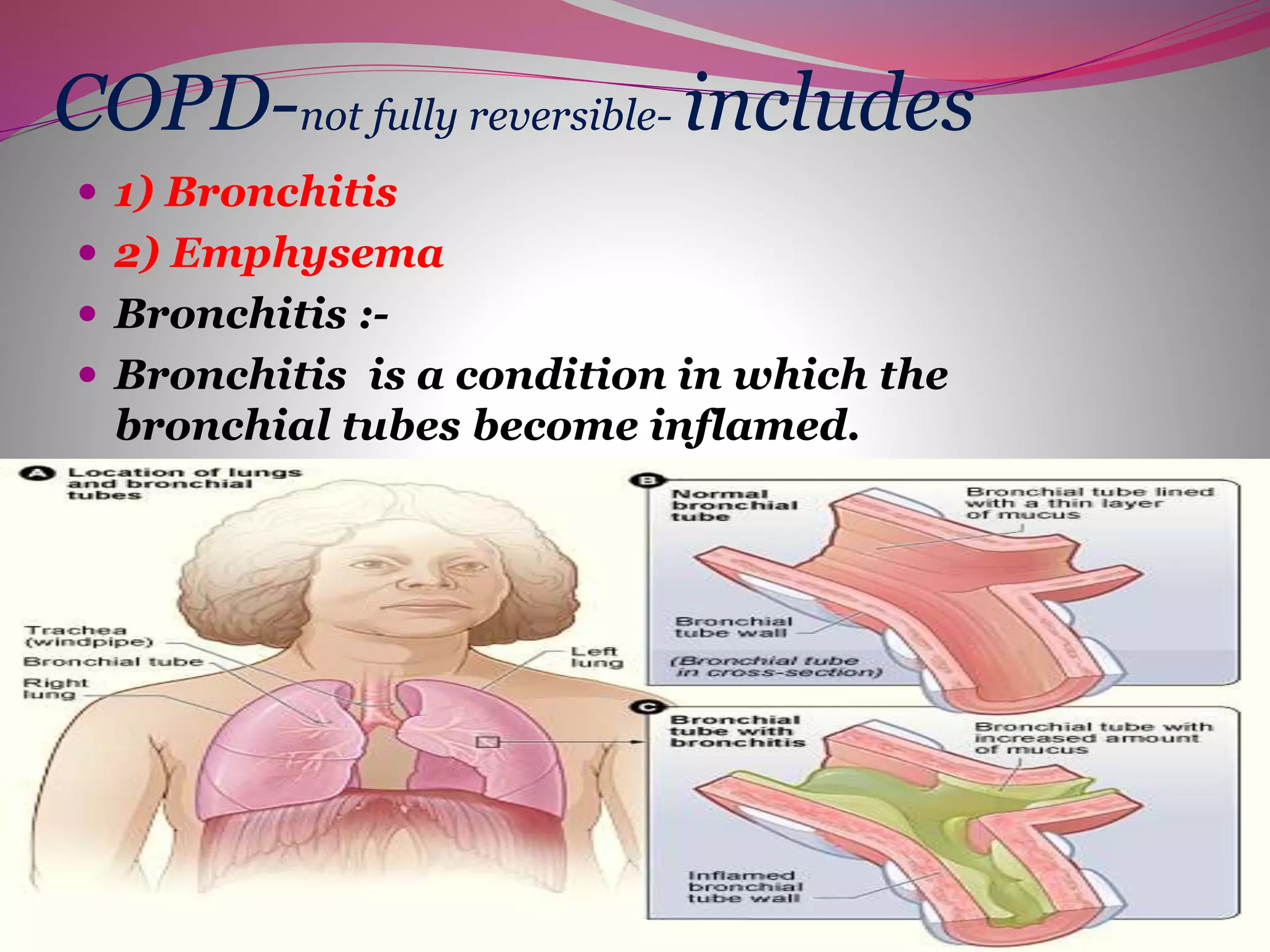
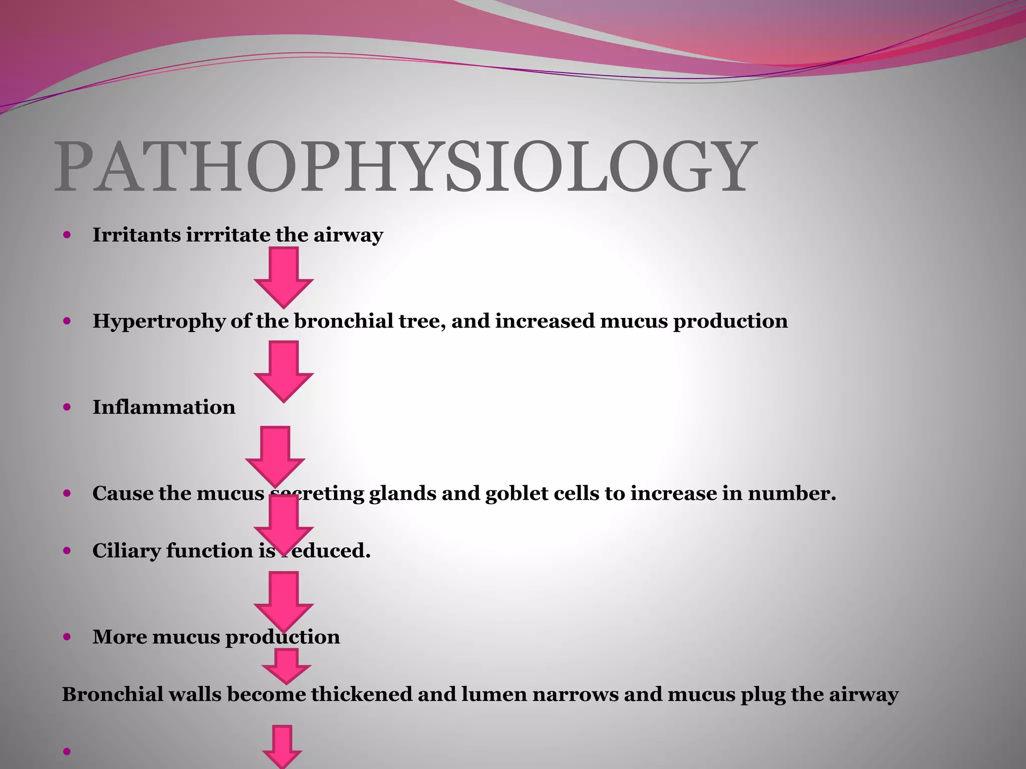
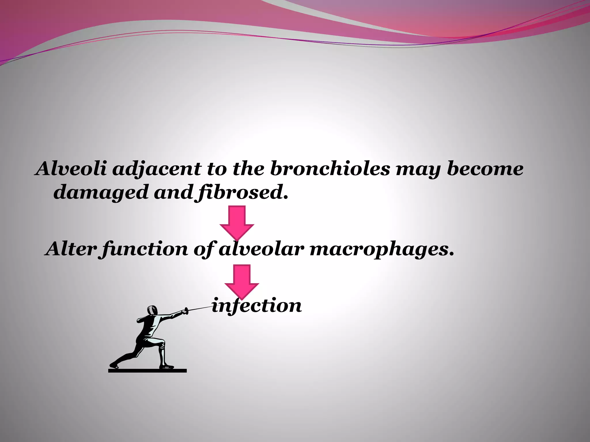
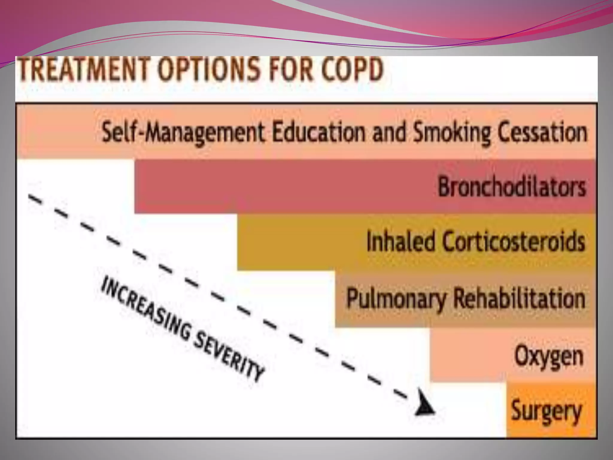
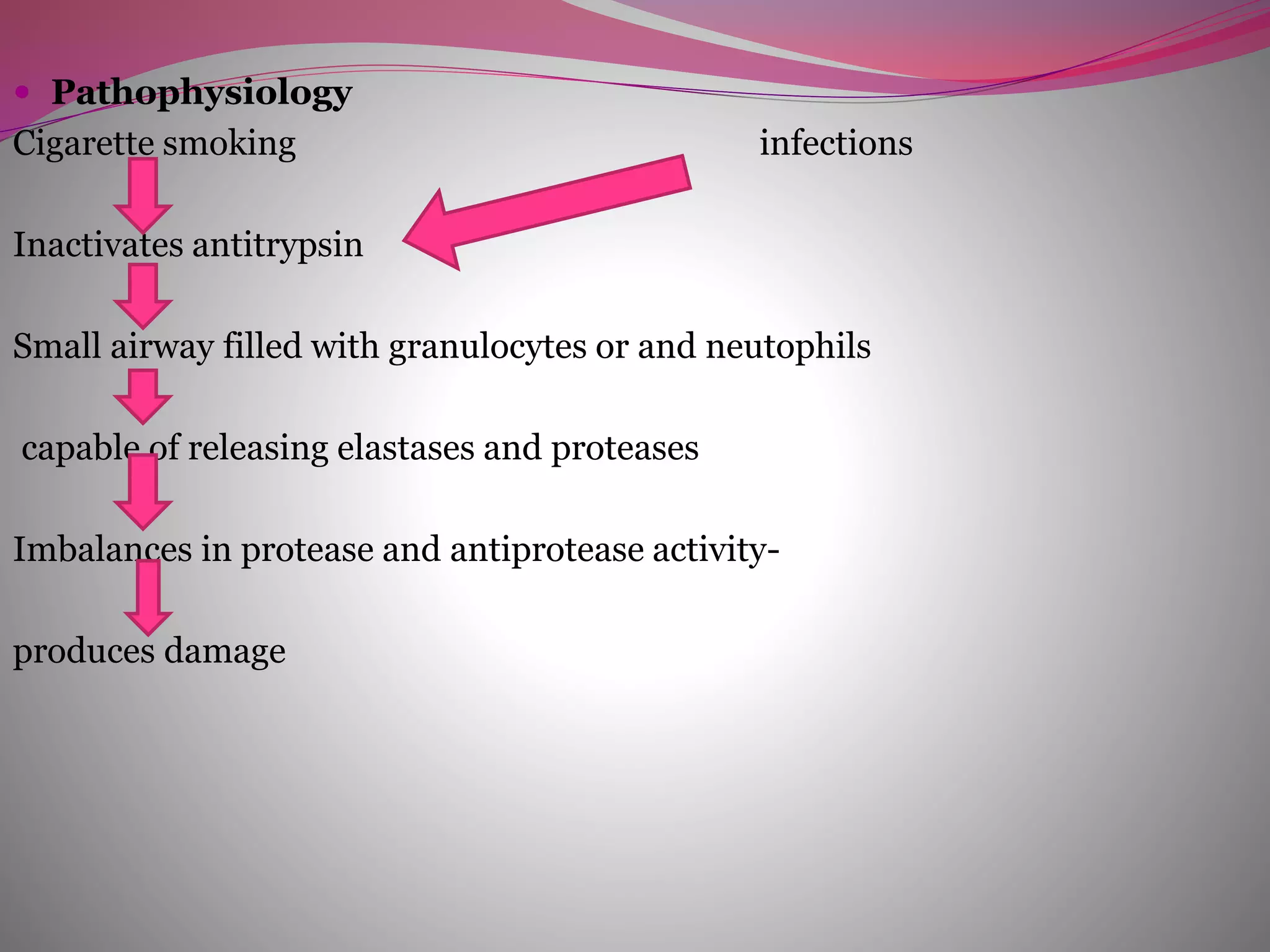
“Everyone is responsible for
his/her own looks after 40”
OBSTRUCTIVE PULMONARY
DISORDERS
DICKSON AKANKWATSA {BSN 3}-
BISHOP STUART UNIVERSITY
cvdicksonakankwatsa@outlook.com
Divided into two:
Fully reversible disorders
{asthma}, and
Non fully reversible/ partially
reversible{ COPD}
Causes
 1)Smoking
 2) Occupational exposures- exposure to
workplace dusts found in coal mining, gold
mining, and the cotton textile industry and
chemicals such as cadmium, and fumes from
welding have been implicated in the
development of airflow obstruction.
 3) Air pollution
 4) sudden airway constriction in response to
inhaled irritants,
 5) Bronchial hyperresponsiveness, is a
characteristic of asthma.
 6) Genetics-Alpha 1-antitrypsin deficiency
COPD
 In COPD, less air flows in and out of the
airways because of one or more of the
following:
 The airways and air sacs lose their elastic
quality.
 The walls between many of the air sacs are
destroyed.
 The walls of the airways become thick and
inflamed.
 The airways make more mucus than usual,
which tends to clog them.
COPD-not fully reversible- includes
 1) Bronchitis
 2) Emphysema
 Bronchitis :-
 Bronchitis is a condition in which the
bronchial tubes become inflamed.
BRONCHITIS
 Chronic bronchitis:
 It is defined as the presence of cough and
sputum production for atleast 3 months.
PATHOPHYSIOLOGY
 Irritants irrritate the airway
 Hypertrophy of the bronchial tree, and increased mucus production
 Inflammation
 Cause the mucus secreting glands and goblet cells to increase in number.
 Ciliary function is reduced.
 More mucus production
Bronchial walls become thickened and lumen narrows and mucus plug the airway

Alveoli adjacent to the bronchioles may become
damaged and fibrosed.
Alter function of alveolar macrophages.
infection
Acute signs and symptoms
 sore throat,
 fatigue (tiredness),
 fever, body aches,
 stuffy or runny nose,
 Vomiting & Diarrhea
 persistent cough
 cough may produce clear mucus
 shortness of breath
Chronic symptoms
 coughing,
 wheezing, and
 chest discomfort.
 The coughing may produce large amounts
of mucus. This type of cough often is called
a smoker's cough.
Diagnostic evaluation
 History - medical history
 •Whether you've recently had a cold or the
flu
 •Whether you smoke or spend time around
others who smoke
 •Whether you've been exposed to dust,
fumes, vapors, or air pollution -
 Mucus -to see whether you have a bacterial
infection
 chest x ray-may show consolidation
 lung function tests-brianstorm them
 CBC – significantly, may have neutrophilia,
eosinophilia, reduced Hb levels, etc
MANAGEMENT
 medical management
 surgical management
 nursing management
MEDICAL MANAGEMENT improve ventillation
1. broncho dilators like beta2
agonists(albuterol),anticholinergics(i
pratropium bromide-atrovent).
2. methylxanthines(theophylline,amino
phylline)
3. corticosteroids
4. oxygen administration
SURGICAL MANAGEMENT
 bullectomy
bullae are enlarged airspaces that do not contribute
to ventillation but occupy space in the thorax,these
areas may be surgically excised
 lung volume reduction surgery
 it involves the removal of a portion of the diseased
lung parenchyma.this allows the functional tissue to
expand.
 lung transplantation
NURSING MANAGEMENT
 assessment
 physical examination
 diagnosis
 intervention
Care plan
 impaired gad exchange related to decreased ventillation and
mucous plugs
 ineffective airway clearence related to excessive secretion and
ineffective coughing
 anxiety related to acute breathing difficulties and fear of
suffocation
 activity intolerence related to inadequate oxygenation and
dyspnoe
 imbalanced nutrition less than body requirement related to
reduced appetite,decreased energy level and dyspnoea
 disturbed sleep pattern related to dyspnoea and external stimuli
 risk for infection related to ineffective pulmonary clearence
Empysema
Defined pathologically as
dilatation and destruction of the
lung tissue distal to the terminal
bronchiole. It is classified
according to the site of damage:
classification
 Centri-acinar emphysema. More common .Distension and damage of lung tissue is
concentrated around the respiratory bronchioles; more distal alveolar ducts and
alveoli tend to be well preserve.
 Pan-acinar emphysema. Less common. Distension
and destruction appear to involve the whole of the acinus, and in the extreme form
the lung becomes a mass of bullae.
■ Irregular emphysema. There is scarring and damage affecting the lung
parenchyma patchily without particular regard for acinar structure.

 Pathophysiology
Cigarette smoking infections
Inactivates antitrypsin
Small airway filled with granulocytes or and neutophils
capable of releasing elastases and proteases
Imbalances in protease and antiprotease activity-
produces damage
 Emphysema leads to expiratory airflow limitation
and air trapping. The loss of lung elastic recoil
results in an increase in TLC while the loss of alveoli
results in decreased gas transfer.
Cont…
 VA/Q mismatch occurs partly because of damage and
mucus plugging of smaller airways from chronic
inflammation, and partly because of the rapid
expiratory closure of the smaller airways owing to
loss of elastic recoil from emphysema.
 In summary, three mechanisms have been suggested
for this limitation of airflow in small airways (< 2 mm in
diameter).
 Loss of elasticity and alveolar attachments of airways
due to emphysema. Reduces the elastic recoil and
the airways collapse during expiration.
 Inflammation and scarring cause the small airways to
narrow.
 Mucus secretion which blocks the airways.
 All cause narrowing of the small airways and trapping
of air leading to hyperinflation of the lungs and
breathlessness.
Signs
 Wheeze
 tachypnoea
 prolonged expiration
 Use of accessory muscles
 intercostal in-drawing on inspiration
 pursing of the lips on expiration .
 Chest expansion is poor
 lungs hyper-resonant
 loss of the normal cardiac and liver dullness.
.
Cont…
 Patients who remain responsive to CO2 are usually:
 breathless
 rarely cyanosed.
 Heart failure and edema in terminal events.
Patients who become insensitive to CO2 are often
edematous and cyanosed but not particularly breathless.
Those with hypercapnia may have peripheral
vasodilatation. Severe hypercapnia will lead to confusion
and progressive drowsiness. At this stage papilledema
may be present but is neither specific nor sensitive as a
diagnostic feature
Diagnosis
a) History
b) PFT
c) Spirometry-to find out airflow obstruction.
d) ABG analysis
e) CT scan of the lung.
f) Screening of alpha antitrypsin deficiency
g) X-ray radiography may aid in the
diagnosis.
MANAGEMENT
 medical management
 surgical management as for bronchitis
 nursing management
 Complications
 Respiratory insufficiency
 Respiratory failure
 Pneumonia
 Pneumothorax
 Pulmonary artery hypertension.
Self management of COPD
 STAY AWAY FROM INFECTIONS BY MAINTAINING GOOD HYGIENE
 QUIT SMOKIN
 GEAT A REGULAR BALANCED DIET
• DRINK PLENTY OF PLAIN FRESH WATER ATLEAST 1.5L/DAY
Copd  presentation  dickson bns 3

Copd presentation dickson bns 3

  • 1.
    “Everyone is responsiblefor his/her own looks after 40”
  • 2.
    OBSTRUCTIVE PULMONARY DISORDERS DICKSON AKANKWATSA{BSN 3}- BISHOP STUART UNIVERSITY cvdicksonakankwatsa@outlook.com
  • 3.
    Divided into two: Fullyreversible disorders {asthma}, and Non fully reversible/ partially reversible{ COPD}
  • 4.
    Causes  1)Smoking  2)Occupational exposures- exposure to workplace dusts found in coal mining, gold mining, and the cotton textile industry and chemicals such as cadmium, and fumes from welding have been implicated in the development of airflow obstruction.  3) Air pollution  4) sudden airway constriction in response to inhaled irritants,  5) Bronchial hyperresponsiveness, is a characteristic of asthma.  6) Genetics-Alpha 1-antitrypsin deficiency
  • 5.
    COPD  In COPD,less air flows in and out of the airways because of one or more of the following:  The airways and air sacs lose their elastic quality.  The walls between many of the air sacs are destroyed.  The walls of the airways become thick and inflamed.  The airways make more mucus than usual, which tends to clog them.
  • 6.
    COPD-not fully reversible-includes  1) Bronchitis  2) Emphysema  Bronchitis :-  Bronchitis is a condition in which the bronchial tubes become inflamed.
  • 7.
    BRONCHITIS  Chronic bronchitis: It is defined as the presence of cough and sputum production for atleast 3 months.
  • 8.
    PATHOPHYSIOLOGY  Irritants irrritatethe airway  Hypertrophy of the bronchial tree, and increased mucus production  Inflammation  Cause the mucus secreting glands and goblet cells to increase in number.  Ciliary function is reduced.  More mucus production Bronchial walls become thickened and lumen narrows and mucus plug the airway 
  • 9.
    Alveoli adjacent tothe bronchioles may become damaged and fibrosed. Alter function of alveolar macrophages. infection
  • 10.
    Acute signs andsymptoms  sore throat,  fatigue (tiredness),  fever, body aches,  stuffy or runny nose,  Vomiting & Diarrhea  persistent cough  cough may produce clear mucus  shortness of breath
  • 11.
    Chronic symptoms  coughing, wheezing, and  chest discomfort.  The coughing may produce large amounts of mucus. This type of cough often is called a smoker's cough.
  • 12.
    Diagnostic evaluation  History- medical history  •Whether you've recently had a cold or the flu  •Whether you smoke or spend time around others who smoke  •Whether you've been exposed to dust, fumes, vapors, or air pollution -
  • 13.
     Mucus -tosee whether you have a bacterial infection  chest x ray-may show consolidation  lung function tests-brianstorm them  CBC – significantly, may have neutrophilia, eosinophilia, reduced Hb levels, etc
  • 14.
    MANAGEMENT  medical management surgical management  nursing management
  • 15.
    MEDICAL MANAGEMENT improveventillation 1. broncho dilators like beta2 agonists(albuterol),anticholinergics(i pratropium bromide-atrovent). 2. methylxanthines(theophylline,amino phylline) 3. corticosteroids 4. oxygen administration
  • 16.
    SURGICAL MANAGEMENT  bullectomy bullaeare enlarged airspaces that do not contribute to ventillation but occupy space in the thorax,these areas may be surgically excised  lung volume reduction surgery  it involves the removal of a portion of the diseased lung parenchyma.this allows the functional tissue to expand.  lung transplantation
  • 17.
    NURSING MANAGEMENT  assessment physical examination  diagnosis  intervention
  • 18.
    Care plan  impairedgad exchange related to decreased ventillation and mucous plugs  ineffective airway clearence related to excessive secretion and ineffective coughing  anxiety related to acute breathing difficulties and fear of suffocation  activity intolerence related to inadequate oxygenation and dyspnoe  imbalanced nutrition less than body requirement related to reduced appetite,decreased energy level and dyspnoea  disturbed sleep pattern related to dyspnoea and external stimuli  risk for infection related to ineffective pulmonary clearence
  • 20.
    Empysema Defined pathologically as dilatationand destruction of the lung tissue distal to the terminal bronchiole. It is classified according to the site of damage:
  • 21.
    classification  Centri-acinar emphysema.More common .Distension and damage of lung tissue is concentrated around the respiratory bronchioles; more distal alveolar ducts and alveoli tend to be well preserve.  Pan-acinar emphysema. Less common. Distension and destruction appear to involve the whole of the acinus, and in the extreme form the lung becomes a mass of bullae. ■ Irregular emphysema. There is scarring and damage affecting the lung parenchyma patchily without particular regard for acinar structure. 
  • 22.
     Pathophysiology Cigarette smokinginfections Inactivates antitrypsin Small airway filled with granulocytes or and neutophils capable of releasing elastases and proteases Imbalances in protease and antiprotease activity- produces damage
  • 23.
     Emphysema leadsto expiratory airflow limitation and air trapping. The loss of lung elastic recoil results in an increase in TLC while the loss of alveoli results in decreased gas transfer.
  • 24.
    Cont…  VA/Q mismatchoccurs partly because of damage and mucus plugging of smaller airways from chronic inflammation, and partly because of the rapid expiratory closure of the smaller airways owing to loss of elastic recoil from emphysema.
  • 25.
     In summary,three mechanisms have been suggested for this limitation of airflow in small airways (< 2 mm in diameter).  Loss of elasticity and alveolar attachments of airways due to emphysema. Reduces the elastic recoil and the airways collapse during expiration.  Inflammation and scarring cause the small airways to narrow.  Mucus secretion which blocks the airways.  All cause narrowing of the small airways and trapping of air leading to hyperinflation of the lungs and breathlessness.
  • 26.
    Signs  Wheeze  tachypnoea prolonged expiration  Use of accessory muscles  intercostal in-drawing on inspiration  pursing of the lips on expiration .  Chest expansion is poor  lungs hyper-resonant  loss of the normal cardiac and liver dullness. .
  • 27.
    Cont…  Patients whoremain responsive to CO2 are usually:  breathless  rarely cyanosed.  Heart failure and edema in terminal events. Patients who become insensitive to CO2 are often edematous and cyanosed but not particularly breathless. Those with hypercapnia may have peripheral vasodilatation. Severe hypercapnia will lead to confusion and progressive drowsiness. At this stage papilledema may be present but is neither specific nor sensitive as a diagnostic feature
  • 28.
    Diagnosis a) History b) PFT c)Spirometry-to find out airflow obstruction. d) ABG analysis e) CT scan of the lung. f) Screening of alpha antitrypsin deficiency g) X-ray radiography may aid in the diagnosis.
  • 29.
    MANAGEMENT  medical management surgical management as for bronchitis  nursing management  Complications  Respiratory insufficiency  Respiratory failure  Pneumonia  Pneumothorax  Pulmonary artery hypertension.
  • 30.
    Self management ofCOPD  STAY AWAY FROM INFECTIONS BY MAINTAINING GOOD HYGIENE  QUIT SMOKIN
  • 31.
     GEAT AREGULAR BALANCED DIET • DRINK PLENTY OF PLAIN FRESH WATER ATLEAST 1.5L/DAY