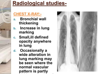
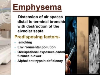
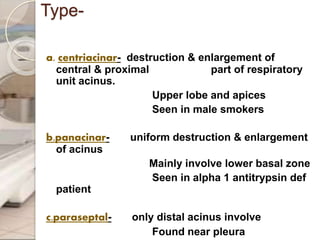
Shree b.m.patil medicalcollege,bijapur
Department of Radiology
PRESENTED BY-
-
Ravi Prakash
Chronic obstructive pulmonary
disease.
Disease state characterized by airflow
limitation that is not fully reversible.
A. Chronic bronchitis- a clinically defined
condition with chronic cough and phlegm.
B. Emphysema- an anatomically defined
condition characterized by destruction &
enlargement of lung alveoli.
Chronic bronchitis:-
condition associated with excessive
tracheobronchial mucus production
to cause cough with expectoration
for at least 3 month of year for
more than 2 year.
Aetiology-
 cigarette smoking
 air pollution
 occupational hazards -
cotton mill , coal mining , rubber
industry , gold mining
 familial factor
 genetic factor
 infection-by rhinovirus , strep
Pathophysiology-
 Hypertrophy & hyperplasia of mucus producing
gland
 Reduction in caliber of air passage
airflow obstruction
Clinical feature-
Cough
Sputum production
Exertional dyspnoea
Breathlessness relatively late onset
Percussion note- resonant over
lung field
Auscultation- vesicular breathing,
rhonchi, crepitation
Blue bloaters
Gold criteria-
GOLD
STAGE
SEVERITY SYMPTOM SPIROMETRY
0 At risk Chronic cough , sputum
production
normal
1 mild With or without chronic cough or
sputum production
FEV1/FVC<0.7
FEV1>=80%predi
cted
11 moderat
e
With or without chronic cough or
sputum production
FEV1/FVC<0.7
50%<=FEV1<80%
predicted
111 severe With or without chronic cough or
sputum production
FEV1/FVC<0.7
30%<=FEV1<50%
predicted
1V Very
severe
With or without chronic cough or
sputum production
FEV1/FVC<0.7
FEV1<=30%predi
cted
Or
Investigation-
PULMONARY FUNCTION TEST
FEV1-reduced
VC-reduced
RV-increased
TLC-increased
Gas transfer-decrease or normal
Arterial blood gas analysis-
pao2-decreased
Paco2-increased
Radiological studies-
CHEST X-RAY:-
a. Bronchial wall
thickening
b. Increase in lung
marking
c. Small,ill defined
opacity anywhere
in lung
d. Occasionally a
wide alteration in
lung marking may
be seen where the
normal vascular
pattern is partly
Linear shadow
Increase peribronchial marking
especially at the base due to
thickened bronchial &
peribronchial tissue
Bronchogram-
 dilatation of
bronchial gland
leading to irregular
out pouching from
bronchial lumen
mostly often seen in
wall of major bronchi.
 abrupt termination of
smaller branch of
bronchi with square
& truncated ending
 bronchiolar spasm
 increased secretion
Complication-
Mismatch of ventilation &
perfusion
Co2 narcosis Respiratory
failure
Hypoxaemia
hypercarbia
Pulmonary
vasoconstriction
Pulmonary
hypertension
Right ventricular
hypertrophy
Right
ventricular
failure
Increased
erythropoisis
Sec.
polycythe
mia
Desaturation
of Hb
cynaosis
Treatment-
1. general measure-
 Smoke caessation
 Regular exercise
 Weight loss
2.bronchodialator-
 Inhalation of b2 agonist-salbutamol 200mcg
Terbutalin 600mcg 6th
hourly
3.glucocorticoides-
 improve the gas exchange to some extent
4.oxygen-
 Supplemental o2 by nasal catheter & through
face mask
 Mainly for exertional or nocturnal hypoxaemia
6.antibiotic-
If any infection is there-
Doxycycline,co-trimoxazole,amoxycillin-
clavulanate,gatifloxacin,iv azithromycin ,
ceftriaxone,cefotaxime
5.Others
 N-acetyl cysteine-mucolytic & antioxidant
Emphysema
Distension of air spaces
distal to terminal bronchiole
with destruction of the
alveolar septa.
Predisposing factors-
 smoking
 Environmental pollution
 Occupational exposure-cadmium,
furnace blower
 Alpha1antitrypsin deficiency
Type-
a. centriacinar- destruction & enlargement of
central & proximal part of respiratory
unit acinus.
Upper lobe and apices
Seen in male smokers
b.panacinar- uniform destruction & enlargement
of acinus
Mainly involve lower basal zone
Seen in alpha 1 antitrypsin def
patient
c.paraseptal- only distal acinus involve
Found near pleura
Type..
1.compensatory-
 Normal lung tissue undergoes compensatory
mechanism for an extensive damage to other
lung or part of the same lung.
 Hyper resonant percussion note.
 Increase breath sound.
2.subcutaneous-
 seen in case of penetrating chest injury
 Rib fracture
 Intercostals tube introduction
3.Mediastinal emphysema-
 Vertical translucent streak , separating & outlining
the soft tissue layers & structure of mediastinum.
 Air often separates the parietal pleura from the
mediastinum & pleura may then be visible as a
thin hair line running parallel to mediastinum.
 Air may be seen in neck also. Due to escape of air
rapidly into mediastinum after rupture of over
distended alveoli.
 On auscultation crunching sound heard.
Pathogenesis-
Cigarette
smoke
Inflammatory
cell
recruitment
neutrophil
macrophage
Serin
e
prote
inase
s
ECM
degradati
on, lung
tissue
destructio
n
Cyste
ine
protei
nases
Proteina
se
inhibitor
s
Clinical feature-
Progressive
Exertional
breathlessness
minimum
cough &
expectoratio
n
Weakness
lethargy
Weight
loss
anorexia
SIGN..
Inspection & palpation-
 Tachypnoeic
 Hypertrophy of accessory muscle of
respiration
 Purse lip breathing
 Chest bowel shaped
 AP diameter- increased
 AP:TD –altered(normal-5:7)
 Angle of Luis-prominent
 Sub costal angle widened(normal 70’)
 Apical impulse is usually invisible or
feeble.
Percussion-
 hyper resonant over lung field area
 cardiac dullness decreases
 liver dullness pushed down or absent if right
side involved
Auscultation-
 Breath sound –decreases
 Prolong expiration
 Pink puffers
Difference In chronic bronchitis &
emphysema.
feature chronic bronchitis emphysema
cough Before dyspnoea starts After dyspnoea starts
sputum Copious , purulent Scanty , mucoid
dyspnoea mild severe
cynosis present absent
infection common Less common
Chest x-ray Increased bronchovascular
marking , cardiomegaly
Feature of hyperinflation ,
bullae,tubular heart
Pulmonary
hypertension
Moderate to severe mild
Diffusing
capacity
normal decreased
Investigation-
PULMONARY FUNCTION TEST
FEV1 -decreases
VC -decreases
FEV1/VC -decreases
PEF -decreases
TLC -increases
RV -increases
CHEST X-RAY-
 Irregular area of
radiolucency related to
tissue loss
 Increase in retero
sternal air space
Alteration in vascular pattern
Presence of over inflation
Increased lung volume
Bullae-
 Multiple & small-
May be localized to a lobe
or may be B/L
Upper lobe more often
affected
 Giant bulla-
Rib interspaced is
widened in region of giant
bulla
Neighboring normal
tissue is compressed with
vessels crowded together
CT SCAN Finding:-
it permit the direct identification of destroyed lung
tissue with high precision.
It shows-
 Area of decreased attenuation without visible walls
 Pruning of pulmonary vessels
 Distortion of pulmonary vessels
 Decrease lung tissue gradient
Centrilobular-
lucent region initially
surrounded by normal
lung parenchyma but
become confluent
along with obliteration
of peripheral vascular
as disease progresses.
Panlobular-
Widespread area of
low attenuation usually
seen accompanied by
diffuse vascular
distortion.
Nuclear imaging-
 Functional evaluation of the lungs can be
carried out by using xenon-133 (133 Xe) lung
ventilation scintigraphy before and after lung-
volume–reduction surgery (LVRS) in patients with
pulmonary emphysema.
Xenon-133 washout curves during lung
scintigraphy exhibit a biphasic pattern:
(1) the first component corresponds to an initial
rapid phase in washout that reflects emptying
of the large airways, and
(2) the second component, reflects a slower
phase of washout that is attributed to gas
elimination in the small airways.
Complication-
pulmonary bullae
Significant Weight loss
Respiratory failure
Pulmonary hypertension-late
Right heart failure-late
Emphysema with chronic bronchitis-
Radiological finding--
 Decrease in size & no. Of small vascular marking
specially in middle & outer 1/3rd of lung
 Main pulmonary arteries are enlarged making hilar
shadow prominent
 May be barrel shaped chest with increase in AP
diameter & anterior bowing of sternum
 Ant mediastinal space is usually increased in depth
 Heart shadow is often long,narrow &
cardiothoracic ratio may change to 1:3 or 1:4.
 Lung fields are seen more translucent
 Increased vol of lung results in flattening of
contour of diaphragm
 Best seen in lateral projection & level is
lowered below the 11th rib.
Treatment-
cessation of smoking
Antibiotics
IV alpha1 AT therapy
Vaccination against
influenza & pneumococcal
Low dose benzodiazepine
i.e. alprazolam
Mechanical ventilatory support-
Noninvasive PPV-
paco2 more than 45 mmhg
Acidosis
Invasive PPV-
Severe respiratory distress
Severe acidosis
Severe hypoxia
NIPPV failure
Surgical-
A. bullectomy
B. Lung volume reduction surgery-
Not done if significant pleural disease
PASPmore than 45 mm mg
C.Lung transplantation-
Age should be less than 65 yrs
Free from liver cardiac renal disease
REFERENCES….
Harrison’s principles of internal medicine
Manual of practical medicine-R Alagappan
Fraser& pare’s diagnosis of diseases of chest
Roentgenlogic diagnosis bysaunder’s
CT & MRI imaging of whole body-Haaga,Tanzien,Gilkeson
Internet
COPD

COPD

  • 1.
    Shree b.m.patil medicalcollege,bijapur Departmentof Radiology PRESENTED BY- - Ravi Prakash
  • 2.
    Chronic obstructive pulmonary disease. Diseasestate characterized by airflow limitation that is not fully reversible. A. Chronic bronchitis- a clinically defined condition with chronic cough and phlegm. B. Emphysema- an anatomically defined condition characterized by destruction & enlargement of lung alveoli.
  • 3.
    Chronic bronchitis:- condition associatedwith excessive tracheobronchial mucus production to cause cough with expectoration for at least 3 month of year for more than 2 year. Aetiology-  cigarette smoking  air pollution  occupational hazards - cotton mill , coal mining , rubber industry , gold mining  familial factor  genetic factor  infection-by rhinovirus , strep
  • 4.
    Pathophysiology-  Hypertrophy &hyperplasia of mucus producing gland  Reduction in caliber of air passage
  • 5.
  • 6.
    Clinical feature- Cough Sputum production Exertionaldyspnoea Breathlessness relatively late onset Percussion note- resonant over lung field Auscultation- vesicular breathing, rhonchi, crepitation Blue bloaters
  • 7.
    Gold criteria- GOLD STAGE SEVERITY SYMPTOMSPIROMETRY 0 At risk Chronic cough , sputum production normal 1 mild With or without chronic cough or sputum production FEV1/FVC<0.7 FEV1>=80%predi cted 11 moderat e With or without chronic cough or sputum production FEV1/FVC<0.7 50%<=FEV1<80% predicted 111 severe With or without chronic cough or sputum production FEV1/FVC<0.7 30%<=FEV1<50% predicted 1V Very severe With or without chronic cough or sputum production FEV1/FVC<0.7 FEV1<=30%predi cted Or
  • 8.
    Investigation- PULMONARY FUNCTION TEST FEV1-reduced VC-reduced RV-increased TLC-increased Gastransfer-decrease or normal Arterial blood gas analysis- pao2-decreased Paco2-increased
  • 9.
    Radiological studies- CHEST X-RAY:- a.Bronchial wall thickening b. Increase in lung marking c. Small,ill defined opacity anywhere in lung d. Occasionally a wide alteration in lung marking may be seen where the normal vascular pattern is partly
  • 10.
    Linear shadow Increase peribronchialmarking especially at the base due to thickened bronchial & peribronchial tissue
  • 11.
    Bronchogram-  dilatation of bronchialgland leading to irregular out pouching from bronchial lumen mostly often seen in wall of major bronchi.  abrupt termination of smaller branch of bronchi with square & truncated ending  bronchiolar spasm  increased secretion
  • 12.
    Complication- Mismatch of ventilation& perfusion Co2 narcosis Respiratory failure Hypoxaemia hypercarbia Pulmonary vasoconstriction Pulmonary hypertension Right ventricular hypertrophy Right ventricular failure Increased erythropoisis Sec. polycythe mia Desaturation of Hb cynaosis
  • 13.
    Treatment- 1. general measure- Smoke caessation  Regular exercise  Weight loss 2.bronchodialator-  Inhalation of b2 agonist-salbutamol 200mcg Terbutalin 600mcg 6th hourly 3.glucocorticoides-  improve the gas exchange to some extent
  • 14.
    4.oxygen-  Supplemental o2by nasal catheter & through face mask  Mainly for exertional or nocturnal hypoxaemia 6.antibiotic- If any infection is there- Doxycycline,co-trimoxazole,amoxycillin- clavulanate,gatifloxacin,iv azithromycin , ceftriaxone,cefotaxime 5.Others  N-acetyl cysteine-mucolytic & antioxidant
  • 15.
    Emphysema Distension of airspaces distal to terminal bronchiole with destruction of the alveolar septa. Predisposing factors-  smoking  Environmental pollution  Occupational exposure-cadmium, furnace blower  Alpha1antitrypsin deficiency
  • 16.
    Type- a. centriacinar- destruction& enlargement of central & proximal part of respiratory unit acinus. Upper lobe and apices Seen in male smokers b.panacinar- uniform destruction & enlargement of acinus Mainly involve lower basal zone Seen in alpha 1 antitrypsin def patient c.paraseptal- only distal acinus involve Found near pleura
  • 17.
  • 18.
    1.compensatory-  Normal lungtissue undergoes compensatory mechanism for an extensive damage to other lung or part of the same lung.  Hyper resonant percussion note.  Increase breath sound. 2.subcutaneous-  seen in case of penetrating chest injury  Rib fracture  Intercostals tube introduction
  • 19.
    3.Mediastinal emphysema-  Verticaltranslucent streak , separating & outlining the soft tissue layers & structure of mediastinum.  Air often separates the parietal pleura from the mediastinum & pleura may then be visible as a thin hair line running parallel to mediastinum.  Air may be seen in neck also. Due to escape of air rapidly into mediastinum after rupture of over distended alveoli.  On auscultation crunching sound heard.
  • 20.
  • 21.
  • 22.
    SIGN.. Inspection & palpation- Tachypnoeic  Hypertrophy of accessory muscle of respiration  Purse lip breathing  Chest bowel shaped  AP diameter- increased  AP:TD –altered(normal-5:7)  Angle of Luis-prominent  Sub costal angle widened(normal 70’)  Apical impulse is usually invisible or feeble.
  • 23.
    Percussion-  hyper resonantover lung field area  cardiac dullness decreases  liver dullness pushed down or absent if right side involved Auscultation-  Breath sound –decreases  Prolong expiration  Pink puffers
  • 24.
    Difference In chronicbronchitis & emphysema. feature chronic bronchitis emphysema cough Before dyspnoea starts After dyspnoea starts sputum Copious , purulent Scanty , mucoid dyspnoea mild severe cynosis present absent infection common Less common Chest x-ray Increased bronchovascular marking , cardiomegaly Feature of hyperinflation , bullae,tubular heart Pulmonary hypertension Moderate to severe mild Diffusing capacity normal decreased
  • 25.
    Investigation- PULMONARY FUNCTION TEST FEV1-decreases VC -decreases FEV1/VC -decreases PEF -decreases TLC -increases RV -increases
  • 26.
    CHEST X-RAY-  Irregulararea of radiolucency related to tissue loss  Increase in retero sternal air space
  • 27.
    Alteration in vascularpattern Presence of over inflation Increased lung volume
  • 29.
    Bullae-  Multiple &small- May be localized to a lobe or may be B/L Upper lobe more often affected  Giant bulla- Rib interspaced is widened in region of giant bulla Neighboring normal tissue is compressed with vessels crowded together
  • 30.
    CT SCAN Finding:- itpermit the direct identification of destroyed lung tissue with high precision. It shows-  Area of decreased attenuation without visible walls  Pruning of pulmonary vessels  Distortion of pulmonary vessels  Decrease lung tissue gradient
  • 31.
    Centrilobular- lucent region initially surroundedby normal lung parenchyma but become confluent along with obliteration of peripheral vascular as disease progresses. Panlobular- Widespread area of low attenuation usually seen accompanied by diffuse vascular distortion.
  • 32.
    Nuclear imaging-  Functionalevaluation of the lungs can be carried out by using xenon-133 (133 Xe) lung ventilation scintigraphy before and after lung- volume–reduction surgery (LVRS) in patients with pulmonary emphysema. Xenon-133 washout curves during lung scintigraphy exhibit a biphasic pattern: (1) the first component corresponds to an initial rapid phase in washout that reflects emptying of the large airways, and (2) the second component, reflects a slower phase of washout that is attributed to gas elimination in the small airways.
  • 33.
    Complication- pulmonary bullae Significant Weightloss Respiratory failure Pulmonary hypertension-late Right heart failure-late
  • 34.
    Emphysema with chronicbronchitis- Radiological finding--  Decrease in size & no. Of small vascular marking specially in middle & outer 1/3rd of lung  Main pulmonary arteries are enlarged making hilar shadow prominent  May be barrel shaped chest with increase in AP diameter & anterior bowing of sternum  Ant mediastinal space is usually increased in depth
  • 35.
     Heart shadowis often long,narrow & cardiothoracic ratio may change to 1:3 or 1:4.  Lung fields are seen more translucent  Increased vol of lung results in flattening of contour of diaphragm  Best seen in lateral projection & level is lowered below the 11th rib.
  • 36.
    Treatment- cessation of smoking Antibiotics IValpha1 AT therapy Vaccination against influenza & pneumococcal Low dose benzodiazepine i.e. alprazolam
  • 37.
    Mechanical ventilatory support- NoninvasivePPV- paco2 more than 45 mmhg Acidosis Invasive PPV- Severe respiratory distress Severe acidosis Severe hypoxia NIPPV failure
  • 38.
    Surgical- A. bullectomy B. Lungvolume reduction surgery- Not done if significant pleural disease PASPmore than 45 mm mg C.Lung transplantation- Age should be less than 65 yrs Free from liver cardiac renal disease
  • 39.
    REFERENCES…. Harrison’s principles ofinternal medicine Manual of practical medicine-R Alagappan Fraser& pare’s diagnosis of diseases of chest Roentgenlogic diagnosis bysaunder’s CT & MRI imaging of whole body-Haaga,Tanzien,Gilkeson Internet