Downloaded 228 times


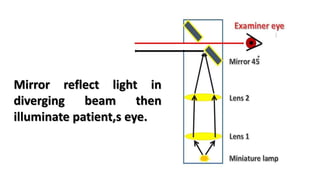

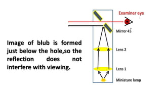
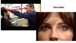
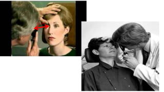
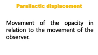


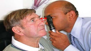

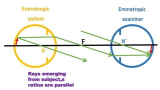

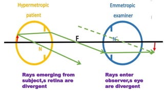
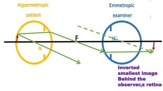
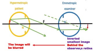
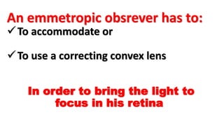
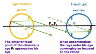
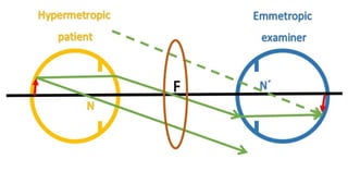


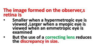


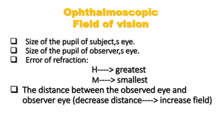


The direct ophthalmoscope allows an examiner to perform a preliminary examination of the internal eye including the retina and optic disc. It works by focusing light through a 45 degree mirror into the patient's eye. The reflected light from the retina passes back through the mirror and forms an erect, magnified image that the examiner can view. The size and clarity of the image provides information about the patient's refractive error and can detect conditions like cataracts, retinal detachments, or tumors. By observing how an opacity moves with the patient's eye movements, its location relative to the pupil can be determined.


