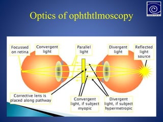
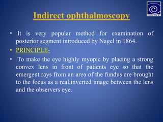
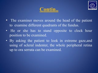
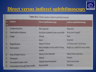
Direct ophthalmoscopy involves examining the retina using an ophthalmoscope held close to the patient's eye, providing a magnified inverted image. Indirect ophthalmoscopy uses a condensing lens placed near the eye to render it highly myopic, producing an upright magnified image seen by the examiner. Both methods allow assessment of the retina but indirect provides a wider field of view and is better for opaque media or uncooperative patients. The document describes the techniques, advantages, and applications of direct and indirect ophthalmoscopy.
![Optic disc
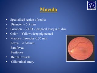
• DISC: LOCATION –nasal to geometric axis
• DIAMETER – 1.5mm [1 disc diameter]
• COLOR – Pale pink
• SHAPE – Circular
• EDGES – Regular
• Termination of all layers except NFL
• CUP: C/D ratio – 0.3 to 0.5](https://image.slidesharecdn.com/presentationretina-231201172157-75bec953/85/Presentation-Retina-pptx-4-320.jpg)


![Hypothalamus short notes on location, function and disorders by Dr. Neha [PT]...](https://cdn.slidesharecdn.com/ss_thumbnails/hypothalamusbydr-260124142231-2b48143d-thumbnail.jpg?width=640&height=640&fit=bounds)


