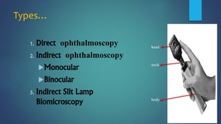
1. Direct ophthalmoscopy allows for a magnified but erect virtual image of the fundus using a light source and lenses.
2. It provides the highest magnification of fundus examination techniques but has a limited field of view and is monocular.
3. The examiner holds the ophthalmoscope and looks through the lens while adjusting focus to examine the different structures of the eye from the anterior segment to the fundus.


![CASE_PRESENTATION_ON_subdural_hematoma(SDH)[1 FINAL PPT]-1.pptx](https://cdn.slidesharecdn.com/ss_thumbnails/casepresentationonsubduralhematomasdh1finalppt-1-260129172522-d405d375-thumbnail.jpg?width=640&height=640&fit=bounds)


