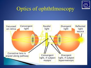
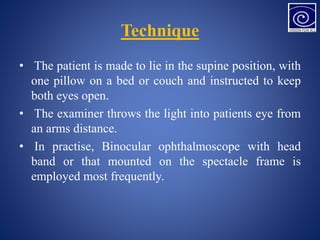
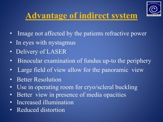
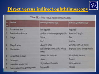
Direct ophthalmoscopy involves examining the retina using an ophthalmoscope held close to the patient's eye, providing a magnified inverted image. Indirect ophthalmoscopy uses a condensing lens placed near the eye to form an erect magnified image, allowing a wider field of view but is more difficult to perform. The document describes the techniques, advantages, and disadvantages of direct and indirect ophthalmoscopy for examining the interior of the eye.


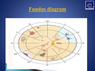
![Optic disc
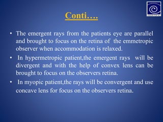
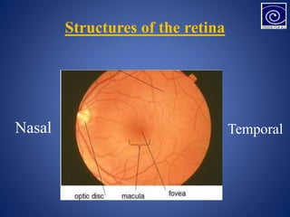
• DISC: LOCATION –nasal to geometric axis
• DIAMETER – 1.5mm [1 disc diameter]
• COLOR – Pale pink
• SHAPE – Circular
• EDGES – Regular
• Termination of all layers except NFL
• CUP: C/D ratio – 0.3 to 0.5](https://image.slidesharecdn.com/presentationretina-231201172503-1fd85b5a/85/Presentation-Retina-pptx-4-320.jpg)