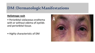
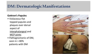
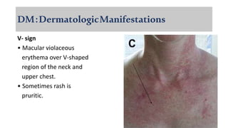
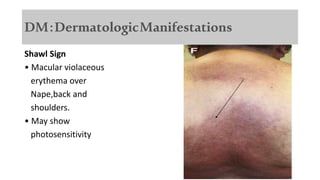
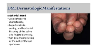
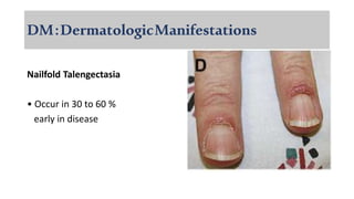
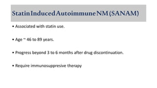
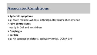
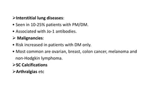
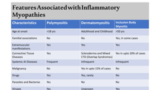
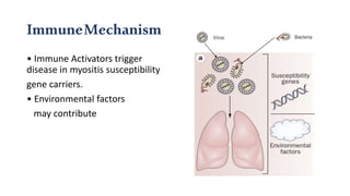
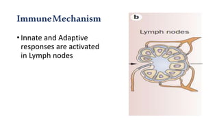
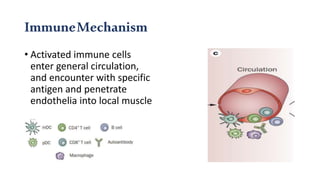
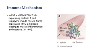
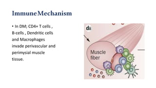
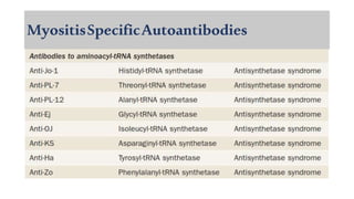
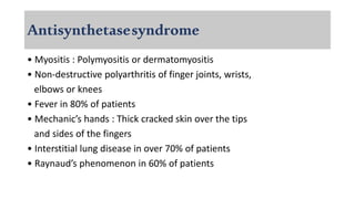
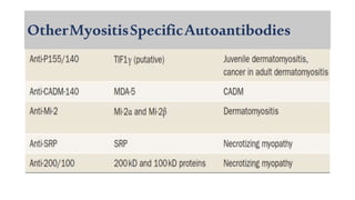
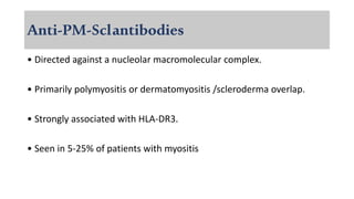
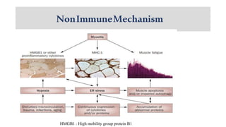
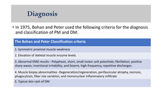
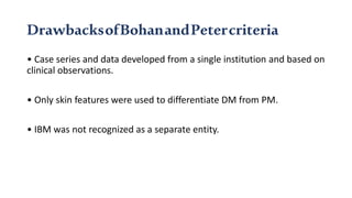
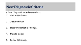
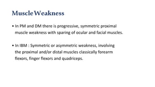
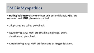
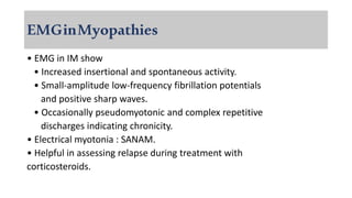
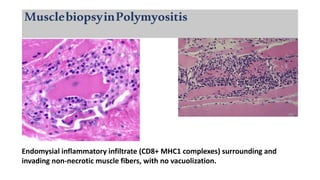
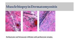
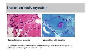
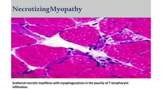
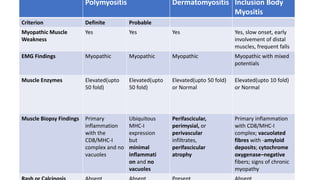
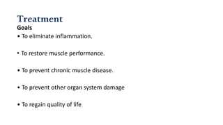
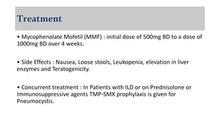
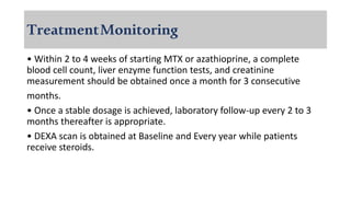
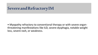
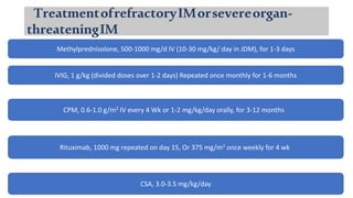
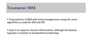
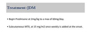
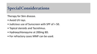
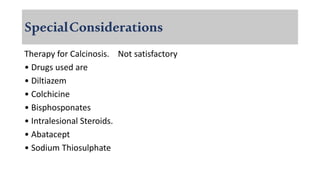
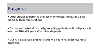
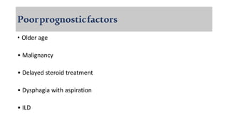
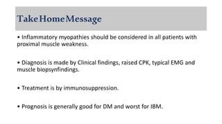
Dermatomyositis is a rare inflammatory myopathy with characteristic skin manifestations and muscular weakness.
Polymyositis is a similar disease without skin lesions.
Amyopathic dermatomyositis: typical cutaneous manifestation of DM without clinical and/or laboratory findings of muscle involvement for at least 6 months after the onset of skin rash.
A brief coverage of all IIM, including major junk of #Polymyositis, #Dermatomyositis #InclusionBodyMyositis and other IIM's.
Includes classification, characteristic features of all and specific features of each of them with diagnosing and approach to management.
NB: This presentation is equipped with animations, which might not work on slideshare
Dermatomyositis (DM) is an inflammatory myopathy characterized by a distinctive rash that often precedes progressive symmetric muscle weakness. The rash may involve areas of the face, eyelids, knuckles, shoulders, and back. Muscle biopsy is required to confirm diagnosis and shows inflammation around blood vessels in the muscle tissue. Treatment involves immunosuppressive drugs like glucocorticoids to improve muscle strength and function. Prognosis is generally good with most patients improving on therapy, though relapses can occur.
Dermatomyositis is a chronic inflammatory disorder of the skin and muscles that is characterized by an autoimmune pathogenesis. It commonly presents with characteristic rashes like Gottron's papules and heliotrope rash as well as proximal muscle weakness. Dermatomyositis can also involve internal organs like the lungs, esophagus and heart. Diagnosis involves assessing clinical features, muscle enzymes, electromyography, muscle/skin biopsies and identifying myositis-specific antibodies. Prognosis depends on the severity and organ involvement, with risks of residual weakness, contractures and death from respiratory or cardiac complications.
This document provides an overview of idiopathic inflammatory myositis, which includes three main types: polymyositis (PM), dermatomyositis (DM), and inclusion body myositis (IBM). PM primarily involves muscle weakness while DM also includes skin involvement. IBM predominantly affects those over 50. Together these conditions represent acquired causes of skeletal muscle weakness. The document discusses their definitions, classifications, epidemiology, clinical features, pathogenesis, associations and treatment considerations.
This document discusses inflammatory myopathies, including types such as polymyositis, dermatomyositis, and immune-mediated necrotizing myopathy. It is characterized by chronic muscle inflammation that is often autoimmune in nature and causes weakness. Diagnosis involves evaluating for proximal muscle weakness, elevated muscle enzymes, EMG findings of myopathy, and muscle biopsy showing inflammation. Skin manifestations may also be present in dermatomyositis. Treatment typically involves glucocorticoids and immunosuppressants like methotrexate or mycophenolate mofetil. The document also presents four clinical cases of patients with suspected inflammatory myopathies and discusses their evaluations, diagnoses, and management considerations.
Polyarteritis nodosa (PAN) is a systemic necrotizing vasculitis that causes inflammation of small and medium arteries. It predominantly affects the arteries of the kidneys, heart, liver, and gastrointestinal tract, causing ischemia and tissue damage. The pathology is characterized by segmental necrotizing inflammation of the arterial walls. PAN is diagnosed based on biopsy of an affected organ showing arteritis or angiography demonstrating aneurysms of small and medium arteries. Treatment involves high-dose glucocorticoids and cyclophosphamide to induce remission, though relapse can occur in 10-20% of cases.
A 60-year-old woman presented with painful, sclerotic hands and fingers due to progressive cutaneous scleroderma. She was started on a compounded topical cream containing ketamine, baclofen, gabapentin, verapamil, and pentoxifylline, which provided significant pain relief and improved sensation within a month. At a 6-month follow up, she had been largely weaned off opioid pain medications. The customized treatment targeted the pathophysiology of the condition and helped manage her debilitating symptoms.
This document discusses multisystem autoimmune disease, also known as systemic lupus erythematosus (SLE). SLE is characterized by the failure to maintain self-tolerance, resulting in an unpredictable and relapsing disease that commonly affects the skin, kidneys, serosal membranes, joints, and heart. Diagnosis requires 4 of several criteria during any observation period, and it is more common in women and black Americans. The pathogenesis involves abnormal self-reactive antibodies against nuclear and cytoplasmic antigens such as Sm/RNP and dsDNA. Complications can include renal disease, skin changes, splenic abnormalities, and Libman-Sacks endocarditis - characterized by nonbacterial vegetations in the heart
Mixed connective tissue disease (MCTD) is a rare autoimmune disorder with features of lupus, scleroderma, rheumatoid arthritis, and polymyositis. It is characterized by high levels of antibodies against ribonucleic proteins. Diagnostic criteria require at least 3 of 5 clinical features plus positive serology. Over time, many patients evolve features meeting criteria for other connective tissue diseases. Common clinical manifestations include Raynaud's phenomenon, arthritis, swollen hands, and lung and heart involvement. Prognosis depends on degree of organ involvement, with 5-year cumulative risks including pulmonary dysfunction in 66% and pericardial disease in 30% of patients.
This document provides an overview of vasculitis in children. It defines vasculitis as an inflammatory destructive process affecting arteries and veins. It discusses the pathogenesis, classification, pathology, clinical features, diagnosis, and treatment of various types of vasculitis in children. The document focuses on defining different types of vasculitis based on the size of blood vessels involved, location of lesions, and pathology. It provides details on the clinical presentation and organ system involvement of conditions like Henoch-Schönlein purpura, Kawasaki disease, polyarteritis nodosa, Wegener's granulomatosis, and Churg-Strauss syndrome. Criteria for diagnosing some common pediatric vasculitides are
This document provides information on autoimmune hepatitis, including:
- It is a chronic hepatitis of unknown etiology that can progress to cirrhosis. It is characterized by the presence of autoimmune antibodies and evidence of hepatitis.
- The two main types are type 1, associated with ANA/SMA positivity, and type 2, associated with LKM1 positivity.
- Treatment involves immunosuppressive drugs like prednisone, either alone or in combination with azathioprine. The goal is to induce and maintain remission.
- Remission is defined as resolution of symptoms and normalization of liver tests and histology. Treatment is then tapered slowly to maintain remission.
This document discusses mixed connective tissue disease (MCTD), including its definition, key diagnostic criteria, common symptoms and organ involvement, treatment approaches, and prognosis. MCTD is characterized by features of lupus, scleroderma, and polymyositis combined with high levels of anti-U1 RNP antibodies. Common symptoms include Raynaud's phenomenon, joint and muscle issues, lung and heart involvement. Treatment focuses on managing symptoms with medications like NSAIDs, corticosteroids, calcium channel blockers, and pulmonary hypertension drugs. Overall mortality is lower than lupus, but progressive pulmonary hypertension can be a major cause of death.
The document discusses amyloidosis of the kidney, including its morphological characteristics, outcomes, and complications. It describes the types and classifications of amyloidosis, the microscopic and macroscopic appearance of amyloid deposits in the kidney, the development stages of renal amyloidosis from latent to nephrotic to uremic, and common complications such as infections. The document contains a detailed plan and sections on the introduction, characteristics, development, complications, and microscopic/macroscopic descriptions of renal amyloidosis.
This document provides information on systemic sclerosis (SSc), a chronic autoimmune disease characterized by thickening and hardening of the skin, and involvement of internal organs. It can present as either limited or diffuse cutaneous forms. Key clinical features include Raynaud's phenomenon, skin thickening and tightening, digital pitting scars, and internal organ involvement such as interstitial lung disease, gastrointestinal issues, and renal crisis. Diagnosis involves clinical examination and the presence of autoantibodies. Differential diagnoses include other conditions presenting with similar skin or vascular changes. Complications can affect multiple organ systems and are a leading cause of mortality.
Christopher Columbus may have suffered from and died of Reiter's arthritis. Reiter's arthritis is a painful inflammatory arthritis that develops after certain bacterial or viral infections, often in the genitourinary or gastrointestinal tracts. Symptoms include joint pain and swelling, eye inflammation, and genital lesions. Treatment focuses on treating underlying infections, reducing pain and inflammation, and managing joint symptoms.
Waterhouse–Friderichsen syndrome (WFS) is defined as adrenal gland failure due to bleeding into the adrenal glands, most commonly caused by the bacterial infection meningococcus. It typically occurs in infants and children under 10 years old. Clinically, it presents with a sudden high fever, rash, shock, and disseminated intravascular coagulation. The adrenal glands hemorrhage and lead to adrenal insufficiency. Treatment involves antibiotics, adrenal support with hydrocortisone, and managing shock. Prevention includes routine meningococcal vaccination in certain groups.
This document provides a review of renal amyloidosis. It begins by defining amyloidosis as a group of diseases caused by the misfolding and accumulation of various proteins. 27 human proteins are known to cause amyloidosis. The kidney is a common site of deposition for several types of amyloidosis. The document reviews the pathogenesis of amyloidosis, determinants of renal deposition, how it causes renal disease, classification, epidemiology including statistics from India, pathology findings including staining techniques, and methods to determine the type of amyloidosis involved.
This document discusses inflammatory myositis, including dermatomyositis (DM), polymyositis (PM), and inclusion body myositis. It is a heterogeneous group of autoimmune disorders that predominantly affect skeletal muscles, resulting in muscle inflammation and weakness. The most common forms are DM, PM, and immune-mediated necrotizing myopathy. Treatment involves systemic glucocorticoids. Refractory cases may require steroid-sparing agents like azathioprine, methotrexate, or rituximab. There is an increased risk of associated malignancy that warrants cancer screening.
This document discusses various pathologies of the kidney including congenital abnormalities, cystic diseases, glomerular diseases, tubular diseases, vascular diseases, and causes of obstruction. It provides descriptions of diseases such as polycystic kidney disease, glomerulonephritis, pyelonephritis, nephrosclerosis, and renal artery stenosis. Diagrams of kidney anatomy and histopathological images are also included.
This document discusses various skin manifestations that are associated with diabetes mellitus. It categorizes the dermatological lesions into those that are associated with but not specific to diabetes, skin alterations due to diabetic complications, dermatological changes associated with neurovascular complications, and dermatologic complications of diabetes treatment. Common conditions discussed include necrobiosis lipoidica diabeticorum, granuloma annulare, diabetic dermopathy, acanthosis nigricans, infections, and manifestations of diabetic foot complications. Prevention and treatment approaches are provided.
This document provides an overview of nephritic and nephrotic syndrome, describing their pathophysiology and clinical features. Nephritic syndrome is characterized by inflammation of the glomeruli, resulting in hematuria, hypertension, and mild proteinuria. Glomerulonephritis causes include post-streptococcal and rapidly progressive crescentic glomerulonephritis. Nephrotic syndrome is caused by increased glomerular permeability, leading to massive proteinuria, hypoalbuminemia, edema, and hyperlipidemia. Specific causes discussed include minimal change disease, membranous nephropathy, focal segmental glomerulosclerosis, and membranoproliferative glomerulone
IgA nephropathy is characterized by predominant IgA deposition in the glomerular mesangium. It is the most common cause of glomerulonephritis worldwide. Primary IgA nephropathy occurs most commonly in the second and third decades of life and presents with macroscopic hematuria, asymptomatic hematuria with mild proteinuria, or nephrotic syndrome. Treatment involves controlling blood pressure with ACE inhibitors or ARBs, treating any proteinuria or hematuria, and considering corticosteroids for progressive disease. The pathogenesis involves abnormal IgA immune responses leading to pathogenic IgA deposition in the kidney and subsequent glomerular inflammation and injury.
This document discusses idiopathic inflammatory myopathies (IIM), which are rare diseases characterized by symmetrical proximal muscle weakness, elevated skeletal muscle enzymes, and characteristic electromyography and muscle biopsy findings. IIM includes dermatomyositis (DM), polymyositis (PM), necrotizing autoimmune myopathy (NAM), and inclusion body myositis. It most commonly affects women aged 50-59 and can cause difficulty with activities like lifting arms or climbing stairs due to proximal muscle weakness. Investigations may show elevated muscle enzymes or skin rash in DM cases, while management involves immunosuppressive drugs.
Refractory ascites is ascites that cannot be mobilized or recurs early after paracentesis despite medical therapy. It is most commonly caused by cirrhosis of the liver and is characterized by progression of portal hypertension and systemic vasodilation, as well as increased sodium reabsorption in the kidneys. Treatment involves repeated large volume paracentesis with albumin supplementation to prevent circulatory dysfunction, use of diuretics if urinary sodium excretion is over 30 mmol/day, placement of transjugular intrahepatic portosystemic shunts to reduce portal pressure in selected patients, and liver transplantation.
Clinical diagnosis of systemic lupus erythematosus Samar Tharwat
The document discusses the classification criteria for systemic lupus erythematosus (SLE) including the original ACR criteria from 1971 and revised versions, as well as the SLICC criteria. It provides details on the individual criteria such as malar rash, discoid rash, photosensitivity, and the differences between the ACR and SLICC criteria. The document also discusses assessing disease activity using measures like SLEDAI, organ system involvement, damage accumulation over time, and quality of life assessments like the SF-36.
Voluntary activity values are also called Motor unit action potentials (MUAPs).
In PM and DM, EMG shows :
- Short, small, polyphasic MUAPs.
- Increased insertional and spontaneous activity.
In IBM, EMG shows :
- Short, small, polyphasic MUAPs.
- Increased insertional activity.
- Complex repetitive discharges and myotonic discharges may be seen.
EMG is helpful in differentiating myopathic from neuropathic process and also helps in monitoring disease progression and response to treatment.
Dermatomyositis (DM) is an autoimmune disease characterized by inflammation and damage of the skin and muscles. It can affect both children and adults. Pulmonary involvement occurs in 20% of patients and is a major cause of morbidity and mortality. In adults, DM is associated with underlying malignancy in 10-20% of cases. Diagnosis involves examination of the skin and muscles for characteristic findings as well as screening for internal cancers and pulmonary involvement. Treatment involves screening and management of extracutaneous manifestations along with topical and systemic therapies to control skin and muscle inflammation.
1) Idiopathic inflammatory myopathies (IIMs) are a heterogeneous group of autoimmune muscle disorders characterized by chronic muscle weakness and fatigue, and can involve other organs.
2) The main types of IIMs are dermatomyositis, polymyositis, immune-mediated necrotizing myopathy, antisynthetase syndrome, and inclusion body myositis.
3) Diagnosis involves assessing symptoms, muscle enzyme levels, autoantibody testing, electromyography, and muscle biopsy findings. MRI and ultrasound can also help evaluate muscle inflammation and damage.
Exploring Polymyositis and Dermatomyositis: Understanding the Intricacies of ...muhammaduzair780907
Introduction
Polymyositis and Dermatomyositis stand as enigmatic entities within the spectrum of autoimmune diseases, characterized by their unique pathophysiology and clinical manifestations. In this extensive presentation, we embark on a journey to dissect the intricacies of these conditions, unraveling their underlying mechanisms, exploring diagnostic modalities, delineating treatment approaches, and delving into the lived experiences of individuals affected by these disorders.
Understanding Polymyositis and Dermatomyositis
Polymyositis and Dermatomyositis represent autoimmune disorders primarily affecting skeletal muscles and skin, characterized by inflammation and tissue damage. While both conditions share certain similarities, they also exhibit distinct features.
Etiology and Pathogenesis
The exact etiology of Polymyositis and Dermatomyositis remains elusive, but a combination of genetic predisposition, environmental triggers, and immune dysregulation is thought to play a role. In Polymyositis, the immune system targets the muscle fibers, leading to inflammation and muscle weakness. Dermatomyositis, on the other hand, involves not only muscle inflammation but also skin involvement, presenting with characteristic skin rashes.
Clinical Manifestations
The clinical presentation of Polymyositis and Dermatomyositis varies widely among individuals. Common symptoms include muscle weakness, fatigue, skin rashes, difficulty swallowing, and joint pain. Dermatomyositis is often associated with characteristic skin findings such as the classic heliotrope rash and Gottron's papules.
Diagnostic Evaluation
Diagnosing Polymyositis and Dermatomyositis can be challenging and often requires a combination of clinical evaluation, laboratory tests, and imaging studies. Serum muscle enzymes such as creatine kinase (CK) and aldolase may be elevated in both conditions. Electromyography (EMG) and muscle biopsy are valuable tools to confirm the diagnosis and assess the extent of muscle involvement.
Treatment Strategies
The management of Polymyositis and Dermatomyositis aims to control inflammation, alleviate symptoms, and prevent complications. Corticosteroids, such as prednisone, are the mainstay of treatment, often combined with immunosuppressive agents such as methotrexate or azathioprine for long-term management. Intravenous immunoglobulin (IVIG) and biologic agents may be considered in refractory cases.
Prognosis and Complications
The prognosis of Polymyositis and Dermatomyositis varies depending on factors such as disease severity, response to treatment, and the presence of complications. Early diagnosis and aggressive management can improve outcomes and reduce the risk of long-term disability. Complications may include muscle atrophy, joint contractures, interstitial lung disease, and malignancy.
Research Advances
Ongoing research efforts continue to expand our understanding of the pathogenesis of Polymyositis and Dermatomyositis and identify novel
Dermatomyositis is a rare chronic autoimmune disorder characterized by inflammation of the skin and muscles. It commonly presents with a reddish rash around the eyelids called heliotrope rash and red papules over the knuckles. Symptoms include proximal muscle weakness, skin changes like rashes and discoloration, and in some cases lung or heart involvement. Diagnosis involves diagnostic criteria of rash and muscle symptoms plus elevated enzymes and muscle biopsy. Treatment focuses on immunosuppressants like corticosteroids and azathioprine to reduce inflammation.
Dermatomyositis is part of a group of rare diseases called the inflammatory myopathies that involve chronic (long-standing) muscle inflammation, muscle weakness, and in some cases, muscle pain. Myopathy is a general term used to describe a number of conditions affecting the muscles.
Meningioma is a type of tumor that arises from the meninges, the membranes surrounding the brain and spinal cord. It is the most common non-cancerous brain tumor, accounting for 20% of all primary brain tumors. The standard treatment is surgical resection if the tumor is symptomatic or growing in size. For inoperable or recurrent tumors, radiation therapy and medical therapies may be options, though medical therapies have limited effectiveness. The prognosis depends on the tumor grade, with lower grade tumors having better outcomes.
Dermatomyositis is an inflammatory myopathy that affects the skin and muscles. It is characterized by progressive proximal muscle weakness, elevated muscle enzymes, abnormal electromyography and muscle biopsy findings. The cause is unknown but genetic, immunological, infectious and environmental factors may play a role. Treatment involves sun protection, immunosuppressants like hydroxychloroquine and methotrexate, and corticosteroids like prednisone to control muscle and skin symptoms.
Dermatomyositis is an inflammatory myopathy that affects the skin and muscles. It is characterized by progressive proximal symmetrical muscle weakness, elevated muscle enzyme levels, abnormal electromyography and muscle biopsy findings. The cause is unknown but may involve genetic, immunological, infectious or environmental factors. Diagnosis involves assessing muscle symptoms, rashes and laboratory tests showing muscle inflammation. Treatment focuses on sun protection, immunosuppressants like hydroxychloroquine and methotrexate, and corticosteroids. Lifestyle measures like diet, activity and sun avoidance are also important.
- Muscle biopsy is the most definitive way to diagnose inclusion body myositis in this case. Inclusion body myositis is characterized by asymmetric weakness, fatigue is uncommon, and dysphagia is common.
- The patient has weakness affecting the shoulders, hips, and finger flexors over 4 months with difficulty swallowing liquids. A muscle biopsy would show the pathognomonic vacuoles and filamentous inclusions.
This document discusses immune-mediated myopathies and provides details on muscle weakness patterns, diagnostic evaluations including autoantibodies, and features of specific conditions like dermatomyositis and antisynthetase syndrome. Muscle weakness in these conditions is typically symmetric, proximal, and involves shoulder and hip girdle muscles. Diagnostic tests include muscle enzymes, electrodiagnostic studies, muscle biopsy, and muscle MRI. Dermatomyositis is characterized by skin rashes and may be associated with cancers. Antisynthetase syndrome can cause myositis and interstitial lung disease and is associated with specific autoantibodies.
Inflammatory myopathies are rare diseases characterized by symmetric proximal muscle weakness. The main types are polymyositis, dermatomyositis, juvenile dermatomyositis, myositis associated with other conditions, and inclusion body myositis. Diagnosis involves muscle biopsy showing inflammation, elevated muscle enzymes, EMG changes, and sometimes autoantibodies. Treatment begins with glucocorticoids and may include immunosuppressants if weak response. Prognosis depends on type, with dermatomyositis having best outlook.
This document provides an overview of the approach to diagnosing and classifying muscle disorders. It discusses taking a clinical history and performing a physical exam to identify symptoms and patterns of weakness. Key investigations include blood tests of enzymes like CK, electromyography and muscle biopsy. Major classifications of myopathies covered are immune-mediated myopathies like dermatomyositis and polymyositis, necrotizing myopathies, inclusion body myositis, and others. Treatment typically involves immunosuppressants like corticosteroids and newer targeted immunotherapies depending on antibody status and subtype.
Mixed connective tissue disease (MCTD) is characterized by features of systemic lupus erythematosus, systemic sclerosis, polymyositis, and rheumatoid arthritis. It is defined by very high levels of anti-U1RNP antibodies. Clinical features include Raynaud's phenomenon, joint and muscle involvement, lung and heart disease, gastrointestinal issues, and kidney disease. Diagnosis requires clinical and serological criteria including high titers of anti-U1RNP antibodies. Treatment depends on organ system involvement but may include analgesics, steroids, immunosuppressants, and calcium channel blockers. Prognosis is variable depending on degree of organ involvement.
Multiple myeloma is a malignant proliferation of plasma cells that commonly affects bone. It causes bone pain, fractures, renal failure, anemia, and susceptibility to infection. The cause is unknown but genetic factors may be involved. Myeloma cells interact with bone marrow to increase osteoclast activity, causing lytic bone lesions. Diagnosis requires bone marrow plasmocytosis, serum/urine monoclonal protein, and end-organ damage. Risk is stratified using beta-2 microglobulin and FISH. Treatment involves induction, consolidation, and maintenance therapies such as bortezomib, lenalidomide, and dexamethasone combinations. Supportive care focuses on complications like hypercalcemia, fractures, and anemia
The document discusses various types of inflammatory myopathies (myositis):
1) Polymyositis causes symmetric proximal muscle weakness and inflammation within muscle fibers. Symptoms include fatigue and a skin rash.
2) Dermatomyositis causes similar muscle symptoms but also features a characteristic rash on the skin. It can also involve other organs.
3) Inclusion body myositis typically causes asymmetric weakness of wrist, finger, and thigh muscles. It involves degeneration in addition to immune dysfunction.
4) Overlap myositis combines myopathy with connective tissue diseases like lupus. Diagnosis involves blood tests, muscle enzyme levels, muscle biopsy and MRI or EMG. Treatment
Skin cancers or cutaneous malignancies including Basal cell carcinoma, Squamous cell carcinoma and Melanoma and with a brief introduction of skin as an organ itself.
This document provides an outline and overview of multiple myeloma. It begins with definitions of multiple myeloma and the monoclonal proteins involved. It then discusses the epidemiology, etiology, pathogenesis, clinical manifestations including bone disease, renal failure, neurologic symptoms, and more. It covers the workup, diagnosis involving bone marrow plasmacytosis and monoclonal proteins. It concludes with sections on prognosis, standard therapeutic agents, treatment of relapsed multiple myeloma, supportive care, monitoring response, and reference materials.
This document provides information on occupational lung diseases. It begins with a brief history of occupational lung diseases dating back to Roman times. It then defines occupational lung diseases and pneumoconiosis. The document classifies and describes various types of pneumoconiosis including anthracosis, silicosis, asbestosis, and berylliosis. For each type, it discusses associated occupations, pathogenesis, clinical features, diagnosis, and management. The document provides detailed information on the pathogenesis, clinical presentation, radiographic findings, and complications of major occupational lung diseases like silicosis and asbestosis.
Does Over-Masturbation Contribute to Chronic Prostatitis.pptxwalterHu5
In some case, your chronic prostatitis may be related to over-masturbation. Generally, natural medicine Diuretic and Anti-inflammatory Pill can help mee get a cure.
- Video recording of this lecture in English language: https://youtu.be/kqbnxVAZs-0
- Video recording of this lecture in Arabic language: https://youtu.be/SINlygW1Mpc
- Link to download the book free: https://nephrotube.blogspot.com/p/nephrotube-nephrology-books.html
- Link to NephroTube website: www.NephroTube.com
- Link to NephroTube social media accounts: https://nephrotube.blogspot.com/p/join-nephrotube-on-social-media.html
Promoting Wellbeing - Applied Social Psychology - Psychology SuperNotesPsychoTech Services
A proprietary approach developed by bringing together the best of learning theories from Psychology, design principles from the world of visualization, and pedagogical methods from over a decade of training experience, that enables you to: Learn better, faster!
These lecture slides, by Dr Sidra Arshad, offer a quick overview of the physiological basis of a normal electrocardiogram.
Learning objectives:
1. Define an electrocardiogram (ECG) and electrocardiography
2. Describe how dipoles generated by the heart produce the waveforms of the ECG
3. Describe the components of a normal electrocardiogram of a typical bipolar lead (limb II)
4. Differentiate between intervals and segments
5. Enlist some common indications for obtaining an ECG
6. Describe the flow of current around the heart during the cardiac cycle
7. Discuss the placement and polarity of the leads of electrocardiograph
8. Describe the normal electrocardiograms recorded from the limb leads and explain the physiological basis of the different records that are obtained
9. Define mean electrical vector (axis) of the heart and give the normal range
10. Define the mean QRS vector
11. Describe the axes of leads (hexagonal reference system)
12. Comprehend the vectorial analysis of the normal ECG
13. Determine the mean electrical axis of the ventricular QRS and appreciate the mean axis deviation
14. Explain the concepts of current of injury, J point, and their significance
Study Resources:
1. Chapter 11, Guyton and Hall Textbook of Medical Physiology, 14th edition
2. Chapter 9, Human Physiology - From Cells to Systems, Lauralee Sherwood, 9th edition
3. Chapter 29, Ganong’s Review of Medical Physiology, 26th edition
4. Electrocardiogram, StatPearls - https://www.ncbi.nlm.nih.gov/books/NBK549803/
5. ECG in Medical Practice by ABM Abdullah, 4th edition
6. Chapter 3, Cardiology Explained, https://www.ncbi.nlm.nih.gov/books/NBK2214/
7. ECG Basics, http://www.nataliescasebook.com/tag/e-c-g-basics
Here is the updated list of Top Best Ayurvedic medicine for Gas and Indigestion and those are Gas-O-Go Syp for Dyspepsia | Lavizyme Syrup for Acidity | Yumzyme Hepatoprotective Capsules etc
TEST BANK For Community Health Nursing A Canadian Perspective, 5th Edition by...Donc Test
TEST BANK For Community Health Nursing A Canadian Perspective, 5th Edition by Stamler, Verified Chapters 1 - 33, Complete Newest Version Community Health Nursing A Canadian Perspective, 5th Edition by Stamler, Verified Chapters 1 - 33, Complete Newest Version Community Health Nursing A Canadian Perspective, 5th Edition by Stamler Community Health Nursing A Canadian Perspective, 5th Edition TEST BANK by Stamler Test Bank For Community Health Nursing A Canadian Perspective, 5th Edition Pdf Chapters Download Test Bank For Community Health Nursing A Canadian Perspective, 5th Edition Pdf Download Stuvia Test Bank For Community Health Nursing A Canadian Perspective, 5th Edition Study Guide Test Bank For Community Health Nursing A Canadian Perspective, 5th Edition Ebook Download Stuvia Test Bank For Community Health Nursing A Canadian Perspective, 5th Edition Questions and Answers Quizlet Test Bank For Community Health Nursing A Canadian Perspective, 5th Edition Studocu Test Bank For Community Health Nursing A Canadian Perspective, 5th Edition Quizlet Test Bank For Community Health Nursing A Canadian Perspective, 5th Edition Stuvia Community Health Nursing A Canadian Perspective, 5th Edition Pdf Chapters Download Community Health Nursing A Canadian Perspective, 5th Edition Pdf Download Course Hero Community Health Nursing A Canadian Perspective, 5th Edition Answers Quizlet Community Health Nursing A Canadian Perspective, 5th Edition Ebook Download Course hero Community Health Nursing A Canadian Perspective, 5th Edition Questions and Answers Community Health Nursing A Canadian Perspective, 5th Edition Studocu Community Health Nursing A Canadian Perspective, 5th Edition Quizlet Community Health Nursing A Canadian Perspective, 5th Edition Stuvia Community Health Nursing A Canadian Perspective, 5th Edition Test Bank Pdf Chapters Download Community Health Nursing A Canadian Perspective, 5th Edition Test Bank Pdf Download Stuvia Community Health Nursing A Canadian Perspective, 5th Edition Test Bank Study Guide Questions and Answers Community Health Nursing A Canadian Perspective, 5th Edition Test Bank Ebook Download Stuvia Community Health Nursing A Canadian Perspective, 5th Edition Test Bank Questions Quizlet Community Health Nursing A Canadian Perspective, 5th Edition Test Bank Studocu Community Health Nursing A Canadian Perspective, 5th Edition Test Bank Quizlet Community Health Nursing A Canadian Perspective, 5th Edition Test Bank Stuvia
share - Lions, tigers, AI and health misinformation, oh my!.pptxTina Purnat
• Pitfalls and pivots needed to use AI effectively in public health
• Evidence-based strategies to address health misinformation effectively
• Building trust with communities online and offline
• Equipping health professionals to address questions, concerns and health misinformation
• Assessing risk and mitigating harm from adverse health narratives in communities, health workforce and health system
Osteoporosis - Definition , Evaluation and Management .pdfJim Jacob Roy
Osteoporosis is an increasing cause of morbidity among the elderly.
In this document , a brief outline of osteoporosis is given , including the risk factors of osteoporosis fractures , the indications for testing bone mineral density and the management of osteoporosis
8 Surprising Reasons To Meditate 40 Minutes A Day That Can Change Your Life.pptxHolistified Wellness
We’re talking about Vedic Meditation, a form of meditation that has been around for at least 5,000 years. Back then, the people who lived in the Indus Valley, now known as India and Pakistan, practised meditation as a fundamental part of daily life. This knowledge that has given us yoga and Ayurveda, was known as Veda, hence the name Vedic. And though there are some written records, the practice has been passed down verbally from generation to generation.
3. Introduction
• Inflammatory Myopathies are sporadic disorders representing the
largest group of acquired and potentially treatable causes of skeletal
muscle weakness.
• Annual incidence ~ 1 in 100,000
• Women > Men , Polymyositis (PM) and Dermatomyositis (DM)
• Men > Women , Inclusion Body Myositis (IBM)
4. Age at onset for Inflammatory myopathies is different.
• PM > 18 years.
• DM affects both Juvenile population and Adulthood.
• IBM affects persons aged >50 years.
5. History
• In 1888 the first American biopsy documented polymyositis in ruling
out Trichinella.
• Heliotrope rash was first described in 1875 in France.
• In 1930 Gottron’s reported skin lesions .
• In 1967 the pathology of IBM was described.
6. Classification
• There is no internationally accepted classification system for
Inflammatory myopathies.
• For discussion purpose IM’s can be classified as :-
Polymyositis.
Dermatomyositis and Juvenile Dermatomyositis.
Inclusion Body Myositis.
Autoimmune Necrotising Myopathies.
Myositis associated with Collagen Vascular disorder.
Myositis associated with malignancy and Juvenile Dermatomyositis.
7. ClinicalFeatures
Polymyositis
• A rare, subacute inflammatory myopathy affecting adults and rarely
children.
• Progressive, symmetric proximal muscle weakness with sparing of
ocular and facial muscles.
• Pharyngeal and Neck Flexors, also involved causing dysphagia and
Head drop.
8. • Associated with Myalgia and Muscle tenderness in some cases
• In Advanced and Acute cases Respiratory muscles may be affected.
• Severe weakness if untreated leads to muscle wasting.
• Sensations and DTR’s remain intact.
14. DM:DermatologicManifestations
Mechanic’s Hand
• Also considered
characteristic.
• hyperkeratosis,
scaling, and horizontal
fissuring of the palms
and fingers bilaterally.
• Can be a manifestation
of the Antisynthetase
syndrome.
16. AmyopathicandHypo-myopathic
Dermatomyositis
• Amyopathic DM : present with typical dermatological manifestations
of DM, with normal muscle strength, enzyme levels and EMG findings.
• Hypo-myopathic DM : Dermatological manifestations of DM with no
muscle weakness with mild elevation in Enzymes and Abnormal EMG &
Biopsy.
17. JuvenileDermatomyositis
Presents with clinical features similar to dermatomyositis with
following differences
Age of onset ~ 7 years
Less incidences of Malignancy and ILD.
Increased risk for Vasculopathy, Lipodystrophy and calcinosis.
Associations: SOJIA ; Macrophage activation syndrome
Less mortality.
18. InclusionBodyMyositis
• Symmetric or asymmetric weakness.
• Insidious onset.
• After 50 years, Male to female ratio of 3:1.
• Proximal and/or distal muscle involvement.
• Classically forearm flexors, finger flexors and Quadriceps.
• Facial and Pharyngeal muscles are more commonly involved than PM/DM.
19. Immune-MediatedNecrotizingMyopathy
• Subacute progressive proximal muscle weakness without a rash.
• Female to Male ratio 2:1.
• Generally develops more rapidly than with PM, and is markedly
severe.
• Associated myalgia and dysphagia can be seen.
• Associated with Paraneoplastic syndrome and Statin use.
20. ParaneoplasticNM
• Rare, rapid progression, association with Adenocarcinoma.
• Age > 40 years.
• Presentation in Fall or Winter.
• Antibodies against signal recognition particles (SRP) seen.
• Coexisting cardiomyopathy and ILD maybe present.
• Muscle Biopsy : Necrotic fibres infiltrated by macrophages and rare
T- cells
22. AssociatedConditions
Systemic symptoms
e.g. fever, malaise ,wt. loss, arthralgia, Raynaud’s phenomenon
Joint contractures
mostly in DM and in children
Dysphagia
Cardiac
e.g. AV conduction defects, tachyarrythmias, DCMP, CHF
23. Interstitial lung diseases:
• Seen in 10-25% patients with PM/DM.
• Associated with Jo-1 antibodies.
Malignancies:
• Risk increased in patients with DM only.
• Most common are ovarian, breast, colon cancer, melanoma and
non-Hodgkin lymphoma.
SC Calcifications
Arthralgias etc
25. Characteristics Polymyositis Dermatomyositis Inclusion Body
Myositis
Age at onset >18 yrs Adulthood and Childhood >50 yrs
Familial associations No No Yes, in some cases
Extramuscular
manifestations
Yes Yes Yes
Connective Tissue
Diseases
Yes Scleroderma and Mixed
CTD (Overlap Syndromes)
Yes in upto 20% of cases
Systemic AI Diseases Frequent Infrequent Infrequent
Malignancy No Yes in upto 15% of cases No
Drugs Yes Yes, rarely No
Parasites and Bacterias Yes No No
Viruses Yes Unproven Yes
FeaturesAssociatedwithInflammatory
Myopathies
26. Pathogenesis
• Both immune and non immune mechanism are
involved in pathogenesis.
• Immune mechanism involve T-cell, B-cells,Dendritic cells,
macrophages, cytokines and Antibodies.
• Non immune mechanisms involve endoplasmic reticulum stress ,
hypoxia and Autophagy.
29. ImmuneMechanism
• Activated immune cells
enter general circulation,
and encounter with specific
antigen and penetrate
endothelia into local muscle
30. ImmuneMechanism
• In PM and IBM CD8+ Tcells
expressing perforin 1 and
Granzyme invade muscle fibres
expressing MHC 1 molecule
leading to muscle inflammation
and necrosis ( In IBM).
31. ImmuneMechanism
• In DM; CD4+ T cells ,
B-cells , Dendritic cells
and Macrophages
invade perivascular and
perimysial muscle
tissue.
34. Antisynthetasesyndrome
• Myositis : Polymyositis or dermatomyositis
• Non-destructive polyarthritis of finger joints, wrists,
elbows or knees
• Fever in 80% of patients
• Mechanic’s hands : Thick cracked skin over the tips
and sides of the fingers
• Interstitial lung disease in over 70% of patients
• Raynaud’s phenomenon in 60% of patients
36. Anti-PM-Sclantibodies
• Directed against a nucleolar macromolecular complex.
• Primarily polymyositis or dermatomyositis /scleroderma overlap.
• Strongly associated with HLA-DR3.
• Seen in 5-25% of patients with myositis
38. Diagnosis
• In 1975, Bohan and Peter used the following criteria for the diagnosis
and classification of PM and DM.
The Bohan and Peter Classification criteria
1. Symmetric proximal muscle weakness
2. Elevation of skeletal muscle enzyme levels.
3. Abnormal EMG results - Polyphasic, short, small motor unit potentials; fibrillation; positive
sharp waves; insertional irritability; and bizarre, high-frequency, repetitive discharges.
4. Muscle biopsy abnormalities -Degeneration/regeneration, perifascicular atrophy, necrosis,
phagocytosis, fiber size variation, and mononuclear inflammatory infiltrate
5. Typical skin rash of DM
39. DrawbacksofBohanandPetercriteria
• Case series and data developed from a single institution and based on
clinical observations.
• Only skin features were used to differentiate DM from PM.
• IBM was not recognized as a separate entity.
41. MuscleWeakness
• In PM and DM there is progressive, symmetric proximal
muscle weakness with sparing of ocular and facial muscles.
• In IBM : Symmetric or asymmetric weakness, involving
the proximal and/or distal muscles classically forearm
flexors, finger flexors and quadriceps.
42. MuscleEnzymes
• In PM, Creatine Kinase is always elevated upto 5 – 50 times the ULN.
• In DM, CK is elevated upto 50 times the ULN, in 90% of patients.
While in others CK levels may be normal.
• In IBM, CK levels are elevated upto 10 times of UNL or
may be Normal.
• Necrotising Myopathy, CK is increased 10 times or more
43. EMGinMyopathies
• In acute myopathies EMG must be conducted about
3 weeks from the onset of symptoms to ensure good sensitivity.
• EMG of the muscle is done in two situations : at rest and at
voluntary activity.
• The Resting values also called spontaneous activity (not seen in
normal muscles) are of three types.
1.Fibrillations and Positive waves
2.High frequency discharges
3.Myotonic Discharges
45. EMGinMyopathies
• High frequency discharges (Pseudomyotonic or Complex repetitive
discharges)
Rhythmic electric
potential with abrupt
beginning and end
typical of long standing
denervation.
47. EMGinMyopathies
• During Voluntary activity motor unit potentials (MUP) ie. are
recorded and MUP phase are studied
• ≥5, phases are called polyphasic.
• Acute myopathy: MUP are small in amplitude, short
duration and polyphasic.
• Chronic myopathy: MUP are large and of longer duration.
48. EMGinMyopathies
• EMG in IM show
• Increased insertional and spontaneous activity.
• Small-amplitude low-frequency fibrillation potentials
and positive sharp waves.
• Occasionally pseudomyotonic and complex repetitive
discharges indicating chronicity.
• Electrical myotonia : SANAM.
• Helpful in assessing relapse during treatment with
corticosteroids.
51. Inclusionbodymyositis
Basophilic rimmed vacuoles Vacuole filled with granules
Vacuolated muscle fibres infiltrated with CD8/MHC-1complexes. Beta-amyloid deposits and
cytochrome oxidase negative fibres may be seen.
53. Polymyositis Dermatomyositis Inclusion Body
Myositis
Criterion Definite Probable
Myopathic Muscle
Weakness
Yes Yes Yes Yes, slow onset, early
involvement of distal
muscles, frequent falls
EMG Findings Myopathic Myopathic Myopathic Myopathic with mixed
potentials
Muscle Enzymes Elevated(upto
50 fold)
Elevated(upto
50 fold)
Elevated(upto 50 fold)
or Normal
Elevated(upto 10 fold)
or Normal
Muscle Biopsy Findings Primary
inflammation
with the
CD8/MHC-I
complex and no
vacuoles
Ubiquitous
MHC-I
expression
but
minimal
inflammati
on and no
vacuoles
Perifascicular,
perimysial, or
perivascular
infiltrates,
perifascicular
atrophy
Primary inflammation
with CD8/MHC-I
complex; vacuolated
fibres with -amyloid
deposits; cytochrome
oxygenase–negative
fibers; signs of chronic
myopathy
54. OtherInvestigations
• MRI-T1-weighted, T2-weighted, and sequences
using fat suppression techniques and short “tau” inversion recovery
(aka STIR), provides useful information to:
– diagnose myositis
– monitor treatment response,
– and identify a muscle site for biopsy.
• Muscle biopsy guided by MRI contains significantly more
inflammatory cells than the biopsy taken from MRI non-affected sites.
55. STIR imaging show
fibrosis or diffuse or
patchy signal symmetric
increase in the proximal
muscles and
intramuscular fascia
indicative of muscle
oedema due to
inflammation.
56. Other Investigations
Malignancy work up (for DM and PM)
• Chest , Abdomen and Pelvic CT scans
• Mammogram, Breast and Pelvic examination in women
• Colonoscopy in patients – Age >50 years or in those with
GI symptoms.
57. Other Investigations
In patients with IM:
• Chest X-ray, PFT and HRCT – especially in Jo1 positive
patients to rule out ILD.
• ECG- for myocardial involvement
• Echocardiogram- in patients with symptoms and signs of
heart failure.
• Video fluoroscopy- to objectively assess swallowing in
patients with dysphagia.
58. Treatment
Goals
• To eliminate inflammation.
• To restore muscle performance.
• To prevent chronic muscle disease.
• To prevent other organ system damage
• To regain quality of life
61. InitialTreatment
Approach Prednisone, 0.5-1 mg/kg/day.
for 2-4 wk
Concurrent with:
MTX, 15 mg/wk, to
Target dose of 25
mg/wk
Azathioprine, 2
mg/kgIBW
Twice daily
Mycophenolate mofetil,
500
mg
Twice daily. Increase by
500
mg/wk
until 1000 mg twice daily
62. InitialTreatment
Approach Prednisone, 0.5-1 mg/kg/day.
for 2-4 wk
Concurrent with:
MTX, 15 mg/wk, to
Target dose of 25
mg/wk
Azathioprine, 2
mg/kgIBW
Twice daily
Mycophenolate mofetil,
500
mg
Twice daily. Increase by
500
mg/wk
until 1000 mg twice daily
Taper prednisone every
2 wk
until completed:
60mg/d
40mg/d
30mg/d
25mg/d
20mg/d
17.5mg/d
15mg/d
12.5mg/d
10mg/d
7.5mg/d
5mg/d
2.5mg/d
63. Treatment
Initiate treatment with high dose corticosteroids and steroid sparing
agents.
• Prednisolone – Dose 0.5 to 1 mg/kg/day for 2- 4 weeks and then
taper off.
• Duration : 3 to 6 months.
• If no objective benefits after 3 months of high dose therapy switch
over to next immunosuppresive drug.
• Long term use of steroids may lead to steroid myopathy.
64. Treatment
• Azathioprine : Dose 2 mg/kg/day for 3 months.
• Before starting Azathioprine patient’s thiopurinemethyl- transferase
levels should be checked, otherwise it may lead to myelosuppression.
• Side Effects : Nausea, Loose stools, fever, and Liver toxicity
65. Treatment
• Methotrexate : Initial dose of 15 mg orally once weekly to a target
dose of 25 mg once weekly within 3 to 6 months.
• Folic acid supplementation to be given along.
• Side Effects : BM suppression, Liver toxicity, Pneumonitis.
• Should be used cautiously in patients with ILD
66. Treatment
• Mycophenolate Mofetil (MMF) : initial dose of 500mg BD to a dose of
1000mg BD over 4 weeks.
• Side Effects : Nausea, Loose stools, Leukopenia, elevation in liver
enzymes and Teratogenicity.
• Concurrent treatment : In Patients with ILD or on Prednisolone or
Immunosuppressive agents TMP-SMX prophylaxis is given for
Pneumocystis.
67. TreatmentMonitoring
• Within 2 to 4 weeks of starting MTX or azathioprine, a complete
blood cell count, liver enzyme function tests, and creatinine
measurement should be obtained once a month for 3 consecutive
months.
• Once a stable dosage is achieved, laboratory follow-up every 2 to 3
months thereafter is appropriate.
• DEXA scan is obtained at Baseline and Every year while patients
receive steroids.
68. SevereandRefractoryIM
• Myopathy refractory to conventional therapy or with severe organ-
threatening manifestations like ILD, severe dysphagia, notable weight
loss, severe rash, or weakness.
69. TreatmentofrefractoryIMorsevereorgan-
threateningIM
Methylprednisolone, 500-1000 mg/d IV (10-30 mg/kg/ day in JDM), for 1-3 days
IVIG, 1 g/kg (divided doses over 1-2 days) Repeated once monthly for 1-6 months
CPM, 0.6-1.0 g/m2 IV every 4 Wk or 1-2 mg/kg/day orally, for 3-12 months
Rituximab, 1000 mg repeated on day 15, Or 375 mg/m2 once weekly for 4 wk
CSA, 3.0-3.5 mg/kg/day
70. Treatment:IBM
• Treat patients of IBM with immunosuppression using the same
algorithms as used for DM and PM
• Goal is to suppress muscle inflammation, although the disease
typically is resistant to standardimmunotherapy.
71. Treatment:JDM
• Begin Prednisone at 2mg/kg to a max of 60mg/day.
• Subcutaneous MTX, at 15 mg/m2 once weekly is added at the onset.
72. SpecialConsiderations
Therapy for Skin disease.
• Avoid UV rays.
• Judicious use of Sunscreen with SPF of > 50.
• Topical steroids and Tacrolimus.
• Hydroxychloroquine at 200mg BD.
• For refractory cases MMF can be used.
73. SpecialConsiderations
Therapy for Calcinosis. Not satisfactory
• Drugs used are
• Diltiazem
• Colchicine
• Bisphosponates
• Intralesional Steroids.
• Abatacept
• Sodium Thiosulphate
74. Prognosis
• Older studies (before the availability of steroids) revealed a 50%
mortality from complications.
• Current estimates of mortality, excluding patients with malignancy, is
less than 10% at 5 years after initial diagnosis.
• DM has a favorable prognosis among all, IBM has least favorable
prognosis.
76. TakeHomeMessage
• Inflammatory myopathies should be considered in all patients with
proximal muscle weakness.
• Diagnosis is made by Clinical findings, raised CPK, typical EMG and
muscle biopsynfindings.
• Treatment is by immunosuppression.
• Prognosis is generally good for DM and worst for IBM.