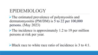
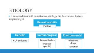
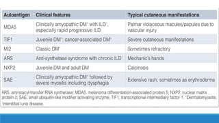
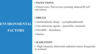
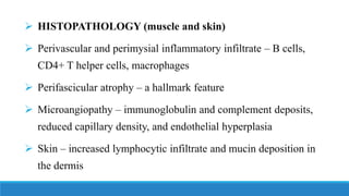
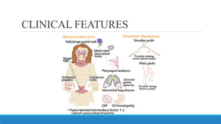
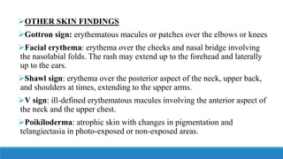
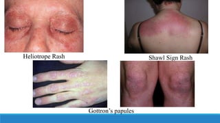
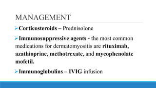
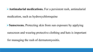
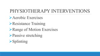
DERMATOMYOSITIS
PRESENTED BY:
PURVA PAI
MPT 2ND YEAR
INTRODUCTION
➢Dermatomyositis is a rare acquired immune-
mediated muscle disease characterized by muscle
weakness and skin rash.
➢It is classified as one of the idiopathic inflammatory
myopathies (IIM) with some clinical differences and
findings.
➢Along with skin findings and symmetric proximal
skeletal muscle weakness, it affects other organ
systems such as pulmonary, cardiovascular, and
gastrointestinal systems.
➢Clinically amyopathic dermatomyositis (CADM) is
a condition in which patients have characteristic
cutaneous findings of dermatomyositis but do not
have muscle weakness.
➢The condition can affect adults and children.
➢In children, it appears between 5 and 15 years of
age.
➢In adults, it occurs in the late 40s to early 60s.
➢Dermatomyositis affects more females than males
with an incidence ratio of 2 to 3:1
EPIDEMIOLOGY
➢The estimated prevalence of polymyositis and
dermatomyositis (PM/DM) is 5 to 22 per 100,000
persons. (May 2023)
➢The incidence is approximately 1.2 to 19 per million
persons at risk per year.
➢Black race to white race ratio of incidence is 3 to 4:1.
ETIOLOGY
➢It is a condition with an unknown etiology but has various factors
implicating it.
Dermatomyositis
Factors
Genetic
HLA antigens
Immunological
Autoantibodies
(myositis-
specific)
Environmental
Infections,
drugs,
radiation
GENETIC
FACTORS
➢Patients with particular HLA haplotypes are at higher
risk.
➢HLA-A*68 North American Whites
➢HLA-DRB1*0301 African Americans
➢HLA-DQA1*0104
➢HLA-DRB1*07
➢DQA1*05
➢DQB1*02
➢DRB1*03-DQA1*05-DQB1*02 haplotypes are
strongly associated with the development of interstitial
lung disease.
→
→
→
→
Han Chinese
UK
IMMUNOLOGICAL
FACTORS
➢Myositis-specific autoantibodies
➢Antisynthetase (Jo-1) – immunological
marker
➢anti-Mi-2
➢anti-MDA-5
➢anti-TIF-1-g
➢anti-NXP-2
➢anti-SAE
➢ Cancer-related and cutaneous skin lesion-
associated antibodies
ENVIRONMENTAL
FACTORS
➢INFECTIONS
➢Enterovirus, Parvovirus (causing induced B cell
activation)
➢DRUGS
➢Antineoplastic drugs – cyclophosphamide
➢Ant-infectious agents – penicillin, isoniazid
➢NSAIDS – diclofenac
➢Statins
➢RADIATION
➢ High-intensity ultraviolet radiation (more frequently
in women)
PATHOPHYSIOLOGY
➢Humoral-mediated attack directed against the muscle capillaries and the endothelium
of arterioles.
➢Activation of completer factor-3 (C3), which forms C3b and C4b, followed by the
formation of the neoantigen C3bNEO and the C5b-C9 membrane attack complex
(MAC)
➢The membrane attack complex deposits on vascular walls and causes inflammation.
➢Hypoxic injury to the muscle fibers ensues, leading to atrophy of muscle fibers,
particularly the fibers at the periphery that are the most remote and away from the
vascular supply.
➢Over time, the capillary density reduces, and muscle fibers start to undergo necrosis
and degeneration.
➢ HISTOPATHOLOGY (muscle and skin)
➢ Perivascular and perimysial inflammatory infiltrate – B cells,
CD4+ T helper cells, macrophages
➢ Perifascicular atrophy – a hallmark feature
➢ Microangiopathy – immunoglobulin and complement deposits,
reduced capillary density, and endothelial hyperplasia
➢ Skin – increased lymphocytic infiltrate and mucin deposition in
the dermis
CLINICAL FEATURES
HISTORYAND PHYSICAL
EXAMINATION
➢Insidious or acute onset of the diseased with waxing and
waning course
➢History of Typical Muscular and Cutaneous Signs and
Symptoms
➢Exclude other causes of muscle weakness
➢Detailed review of systems
➢Possible underlying malignancy
➢ MUSCULAR
➢ Gradually progressive symmetric proximal muscle weakness and
difficulty in carrying out activities such as climbing stairs, getting up from
a seated position, lifting objects, combing hair, and raising their head from
a pillow.
➢ Reduced muscle strength in deltoids, hip flexors, and neck flexors.
➢ Mild muscle tenderness
➢ Depressed Deep Tendon Reflexes and Muscle Atrophy in severe cases
➢ Distal muscle strength preserved
➢ CUTANEOUS
➢ Onset may precede or coincide with muscular symptoms
➢ Skin rashes, photosensitivity, changes in pigmentation, and pruritis
➢ Alopecia and nail changes are also seen
➢ Gottron papules - dorsal metacarpophalangeal and interphalangeal joints
may show the presence of overlying erythematous or violaceous papules
with or without scaling or ulceration.
➢ Heliotrope rash - characteristic skin finding of dermatomyositis and
presents with a violaceous, or an erythematous rash affecting the upper
eyelids with or without periorbital edema. This finding may not be
apparent in patients with dark skin patients.
➢OTHER SKIN FINDINGS
➢Gottron sign: erythematous macules or patches over the elbows or knees
➢Facial erythema: erythema over the cheeks and nasal bridge involving
the nasolabial folds. The rash may extend up to the forehead and laterally
up to the ears.
➢Shawl sign: erythema over the posterior aspect of the neck, upper back,
and shoulders at times, extending to the upper arms.
➢V sign: ill-defined erythematous macules involving the anterior aspect of
the neck and the upper chest.
➢Poikiloderma: atrophic skin with changes in pigmentation and
telangiectasia in photo-exposed or non-exposed areas.
➢Holster sign: poikiloderma involving the lateral aspects of the
thighs.
➢Mechanic's hands: hyperkeratotic, cracked horizontal lines on
the palmar and lateral aspects of the fingers.
➢Scalp involvement: diffuse poikiloderma, with scaling and
pruritis.
➢Calcinosis cutis: calcium deposits in the skin
Heliotrope Rash Shawl Sign Rash
Gottron’s papules
➢ JOINTS
➢ Polyarthritis or arthralgia of the small joints of the hands, may present
with joint pain or swelling.
➢ RESPIRATORY
➢ Exertional dyspnea, exercise intolerance, non-productive cough due to
underlying Interstitial Lung Disease. On Auscultation – B/L dry
crackles
➢ Reduced chest movement due to respiratory muscle weakness
➢ ESOPHAGEAL
➢ Dysphagia – due to weakness of muscles of the oropharynx
and upper esophagus
➢ May have symptoms of gastroesophageal reflux
➢OTHER
➢Raynaud’s phenomenon
➢GI Ulcers
➢Cardiac symptoms
➢Fever
➢Malaise
➢Weight loss
PETER/BOHAN(1975) CRITERIA
FOR DIAGNOSIS
1.Symmetric proximal muscle weakness
2.Elevated serum muscle enzymes
3.Myopathic changes in electromyography (EMG)
4.Characteristic muscle biopsy abnormalities
5.Typical rash of dermatomyositis
INVESTIGATIONS
➢Blood analysis-A blood test will give elevated levels of muscle
enzymes that can indicate muscle damage. It can also detect
autoantibodies associated with different symptoms of dermatomyositis
➢Chest X-ray-can check for signs of the type of lung damage that
sometimes occurs with dermatomyositis.
➢Investigations for Malignancy – CA-125, CA-19-9 screening
➢ Electromyography - Needle EMG, changes in the pattern of
electrical activity can confirm a muscle disease.
➢ MRI - assesses inflammation over a large area of muscle.
➢ Skin or muscle biopsy -A skin sample can help confirm the
diagnosis of dermatomyositis. A muscle biopsy might reveal
inflammation in your muscles or other problems, such as
damage or infection.
MANAGEMENT
➢Corticosteroids – Prednisolone
➢Immunosuppressive agents - the most common
medications for dermatomyositis are rituximab,
azathioprine, methotrexate, and mycophenolate
mofetil.
➢Immunoglobulins – IVIG infusion
➢Antimalarial medications. For a persistent rash, antimalarial
medication, such as hydroxychloroquine
➢Sunscreens. Protecting skin from sun exposure by applying
sunscreen and wearing protective clothing and hats is important
for managing the rash of dermatomyositis.
➢Physical therapy- exercises to help maintain and improve
strength and flexibility
➢Speech therapy-If swallowing muscles are affected, speech
therapy can help learn how to compensate for those changes.
➢Diet Management – chewing and swallowing can become
more difficult in later stages. For easy-to-eat foods and changes
in diet.
PHYSIOTHERAPY INTERVENTIONS
➢Aerobic Exercises
➢Resistance Training
➢Range of Motion Exercises
➢Passive stretching
➢Splinting
THANK
YOU

DERMATOMYOSITIS.pdf

  • 1.
  • 2.
    INTRODUCTION ➢Dermatomyositis is arare acquired immune- mediated muscle disease characterized by muscle weakness and skin rash. ➢It is classified as one of the idiopathic inflammatory myopathies (IIM) with some clinical differences and findings.
  • 3.
    ➢Along with skinfindings and symmetric proximal skeletal muscle weakness, it affects other organ systems such as pulmonary, cardiovascular, and gastrointestinal systems. ➢Clinically amyopathic dermatomyositis (CADM) is a condition in which patients have characteristic cutaneous findings of dermatomyositis but do not have muscle weakness.
  • 4.
    ➢The condition canaffect adults and children. ➢In children, it appears between 5 and 15 years of age. ➢In adults, it occurs in the late 40s to early 60s. ➢Dermatomyositis affects more females than males with an incidence ratio of 2 to 3:1
  • 5.
    EPIDEMIOLOGY ➢The estimated prevalenceof polymyositis and dermatomyositis (PM/DM) is 5 to 22 per 100,000 persons. (May 2023) ➢The incidence is approximately 1.2 to 19 per million persons at risk per year. ➢Black race to white race ratio of incidence is 3 to 4:1.
  • 6.
    ETIOLOGY ➢It is acondition with an unknown etiology but has various factors implicating it. Dermatomyositis Factors Genetic HLA antigens Immunological Autoantibodies (myositis- specific) Environmental Infections, drugs, radiation
  • 7.
    GENETIC FACTORS ➢Patients with particularHLA haplotypes are at higher risk. ➢HLA-A*68 North American Whites ➢HLA-DRB1*0301 African Americans ➢HLA-DQA1*0104 ➢HLA-DRB1*07 ➢DQA1*05 ➢DQB1*02 ➢DRB1*03-DQA1*05-DQB1*02 haplotypes are strongly associated with the development of interstitial lung disease. → → → → Han Chinese UK
  • 8.
    IMMUNOLOGICAL FACTORS ➢Myositis-specific autoantibodies ➢Antisynthetase (Jo-1)– immunological marker ➢anti-Mi-2 ➢anti-MDA-5 ➢anti-TIF-1-g ➢anti-NXP-2 ➢anti-SAE ➢ Cancer-related and cutaneous skin lesion- associated antibodies
  • 10.
    ENVIRONMENTAL FACTORS ➢INFECTIONS ➢Enterovirus, Parvovirus (causinginduced B cell activation) ➢DRUGS ➢Antineoplastic drugs – cyclophosphamide ➢Ant-infectious agents – penicillin, isoniazid ➢NSAIDS – diclofenac ➢Statins ➢RADIATION ➢ High-intensity ultraviolet radiation (more frequently in women)
  • 11.
    PATHOPHYSIOLOGY ➢Humoral-mediated attack directedagainst the muscle capillaries and the endothelium of arterioles. ➢Activation of completer factor-3 (C3), which forms C3b and C4b, followed by the formation of the neoantigen C3bNEO and the C5b-C9 membrane attack complex (MAC) ➢The membrane attack complex deposits on vascular walls and causes inflammation. ➢Hypoxic injury to the muscle fibers ensues, leading to atrophy of muscle fibers, particularly the fibers at the periphery that are the most remote and away from the vascular supply. ➢Over time, the capillary density reduces, and muscle fibers start to undergo necrosis and degeneration.
  • 12.
    ➢ HISTOPATHOLOGY (muscleand skin) ➢ Perivascular and perimysial inflammatory infiltrate – B cells, CD4+ T helper cells, macrophages ➢ Perifascicular atrophy – a hallmark feature ➢ Microangiopathy – immunoglobulin and complement deposits, reduced capillary density, and endothelial hyperplasia ➢ Skin – increased lymphocytic infiltrate and mucin deposition in the dermis
  • 13.
  • 14.
    HISTORYAND PHYSICAL EXAMINATION ➢Insidious oracute onset of the diseased with waxing and waning course ➢History of Typical Muscular and Cutaneous Signs and Symptoms ➢Exclude other causes of muscle weakness ➢Detailed review of systems ➢Possible underlying malignancy
  • 15.
    ➢ MUSCULAR ➢ Graduallyprogressive symmetric proximal muscle weakness and difficulty in carrying out activities such as climbing stairs, getting up from a seated position, lifting objects, combing hair, and raising their head from a pillow. ➢ Reduced muscle strength in deltoids, hip flexors, and neck flexors. ➢ Mild muscle tenderness ➢ Depressed Deep Tendon Reflexes and Muscle Atrophy in severe cases ➢ Distal muscle strength preserved
  • 17.
    ➢ CUTANEOUS ➢ Onsetmay precede or coincide with muscular symptoms ➢ Skin rashes, photosensitivity, changes in pigmentation, and pruritis ➢ Alopecia and nail changes are also seen ➢ Gottron papules - dorsal metacarpophalangeal and interphalangeal joints may show the presence of overlying erythematous or violaceous papules with or without scaling or ulceration. ➢ Heliotrope rash - characteristic skin finding of dermatomyositis and presents with a violaceous, or an erythematous rash affecting the upper eyelids with or without periorbital edema. This finding may not be apparent in patients with dark skin patients.
  • 18.
    ➢OTHER SKIN FINDINGS ➢Gottronsign: erythematous macules or patches over the elbows or knees ➢Facial erythema: erythema over the cheeks and nasal bridge involving the nasolabial folds. The rash may extend up to the forehead and laterally up to the ears. ➢Shawl sign: erythema over the posterior aspect of the neck, upper back, and shoulders at times, extending to the upper arms. ➢V sign: ill-defined erythematous macules involving the anterior aspect of the neck and the upper chest. ➢Poikiloderma: atrophic skin with changes in pigmentation and telangiectasia in photo-exposed or non-exposed areas.
  • 19.
    ➢Holster sign: poikilodermainvolving the lateral aspects of the thighs. ➢Mechanic's hands: hyperkeratotic, cracked horizontal lines on the palmar and lateral aspects of the fingers. ➢Scalp involvement: diffuse poikiloderma, with scaling and pruritis. ➢Calcinosis cutis: calcium deposits in the skin
  • 20.
    Heliotrope Rash ShawlSign Rash Gottron’s papules
  • 21.
    ➢ JOINTS ➢ Polyarthritisor arthralgia of the small joints of the hands, may present with joint pain or swelling. ➢ RESPIRATORY ➢ Exertional dyspnea, exercise intolerance, non-productive cough due to underlying Interstitial Lung Disease. On Auscultation – B/L dry crackles ➢ Reduced chest movement due to respiratory muscle weakness
  • 22.
    ➢ ESOPHAGEAL ➢ Dysphagia– due to weakness of muscles of the oropharynx and upper esophagus ➢ May have symptoms of gastroesophageal reflux
  • 23.
    ➢OTHER ➢Raynaud’s phenomenon ➢GI Ulcers ➢Cardiacsymptoms ➢Fever ➢Malaise ➢Weight loss
  • 24.
    PETER/BOHAN(1975) CRITERIA FOR DIAGNOSIS 1.Symmetricproximal muscle weakness 2.Elevated serum muscle enzymes 3.Myopathic changes in electromyography (EMG) 4.Characteristic muscle biopsy abnormalities 5.Typical rash of dermatomyositis
  • 25.
    INVESTIGATIONS ➢Blood analysis-A bloodtest will give elevated levels of muscle enzymes that can indicate muscle damage. It can also detect autoantibodies associated with different symptoms of dermatomyositis ➢Chest X-ray-can check for signs of the type of lung damage that sometimes occurs with dermatomyositis. ➢Investigations for Malignancy – CA-125, CA-19-9 screening
  • 26.
    ➢ Electromyography -Needle EMG, changes in the pattern of electrical activity can confirm a muscle disease. ➢ MRI - assesses inflammation over a large area of muscle. ➢ Skin or muscle biopsy -A skin sample can help confirm the diagnosis of dermatomyositis. A muscle biopsy might reveal inflammation in your muscles or other problems, such as damage or infection.
  • 27.
    MANAGEMENT ➢Corticosteroids – Prednisolone ➢Immunosuppressiveagents - the most common medications for dermatomyositis are rituximab, azathioprine, methotrexate, and mycophenolate mofetil. ➢Immunoglobulins – IVIG infusion
  • 28.
    ➢Antimalarial medications. Fora persistent rash, antimalarial medication, such as hydroxychloroquine ➢Sunscreens. Protecting skin from sun exposure by applying sunscreen and wearing protective clothing and hats is important for managing the rash of dermatomyositis.
  • 29.
    ➢Physical therapy- exercisesto help maintain and improve strength and flexibility ➢Speech therapy-If swallowing muscles are affected, speech therapy can help learn how to compensate for those changes. ➢Diet Management – chewing and swallowing can become more difficult in later stages. For easy-to-eat foods and changes in diet.
  • 30.
    PHYSIOTHERAPY INTERVENTIONS ➢Aerobic Exercises ➢ResistanceTraining ➢Range of Motion Exercises ➢Passive stretching ➢Splinting
  • 31.