Pancytopenia refers to decreases in all peripheral blood cell lineages. The initial evaluation of pancytopenia includes history, physical exam, screening labs including CBC and peripheral smear. This helps identify potential causes and emergencies requiring prompt treatment or hematology referral. Bone marrow testing may identify primary hematologic disorders but is sometimes unhelpful and specialized testing is preferred in some cases involving peripheral cell destruction.
Aplastic anemia is one of the stem cell disorder which leads to pancytopenia in the peripheral blood and decrease production of all cell line in bone marrow. it require bone marrow transplantation to cure the patient.
lupus nephritis is a autoimmune disease, commonly seen in adult and child and the medical or nursing care is also very important for this type of disease condition.
Thrombocytopenia is most frequently encountered Hematological problem in hospitalized patients. The most common causes and differential diagnosis of In-patient and Outpatient presentations of Thrombocytopenia is discussed here. Useful for Internal Medicine Boards . Archer Internal Medicine Board review lectures will be released soon.
Thrombocytopenia is generally defined as platelet count <150 × 109/L. It can occur due to several reasons, like decreased platelet production (e.g., inherited bone marrow failure syndromes, acquired aplastic anemia, leukemia), ineffective platelet production (myelodysplastic syndrome, megaloblastic anemia), increased destruction (ITP, HLH), increased consumption (DIC, TTP, HUS), sequestration (hypersplenism), or may be due to combination of multiple mechanisms described above.
During evaluating a case of thrombocytopenia, the first step is getting a detailed history and doing a proper clinical examination. Then the next step would be checking the other parameters of complete blood count (CBC), especially hemoglobin (Hb) and the total WBC count, complemented by a peripheral smear (PS) examination, which will clear many doubts and will help us pinpointing our diagnostic approach.
Many a times pseudo-thrombocytopenia is encountered in a PS due to platelet clumping by EDTA and can be rectified by collecting blood samples in a citrate or heparin vials or by doing a direct finger prick smear. Any accompanying cytopenia will expand the differential diagnosis and an isolated thrombocytopenia will further narrow it down. Presence of any additional abnormalities of red cells (megaloblasts) or white cells (presence of hyper-segmented neutrophils, atypical lymphoid/myeloid cells) could be present in megaloblastic anemia/MDS, leukemia respectively, while in the presence of fragmented red cells microangiopathic hemolytic anemia should always be ruled out by doing PT and aPTT (DIC, TTP, HUS). In case of isolated thrombocytopenia, the platelet morphology is also important. In many patients in India, especially in eastern region many people have large platelets with their normal platelet count around 100 × 109/L with normal platelet function (Harris platelet syndrome). However, presence of any abnormal platelet morphology along with a low platelet count may indicate a platelet function disorder (large platelets in Bernard Soulier syndrome/ Glanzmann thrombasthenia or small platelets in Wiskott-Aldrich syndrome), especially if encountered in early part of life during evaluation for bleeding symptoms. In case of isolated thrombocytopenia, presence of additional congenital anomalies may point out towards an inherited marrow failure syndrome, e.g. amegakayocytic thrombocytopenia. Exposure to certain drugs may result in isolated low platelet count, e.g., ceftriaxone, piperacillin, heparin. Presence of toxic changes in neutrophils may indicate sepsis related thrombocytopenia. By excluding all these, immune thrombocytopenia (ITP) to be thought as no specific tests or markers are available for this entity and its diagnosis is largely clinical. A further work up complemented by bone marrow examination and in few cases a platelet function test will definitely help in reaching the final diagnosis.
So, summarizing, in the evaluation of a case of thrombocytopenia, all the
A presentation made by Dr Gauhar Mahmood Azeem on the interpretations of a simple CBC and the information it can give us, Various conditions which may cause derangement are mentioned,
Title: Sense of Smell
Presenter: Dr. Faiza, Assistant Professor of Physiology
Qualifications:
MBBS (Best Graduate, AIMC Lahore)
FCPS Physiology
ICMT, CHPE, DHPE (STMU)
MPH (GC University, Faisalabad)
MBA (Virtual University of Pakistan)
Learning Objectives:
Describe the primary categories of smells and the concept of odor blindness.
Explain the structure and location of the olfactory membrane and mucosa, including the types and roles of cells involved in olfaction.
Describe the pathway and mechanisms of olfactory signal transmission from the olfactory receptors to the brain.
Illustrate the biochemical cascade triggered by odorant binding to olfactory receptors, including the role of G-proteins and second messengers in generating an action potential.
Identify different types of olfactory disorders such as anosmia, hyposmia, hyperosmia, and dysosmia, including their potential causes.
Key Topics:
Olfactory Genes:
3% of the human genome accounts for olfactory genes.
400 genes for odorant receptors.
Olfactory Membrane:
Located in the superior part of the nasal cavity.
Medially: Folds downward along the superior septum.
Laterally: Folds over the superior turbinate and upper surface of the middle turbinate.
Total surface area: 5-10 square centimeters.
Olfactory Mucosa:
Olfactory Cells: Bipolar nerve cells derived from the CNS (100 million), with 4-25 olfactory cilia per cell.
Sustentacular Cells: Produce mucus and maintain ionic and molecular environment.
Basal Cells: Replace worn-out olfactory cells with an average lifespan of 1-2 months.
Bowman’s Gland: Secretes mucus.
Stimulation of Olfactory Cells:
Odorant dissolves in mucus and attaches to receptors on olfactory cilia.
Involves a cascade effect through G-proteins and second messengers, leading to depolarization and action potential generation in the olfactory nerve.
Quality of a Good Odorant:
Small (3-20 Carbon atoms), volatile, water-soluble, and lipid-soluble.
Facilitated by odorant-binding proteins in mucus.
Membrane Potential and Action Potential:
Resting membrane potential: -55mV.
Action potential frequency in the olfactory nerve increases with odorant strength.
Adaptation Towards the Sense of Smell:
Rapid adaptation within the first second, with further slow adaptation.
Psychological adaptation greater than receptor adaptation, involving feedback inhibition from the central nervous system.
Primary Sensations of Smell:
Camphoraceous, Musky, Floral, Pepperminty, Ethereal, Pungent, Putrid.
Odor Detection Threshold:
Examples: Hydrogen sulfide (0.0005 ppm), Methyl-mercaptan (0.002 ppm).
Some toxic substances are odorless at lethal concentrations.
Characteristics of Smell:
Odor blindness for single substances due to lack of appropriate receptor protein.
Behavioral and emotional influences of smell.
Transmission of Olfactory Signals:
From olfactory cells to glomeruli in the olfactory bulb, involving lateral inhibition.
Primitive, less old, and new olfactory systems with different path
micro teaching on communication m.sc nursing.pdfAnurag Sharma
Microteaching is a unique model of practice teaching. It is a viable instrument for the. desired change in the teaching behavior or the behavior potential which, in specified types of real. classroom situations, tends to facilitate the achievement of specified types of objectives.
New Directions in Targeted Therapeutic Approaches for Older Adults With Mantl...i3 Health
i3 Health is pleased to make the speaker slides from this activity available for use as a non-accredited self-study or teaching resource.
This slide deck presented by Dr. Kami Maddocks, Professor-Clinical in the Division of Hematology and
Associate Division Director for Ambulatory Operations
The Ohio State University Comprehensive Cancer Center, will provide insight into new directions in targeted therapeutic approaches for older adults with mantle cell lymphoma.
STATEMENT OF NEED
Mantle cell lymphoma (MCL) is a rare, aggressive B-cell non-Hodgkin lymphoma (NHL) accounting for 5% to 7% of all lymphomas. Its prognosis ranges from indolent disease that does not require treatment for years to very aggressive disease, which is associated with poor survival (Silkenstedt et al, 2021). Typically, MCL is diagnosed at advanced stage and in older patients who cannot tolerate intensive therapy (NCCN, 2022). Although recent advances have slightly increased remission rates, recurrence and relapse remain very common, leading to a median overall survival between 3 and 6 years (LLS, 2021). Though there are several effective options, progress is still needed towards establishing an accepted frontline approach for MCL (Castellino et al, 2022). Treatment selection and management of MCL are complicated by the heterogeneity of prognosis, advanced age and comorbidities of patients, and lack of an established standard approach for treatment, making it vital that clinicians be familiar with the latest research and advances in this area. In this activity chaired by Michael Wang, MD, Professor in the Department of Lymphoma & Myeloma at MD Anderson Cancer Center, expert faculty will discuss prognostic factors informing treatment, the promising results of recent trials in new therapeutic approaches, and the implications of treatment resistance in therapeutic selection for MCL.
Target Audience
Hematology/oncology fellows, attending faculty, and other health care professionals involved in the treatment of patients with mantle cell lymphoma (MCL).
Learning Objectives
1.) Identify clinical and biological prognostic factors that can guide treatment decision making for older adults with MCL
2.) Evaluate emerging data on targeted therapeutic approaches for treatment-naive and relapsed/refractory MCL and their applicability to older adults
3.) Assess mechanisms of resistance to targeted therapies for MCL and their implications for treatment selection
Ozempic: Preoperative Management of Patients on GLP-1 Receptor Agonists Saeid Safari
Preoperative Management of Patients on GLP-1 Receptor Agonists like Ozempic and Semiglutide
ASA GUIDELINE
NYSORA Guideline
2 Case Reports of Gastric Ultrasound
Ethanol (CH3CH2OH), or beverage alcohol, is a two-carbon alcohol
that is rapidly distributed in the body and brain. Ethanol alters many
neurochemical systems and has rewarding and addictive properties. It
is the oldest recreational drug and likely contributes to more morbidity,
mortality, and public health costs than all illicit drugs combined. The
5th edition of the Diagnostic and Statistical Manual of Mental Disorders
(DSM-5) integrates alcohol abuse and alcohol dependence into a single
disorder called alcohol use disorder (AUD), with mild, moderate,
and severe subclassifications (American Psychiatric Association, 2013).
In the DSM-5, all types of substance abuse and dependence have been
combined into a single substance use disorder (SUD) on a continuum
from mild to severe. A diagnosis of AUD requires that at least two of
the 11 DSM-5 behaviors be present within a 12-month period (mild
AUD: 2–3 criteria; moderate AUD: 4–5 criteria; severe AUD: 6–11 criteria).
The four main behavioral effects of AUD are impaired control over
drinking, negative social consequences, risky use, and altered physiological
effects (tolerance, withdrawal). This chapter presents an overview
of the prevalence and harmful consequences of AUD in the U.S.,
the systemic nature of the disease, neurocircuitry and stages of AUD,
comorbidities, fetal alcohol spectrum disorders, genetic risk factors, and
pharmacotherapies for AUD.
Title: Sense of Taste
Presenter: Dr. Faiza, Assistant Professor of Physiology
Qualifications:
MBBS (Best Graduate, AIMC Lahore)
FCPS Physiology
ICMT, CHPE, DHPE (STMU)
MPH (GC University, Faisalabad)
MBA (Virtual University of Pakistan)
Learning Objectives:
Describe the structure and function of taste buds.
Describe the relationship between the taste threshold and taste index of common substances.
Explain the chemical basis and signal transduction of taste perception for each type of primary taste sensation.
Recognize different abnormalities of taste perception and their causes.
Key Topics:
Significance of Taste Sensation:
Differentiation between pleasant and harmful food
Influence on behavior
Selection of food based on metabolic needs
Receptors of Taste:
Taste buds on the tongue
Influence of sense of smell, texture of food, and pain stimulation (e.g., by pepper)
Primary and Secondary Taste Sensations:
Primary taste sensations: Sweet, Sour, Salty, Bitter, Umami
Chemical basis and signal transduction mechanisms for each taste
Taste Threshold and Index:
Taste threshold values for Sweet (sucrose), Salty (NaCl), Sour (HCl), and Bitter (Quinine)
Taste index relationship: Inversely proportional to taste threshold
Taste Blindness:
Inability to taste certain substances, particularly thiourea compounds
Example: Phenylthiocarbamide
Structure and Function of Taste Buds:
Composition: Epithelial cells, Sustentacular/Supporting cells, Taste cells, Basal cells
Features: Taste pores, Taste hairs/microvilli, and Taste nerve fibers
Location of Taste Buds:
Found in papillae of the tongue (Fungiform, Circumvallate, Foliate)
Also present on the palate, tonsillar pillars, epiglottis, and proximal esophagus
Mechanism of Taste Stimulation:
Interaction of taste substances with receptors on microvilli
Signal transduction pathways for Umami, Sweet, Bitter, Sour, and Salty tastes
Taste Sensitivity and Adaptation:
Decrease in sensitivity with age
Rapid adaptation of taste sensation
Role of Saliva in Taste:
Dissolution of tastants to reach receptors
Washing away the stimulus
Taste Preferences and Aversions:
Mechanisms behind taste preference and aversion
Influence of receptors and neural pathways
Impact of Sensory Nerve Damage:
Degeneration of taste buds if the sensory nerve fiber is cut
Abnormalities of Taste Detection:
Conditions: Ageusia, Hypogeusia, Dysgeusia (parageusia)
Causes: Nerve damage, neurological disorders, infections, poor oral hygiene, adverse drug effects, deficiencies, aging, tobacco use, altered neurotransmitter levels
Neurotransmitters and Taste Threshold:
Effects of serotonin (5-HT) and norepinephrine (NE) on taste sensitivity
Supertasters:
25% of the population with heightened sensitivity to taste, especially bitterness
Increased number of fungiform papillae
Report Back from SGO 2024: What’s the Latest in Cervical Cancer?bkling
Are you curious about what’s new in cervical cancer research or unsure what the findings mean? Join Dr. Emily Ko, a gynecologic oncologist at Penn Medicine, to learn about the latest updates from the Society of Gynecologic Oncology (SGO) 2024 Annual Meeting on Women’s Cancer. Dr. Ko will discuss what the research presented at the conference means for you and answer your questions about the new developments.
These lecture slides, by Dr Sidra Arshad, offer a quick overview of physiological basis of a normal electrocardiogram.
Learning objectives:
1. Define an electrocardiogram (ECG) and electrocardiography
2. Describe how dipoles generated by the heart produce the waveforms of the ECG
3. Describe the components of a normal electrocardiogram of a typical bipolar leads (limb II)
4. Differentiate between intervals and segments
5. Enlist some common indications for obtaining an ECG
Study Resources:
1. Chapter 11, Guyton and Hall Textbook of Medical Physiology, 14th edition
2. Chapter 9, Human Physiology - From Cells to Systems, Lauralee Sherwood, 9th edition
3. Chapter 29, Ganong’s Review of Medical Physiology, 26th edition
4. Electrocardiogram, StatPearls - https://www.ncbi.nlm.nih.gov/books/NBK549803/
5. ECG in Medical Practice by ABM Abdullah, 4th edition
6. ECG Basics, http://www.nataliescasebook.com/tag/e-c-g-basics
TEST BANK for Operations Management, 14th Edition by William J. Stevenson, Ve...kevinkariuki227
TEST BANK for Operations Management, 14th Edition by William J. Stevenson, Verified Chapters 1 - 19, Complete Newest Version.pdf
TEST BANK for Operations Management, 14th Edition by William J. Stevenson, Verified Chapters 1 - 19, Complete Newest Version.pdf
1. DEFINITION OF PANCYTOPENIA
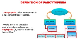
*Pancytopenia refers to decreases in
all peripheral blood lineages.
*Many disorders that cause
pancytopenia can also cause
bicytopenia (ie, decreases in only
two cell lines)
2. Individual laboratories typically establish their own reference ranges for
Hemoglobin/Hematocrit, WBC count, and platelet count.
These institutional cutoff values supersede published reference standards
such as those published by the WHO:
1. RBCs – Hemoglobin ,Hb <12 for non pregnant ,women and < 13 for men
2. WBCs– Because neutrophils constitute the majority of leukocytes in the peripheral blood
and bone marrow, nearly all cases of leukopenia manifest as neutropenia Absolute
neutrophil count (ANC) < 1800
3. Platelet count < 150.000
3. MECHANISMS OF PANCYTOPENIA
Pancytopenia may be caused by one or more of the following mechanisms :
1. Bone marrow infiltration/replacement
a) Hematologic malignancies (eg, leukemia, lymphoma, multiple myeloma,
myelodysplastic syndromes),
b) Metastatic cancer
c) Myelofibrosis
d) Infectious diseases (eg, miliary tuberculosis, fungal infections).
2. Bone marrow aplasia
a) Nutritional disorders (eg, deficiencies of vitamin B12 or folate),
b) Aplastic anemia
c) Infectious diseases (eg, HIV, viral hepatitis, parvovirus B19),
d) Immune destruction
e) Medications .
4. 3. Blood cell destruction or sequestration
a) Excessive blood cell destruction occurs in DIC, TTP, and
ineffective hematopoiesis (eg, myelodysplastic syndromes,
megaloblastic disorders)
b) Excessive sequestration may be due to hypersplenism (eg,
from liver cirrhosis, storage diseases, lymphoma, or
autoimmune disorders).
MECHANISMS OF PANCYTOPENIA
5. Some diseases may cause pancytopenia by multiple mechanisms.
As an example, a lymphoma may infiltrate the bone marrow, cause
hypersplenism, induce immune destruction of blood cells, and require
treatment with cytotoxic agents.
Similarly, Crohn disease may impair absorption of iron, folate, and vitamin
B12; induce an inflammatory state that exacerbates anemia; require partial
bowel resection that affects absorption of nutrients and calories; and
require treatment with myelosuppressive agents.
MECHANISMS OF PANCYTOPENIA
8. Emergencies associated with pancytopenia
Clinical stabilization is the highest priority for the
patient with pancytopenia who is clinically unstable.
Immediate hospitalization may be required to :
Control life-threatening infections
Provide blood product support
Manage other medical emergencies.
9. Pancytopenia associated with the following clinical situations will require
immediate hematology consultation and/or hospitalization:
1. Neutropenia:
a) Absolute neutrophil count (ANC) <1000/microL with fever and/or other evidence of infection or other
acute illness.
b) New diagnosis of moderate or severe neutropenia (ie, ANC <1000/microL and <500/microL,
respectively).
2. Symptomatic anemia (eg, myocardial ischemia, hypotension).
3. Thrombocytopenia:
a) New finding of platelets <10,000/microL
b) Clinically significant bleeding with platelets <50,000/microL.
4. Suspected DIC, TTP, HUS, or other thrombotic microangiopathy because of
schistocytes on peripheral blood smear accompanied by elevated LDH.
Emergencies associated with pancytopenia
10. 5. Suspected acute leukemia:
a) New diagnosis (eg, circulating blasts).
b) Medical emergencies associated with leukemia (eg, DIC from acute promyelocytic leukemia,
tumor lysis syndrome).
6. Suspected severe aplastic anemia (ANC <500/microL, platelets
<20,000/microL, anemia with reticulocyte count <20,000/microL) or other bone
marrow failure syndrome.
7. Suspected hemophagocytic lymphohistiocytosis (HLH) because of
unexplained fever, hepatomegaly, lymphadenopathy, and/or neurologic
symptoms in association with very high serum ferritin, liver function
abnormalities, and/or coagulopathy.
Emergencies associated with pancytopenia
11. 5. Metabolic emergencies in the setting of pancytopenia:
a) Hypercalcemia with symptoms (eg, delirium, abdominal pain,
dehydration) associated with the cause of pancytopenia (eg,
multiple myeloma, metastatic cancer, adult T cell
leukemia/lymphoma).
b) Acute renal failure (eg, hyperkalemia, dehydration, fluid
overload) associated with the cause of pancytopenia (eg,
multiple myeloma, tumor lysis syndrome).
c) Hyperuricemia with renal failure associated with the cause of
pancytopenia.
Emergencies associated with pancytopenia
12. INITIAL EVALUATION
While there are numerous possible causes of pancytopenia, the
differential diagnosis should narrow following :
1. Initial history
2. Physical examination
3. Screening laboratory studies
4. Examination of the peripheral blood smear .
Initial testing should also identify emergency situations and determine
the need for (and urgency of) hematology referral
.
13. History
●Time course and clinical severity – Prior laboratory results (when available) and
severity and duration of symptoms should be evaluated.
●Symptoms associated with cytopenias
Examples include:
Recurrent, severe, or unusual infections that may be due to
leukopenia/neutropenia
Fatigue, dyspnea, chest pain, hemodynamic instability, or claudication due to
anemia
Bleeding or easy bruising due to thrombocytopenia or DIC
Constitutional symptoms, including fevers, night sweats, and/or weight loss
Nausea, vomiting, and jaundice that may be associated with liver disease
Chest pain, hemodynamic instability, severe bleeding, life-threatening
infections, and other medical emergencies may require immediate hospitalization
for clinical stabilization .
14. ●Other medical conditions
Almost any comorbid medical condition or surgical procedure can
contribute to or exacerbate cytopenias.
As an example, a history of Crohn disease is relevant because the IBD
and previous surgeries may affect the patient’s nutritional status and
impair absorption of essential nutrients and vitamins (eg, iron,
folate, vitamin B12), while the inflammatory state may exacerbate
anemia, and therapeutic agents may suppress bone marrow function.
History
15. History
●Problematic medications
Many medications (including prescription and over-the-counter
medications, health supplements, and home or folk remedies) may
cause or contribute to cytopenias .
The relationship between the onset of pancytopenia and the
administration of medications should be defined as much as
possible.
Some medications (eg, cytotoxic or immunosuppressive agents)
cause predictable decreases in blood counts that are generally
reversible if the agent is reduced or stopped.
Other medications are idiosyncratically or less commonly
17. History
●Personal and occupational exposures
Certain personal habits (eg, alcohol consumption, diet),
infection history (eg, HIV, viral hepatitides), exposure to toxic
agents at work or home (eg, organic solvents), and travel
history (eg, exposure to malaria, leishmania) may also be
relevant.
18. Physical findings
●Rashes that may be related to drug reactions, rheumatologic disorders,
infections, and malignancies
●Oral lesions; as examples, thrush suggests immune compromise; oral
ulcers may be seen in diseases such as SLE
●Lymphadenopathy and/or splenomegaly
●Jaundice and stigmata of liver disease
19. Laboratory studies
1. CBC, with white blood cell differential count and red
blood cell indices
2. Peripheral blood smear, which may reveal
abnormalities that would not be detected by automated
methods.
3. Reticulocyte count.
a) An absolute reticulocyte count (ARC) <20,000 indicates a
marked decrease in RBCs production and suggests a
hypoproliferative condition.
4. PT and PTT.
a) Coagulopathies in the setting of pancytopenia generally
require prompt evaluation and referral.
5. Serum chemistry tests, including electrolytes, renal
and liver function tests, lactate dehydrogenase,
calcium, and uric acid.
20. Hematology referral
• Referral to a hematologist for purposes of diagnosis (eg, examination
of the peripheral blood smear, bone marrow studies, interpretation
of specialized molecular or flow cytometry results) and/or
management is nearly always appropriate, unless an etiology is
promptly identified that can be readily managed by the non-specialist
clinician (eg, vitamin B12 or folate deficiency, alcoholic liver
cirrhosis with congestive splenomegaly).
• The urgency of referral to a hematologist is influenced by the severity
and trajectory of cytopenias, clinical stability, medical
complications, and the need for urgent treatment.
21. • Emergencies – Immediate hematology
evaluation should be performed for the
emergency situations .
• Clinical stability – Referral is less urgent
(eg, can occur within days to weeks) if the
patient is asymptomatic, blood counts
are stable and near normal, and there
are no medical emergencies.
•
Serial outpatient evaluation of CBCs and a
review of the peripheral blood smear may be
appropriate in select cases of asymptomatic,
mild pancytopenia.
The case should be discussed with a
hematologist if there is uncertainty over the
urgency of referral.
Neutropenia (new diagnosis or associated with
fever/infection)
Symptomatic anemia (eg, cardiac ischemia,
hemodynamic instability, worsening
congestive heart failure)
Thrombocytopenia (platelets <10,000/microL,
or <50,000/microL associated with bleeding)
Disseminated intravascular coagulation
Abnormal peripheral blood smear (eg,
microangiopathy, blasts)
Severe aplastic anemia
Hemophagocytic lymphohistiocytosis
Metabolic emergencies (eg, symptomatic
hypercalcemia, hyperkalemia, tumor lysis
syndrome)
Emergencies associated with pancytopenia
Hematology referral
22. SUBSEQUENT EVALUATION
Potential explanations for pancytopenia should emerge from the initial
history, physical examination, screening laboratory studies, and
review of the peripheral blood smear .
While a single underlying diagnosis should be sought, more than one
potential cause or contributor to pancytopenia may be identified.
23. Bone marrow and other specialized evaluation
• Bone marrow aspirate and biopsy is useful in many, but not
all, patients with pancytopenia.
It is especially important in patients for whom a primary
hematologic disorder is suspected as the cause of
pancytopenia (eg, acute leukemia, aplastic anemia,
multiple myeloma) or when the cause of pancytopenia
remains elusive after the initial evaluation.
24. •In certain situations, a bone marrow biopsy may be
unhelpful or even distracting and confounding.
•As an example, a bone marrow biopsy performed just
days after discontinuation of a suspect medication
may show a "maturation arrest" (ie, recovery of bone
marrow cells only up to an immature stage of
differentiation) that may be morphologically
indistinguishable from acute leukemia.
Bone marrow and other specialized evaluation
25. .
• Similarly, recent treatment with recombinant hematopoietic
growth factors may induce a bone marrow morphology that is
indistinguishable from certain myeloproliferative neoplasms or
inflammatory conditions.
• In such situations it may be preferable to delay the biopsy by
days to weeks.
Bone marrow and other specialized evaluation
26. •Bone marrow biopsies may also be uninformative
in some cases when pancytopenia is thought to be
due to peripheral blood cell destruction or
sequestration (eg, suspected TTP, cirrhosis with
hypersplenism); clinical evaluation and/or other
specialized testing is generally more useful and
definitive in such cases.
Bone marrow and other specialized evaluation
27. • The hematologist who will perform the procedure should communicate
with a laboratory technician and/or pathologist to properly prepare the
specimens (eg, place fresh aspirate material in appropriate
anticoagulated medium for flow cytometry or molecular studies, and
put biopsy specimens in proper fixative), and to order the appropriate
tests.
• The bone marrow aspirate and biopsy specimens will undergo
microscopic examination by a hematopathologist, pathologist, and/or
hematologist; review by both the pathologist and involved clinicians
can be invaluable in interpreting the bone marrow morphology in the
context of the clinical presentation.
Bone marrow and other specialized evaluation
28. • The differential diagnosis of pancytopenia in a given patient will inform
further specialized testing of the bone marrow and/or peripheral
blood (eg, flow cytometry, cytogenetics, molecular studies, microbiologic
cultures).
• As an example, direct antiglobulin testing and flow cytometry may be
needed to confirm the diagnosis of paroxysmal nocturnal
hemoglobinuria.
Bone marrow and other specialized evaluation
29. • Cytogenetic testing (fluorescent in situ hybridization [FISH] or
karyotype) of bone marrow or peripheral blood may be required for
confirmation of the diagnosis of many hematologic malignancies
(eg, leukemias, myelodysplastic syndromes, myeloproliferative
neoplasms, lymphomas).
• Molecular analysis is increasingly important in the diagnosis and risk
stratification of many cancers, including hematologic malignancies.
Bone marrow and other specialized evaluation
32. Specific clinical scenarios
• Coagulopathy — The finding of elevated PT and/or PTT in the setting
of pancytopenia should focus immediate attention on determining if
microangiopathic hemolytic anemia (MAHA) is present .
This requires urgent examination of the peripheral blood smear by a
hematologist or suitably experienced laboratory personnel for the presence of
schistocytes with thrombocytopenia.
The presence of MAHA may raise the possibility of DIC that may be due to
sepsis, acute promyelocytic leukemia, or other causes.
• If MAHA is not found, other explanations should be sought for the
abnormal PT and/or PTT (eg, liver disease, vitamin K deficiency,
medications).
This evaluation may require mixing studies and/or specific coagulation
factor tests to distinguish the presence of factor inhibitors from effects of
medications, liver disease, or vitamin K deficiency.
33. Specific clinical scenarios
• Abnormal cells on blood smear
• Abnormal cells on the peripheral smear should be examined by an experienced clinician to distinguish
hematologic malignancies (eg, leukemia, lymphoma, myelodysplastic syndrome) from other disorders,
such as infections (eg, atypical lymphocytes associated with viral or other infections), marrow
replacement disorders (eg, myelofibrosis, metastatic cancer, multiple myeloma), and megaloblastic
conditions.
• Malignant disorders — Examples of abnormal malignant cells on the blood smear of a pancytopenic
patient include:
1. Circulating blasts associated with various leukemias; a substantial proportion of adults with
pancytopenia are found to have acute leukemias, hairy cell leukemia, or other hematologic
malignancies .
2. Dysplastic leukocytes, including pseudo-Pelger-Huët cells or reduced neutrophil cytoplasmic granules
in myelodysplastic syndromes .
3. Immature myeloid cells, such as promyelocytes, myelocytes, and metamyelocytes that may reflect an
underlying myeloproliferative neoplasm (MPN), such as primary myelofibrosis .
4. Leukoerythroblastic findings, including nucleated red blood cells associated with myelofibrosis or
other MPNs .
Confirmation of the nature of such abnormal cells will require further specialized testing including:
1. Bone marrow aspirate and biopsy
2. Flow cytometry of peripheral blood and/or bone marrow
3. Cytogenetic testing (fluorescent in situ hybridization [FISH] or karyotype) of bone marrow or peripheral blood
34. Specific clinical scenarios
•
Abnormal cells on blood smear —
•
Non-malignant cells — Abnormalities of granulocytes (eg, hypersegmented neutrophils),
lymphocytes (eg, atypical lymphocytes associated with viral or other infections), and red blood cells
(RBCs) (eg, ovalomacrocytes) may indicate disorders other than hematologic malignancies.
•
Examples include:
1. Hypersegmented neutrophils (ie, five or more nuclear lobes) in association with
ovalomacrocytes (ie, enlarged, ovoid RBCs) suggest a megaloblastic disorder .
The most common causes of these findings are deficiencies of folate and/or vitamin B12. The
appearance of the peripheral blood smear is indistinguishable between these two vitamin
deficiencies, and establishing the diagnosis requires specific testing. It is important to note that serum
folate levels may quickly normalize after feeding a malnourished patient, but RBC folate will more
accurately reflect the prior state. .
1. Atypical lymphocytes (lymphoid cells with generous and malleable cytoplasm, often indented
by surrounding red cells) can be seen following viral infections such as infectious mononucleosis,
and may be associated with pancytopenia due to bone marrow suppression, hypersplenism, and
other .
2. Leukoerythroblastic appearance of the blood smear, with RBC teardrops, nucleated RBCs,
and microangiopathic hemolytic anemia (MAHA), may be associated bone marrow infiltration
caused by myelofibrosis or metastatic cancer .
3. Schistocytes or other evidence of MAHA may reflect disseminated intravascular coagulation,
35. Specific clinical scenarios
• Hypoproliferative conditions — Reticulocytopenia (ie, <20,000 reticulocytes/microL) may
indicate a hypoproliferative pancytopenia.
The urgency and the pace of further evaluation should be influenced by the severity and
trajectory of the cytopenias, and the presence of symptoms or complications of the
cytopenias. Suspected severe aplastic anemia (absolute neutrophil count [ANC]
<500/microL, platelets <20,000/microL), along with reticulocytopenia requires emergency
evaluation.
Other diagnostic considerations include:
●Deficiencies of essential vitamins or minerals (eg, vitamin B12, folate, or copper)
●Medications (eg, cytotoxic agents, or idiosyncratic drug reactions)
●Bone marrow suppression (eg, alcohol, viral infections)
●Aplastic anemia (eg, autoimmune/idiopathic, which may be associated with paroxysmal
nocturnal hemoglobinuria; or associated with drugs, viral infections, or toxins) [17-19]
●Ineffective hematopoiesis (eg, myelodysplastic syndromes, megaloblastic conditions)
●Bone marrow infiltration/replacement (eg, myelofibrosis, metastatic cancer, storage
diseases) [20]
●Malignancies associated with immune suppression (eg, hairy cell leukemia, T cell large
granular lymphocyte leukemia)
36. Specific clinical scenarios
• Hypoproliferative conditions — Reticulocytopenia (ie, <20,000 reticulocytes/microL)
may indicate a hypoproliferative pancytopenia.
Defining the nature of a hypoproliferative bone marrow condition usually
requires testing the following:
●Serum vitamin B12, folate, and/or copper (as appropriate)
If testing for vitamin B12 and folate is unrevealing, and the history does not
suggest alcohol, infections, or reactions to medications as a cause of
hypoproliferative pancytopenia, further testing may be required:
●Bone marrow aspirate and biopsy, with consideration of
immunohistochemical staining, flow cytometry, and other specialized testing.
●Serologic studies to evaluate viral etiologies or autoimmune illnesses
(perhaps in concert with specialists in infectious diseases or rheumatology).
●Flow cytometry of peripheral blood (eg, for CD59) may be useful when
paroxysmal nocturnal hemoglobinuria (PNH) is associated with aplastic anemia
[21].
37. Specific clinical scenarios
Splenomegaly and/or liver disease
The presence of splenomegaly and pancytopenia suggests hypersplenism
(ie, sequestration and/or excessive destruction of blood cells in an enlarged
spleen).
All cell lineages may be affected.
In many, but not all cases, splenomegaly and hypersplenism are
associated with liver disease.
Conversely, other conditions can cause liver disease and pancytopenia without
splenomegaly.
The extent of cytopenias in hypersplenism is variable, but generally less
severe than that caused by primary bone marrow disorders.
However, splenomegaly and liver disease are associated with many disorders that also
contribute to cytopenias by other mechanisms (eg, malignancies, myelofibrosis,
infections), and the resultant multifactorial cytopenias may be severe.
38. Specific clinical scenarios
• Conditions associated with pancytopenia in the setting of splenomegaly and/or liver disease
include:
1. Liver disease/cirrhosis and portal hypertension
2. Infections (eg, viral infections, malaria, leishmaniasis, endocarditis)
3. Hematologic malignancies (eg, lymphomas, hairy cell leukemia, myeloproliferative neoplasms)
4. Extramedullary hematopoiesis (eg, associated with myelofibrosis or thalassemias)
5. Congestion (eg, right sided congestive heart failure)
6. Inflammation (eg, associated with rheumatoid arthritis [Felty syndrome] or other autoimmune illness,
endocarditis)
7. Primary splenic disease (eg, hemorrhage, thrombosis)
8. Storage diseases (eg, Gaucher disease)
9. Hemophagocytic lymphohistiocytosis
The differential diagnosis of pancytopenia in the setting of splenomegaly is influenced by the presence
of concurrent lymphadenopathy, constitutional symptoms, stigmata of chronic liver disease, and
findings of autoimmune disorders.
As examples:
1. ●Presence of both splenomegaly and lymphadenopathy may suggest an underlying hematologic
malignancy (eg, lymphoma, leukemia), infectious disease, or autoimmune disorder.
2. ●Stigmata of chronic liver disease may suggest pancytopenia caused by hypersplenism from cirrhosis. If
no explanation for the underlying liver disease is readily identified, evaluation of the liver by imaging (eg,
ultrasound, CT scan) and/or biopsy may be warranted.
3. ●Abnormalities of liver function without stigmata of chronic liver disease or splenomegaly may be
associated with infectious diseases (eg, acute viral hepatitis), medications, autoimmune disorders, or
39. Specific clinical scenarios
Lymphadenopathy
Detection of lymphadenopathy (localized or generalized) may provide
important information regarding the underlying cause of pancytopenia.
Potential disorders associated with lymphadenopathy and pancytopenia
include:
Hematologic malignancies (eg, lymphoma, leukemia)
Autoimmune illnesses
Infectious diseases
Aids to the diagnosis of an underlying cause of lymphadenopathy in the
setting of pancytopenia include:
Imaging (eg, CT scan, ultrasound, or PET scan to define the extent of lymphadenopathy,
and as a possible adjunct to biopsy)
Lymph node biopsy (including morphology, flow cytometry, molecular studies)
Flow cytometry of peripheral blood and/or lymph node specimen (eg, to evaluate
hematologic malignancies)
Serologic studies for infectious or autoimmune illnesses
40. Specific clinical scenarios
•
Autoimmune conditions —
Variable degrees of pancytopenia are commonly seen in patients with
previously diagnosed autoimmune illnesses.
RA/Felty syndrome, SLE, and sarcoidosis are often associated with
cytopenias, and some of the treatments for these illnesses (eg, gold
salts, cytotoxic agents) may exacerbate the cytopenias.
Furthermore, autoimmune diseases are often associated with other
conditions that can cause pancytopenia (eg, pernicious anemia,
thyroid disease, T cell large granular lymphocyte leukemia [T-LGL])
[30].
Thus, pancytopenia in the setting of autoimmune illnesses is frequently
multifactorial.
41. Specific clinical scenarios
•
Autoimmune conditions
• An important component of the management of cytopenias in the
setting of known autoimmune illness is identifying conditions that may
be contributing to the cytopenias.
• Examples include:
Associated disorders should be sought and managed (eg, folate deficiency,
vitamin B12 deficiency associated with pernicious anemia, autoimmune thyroid
disease)
Alternative therapeutic agents may be considered (in consultation with the
clinician managing the autoimmune illness) in an effort to lessen bone marrow
suppression and inflammation.
Consideration may be given to splenectomy in some patients with symptomatic
Felty syndrome.
If T-LGL is suspected, evaluation by flow cytometry of peripheral blood or bone
marrow aspirate/biopsy should be considered.
• In some patients, a "forme fruste" of an autoimmune illness may be
suspected, but no firm diagnosis has been established.
42. Specific clinical scenarios
Constitutional symptoms
Pancytopenia may present in the setting of otherwise unexplained fevers, soaking
sweats, and weight loss.
It may be especially important to consider an infectious etiology or
hemophagocytic lymphohistiocytosis in this setting.
Possible causes of pancytopenia associated with constitutional symptoms
include:
1. Infections (viral illness, miliary tuberculosis, fungal infection, endocarditis)
2. Hemophagocytic lymphohistiocytosis (HLH)
3. Hematologic malignancies (eg, lymphoma, leukemia)
4. Autoimmune illnesses
The presence of lymphadenopathy, liver disease, splenomegaly, or other findings
can provide important clues to the nature of the underlying illness, and the
evaluation in these settings is discussed above.
In some patients, constitutional symptoms may be the only apparent clinical
findings.
Establishing the diagnosis in such a setting may be challenging.
If no likely diagnosis presents itself, and especially if the cytopenias are severe or associated with
symptoms and/or complications, a bone marrow aspirate and biopsy with specialized infectious
disease evaluation of the bone marrow specimen (eg, fungal and/or mycobacterial cultures and
stains) should be performed, as appropriate.
43. Specific clinical scenarios
•
Constitutional symptoms
• A high index of suspicion must be maintained for the presence of HLH in the
setting of pancytopenia associated with constitutional symptoms but no
other clinical findings .
• Diagnosis of HLH is supported by the following :
Ferritin – Serum ferritin is usually very high (often >5000 ng/mL) and has high
specificity in children, but not in adults [34]; however, a ferritin <500 ng/mL has
excellent negative predictive value for excluding the diagnosis
Liver function tests (LFTs) – While not one of the diagnostic criteria for HLH, elevated
liver enzymes (AST, ALT, GGT), lactate dehydrogenase (LDH), and bilirubin are
elevated in most patients
Hypofibrinogenemia – This may often be out of proportion to other coagulation
parameters
Triglycerides – Marked elevation of triglycerides is typically seen, especially with
severe liver involvement
Soluble CD25 receptor – Elevated soluble IL-2 receptor alpha (sIL-2R or sCD25)
NK cell function – Low/absent NK cell function/degranulation by flow cytometry (in
children but not adults)
Bone marrow biopsy – Findings of hemophagocytosis and/or infiltration by activated
macrophages
Organomegaly – Splenomegaly and/or hepatomegaly may be present
44. Specific clinical scenarios
Metabolic abnormalities —
Certain metabolic disorders (eg, hypercalcemia, tumor lysis
syndrome, renal failure, hyperuricemia) may be associated with
diseases that also cause pancytopenia, including multiple myeloma,
leukemia, and lymphoma [35,36].
The association of these metabolic complications with pancytopenia may
constitute a medical emergency, and require urgent hospitalization and/or referral
to a hematologist for establishing the diagnosis and initiating prompt management
.
If an underlying diagnosis is known but was not previously associated
with pancytopenia, the clinician must investigate causes for the decline
in blood counts.
The cause(s) will often be multifactorial, but consideration should be given to:
Review of disease status to assess progressive disease or treatment resistance, including
restaging of lymphomas or myeloma (eg, repeat CT or PET-CT scans, serologic evaluation of
multiple myeloma)
Assessment of a fundamental change in the disease (eg, Richter transformation of a
previously diagnosed lymphoma, progression of myelodysplastic syndrome to acute leukemia)
may require repeat bone marrow or lymph node biopsy
45. Specific clinical scenarios
•
Metabolic abnormalities — Certain metabolic disorders (eg,
hypercalcemia, tumor lysis syndrome, renal failure, hyperuricemia) may be
associated with diseases that also cause pancytopenia, including multiple
myeloma, leukemia, and lymphoma [35,36]. The association of these
metabolic complications with pancytopenia may constitute a medical
emergency, and require urgent hospitalization and/or referral to a
hematologist for establishing the diagnosis and initiating prompt
management (table 2).
•
If an underlying diagnosis is known but was not previously associated with
pancytopenia, the clinician must investigate causes for the decline in blood
counts. The cause(s) will often be multifactorial, but consideration should be
given to:
•
●Review of disease status to assess progressive disease or treatment
resistance, including restaging of lymphomas or myeloma (eg, repeat CT or
PET-CT scans, serologic evaluation of multiple myeloma)
•
●Assessment of a fundamental change in the disease (eg, Richter
transformation of a previously diagnosed lymphoma, progression of
myelodysplastic syndrome to acute leukemia) may require repeat bone
marrow or lymph node biopsy
46. Specific clinical scenarios
Suspected medications
When a particular medication is suspected as a cause of pancytopenia,
consideration should be given to discontinuing that medication (or perhaps
reducing the dose) in consultation with other treating clinicians.
This decision will be influenced by the severity of the cytopenias, the trajectory of the
blood counts, clinical symptoms, and the reason why the medication is being
administered.
For cytotoxic or myelosuppressive agents, blood count recovery can
generally be expected within days to weeks.
When such drugs are thought to be the cause of pancytopenia, it may be preferable to
observe the blood counts for one or two weeks rather than immediately performing a
bone marrow biopsy.
When an idiosyncratic reaction is a likely cause of pancytopenia, a
response to discontinuing the medication is less predictable and recovery of
blood counts may be protracted.
The clinical presentation and the appearance of the bone marrow in such situations
may be indistinguishable from idiopathic aplastic anemia.
The clinician’s judgment is needed to distinguish between these diagnostic
47. Specific clinical scenarios
Suspected medications
An immediate bone marrow biopsy may not be helpful when a
medication is suspected to be the cause of pancytopenia.
A finding of aplasia or hypoplasia will not confirm the identity or nature of the
etiologic agent.
Additionally, if a biopsy is performed early in the recovery process, the bone
marrow may erroneously suggest an acute leukemia, because the
recovering cells may exhibit a “maturation arrest” as hematopoiesis has only
progressed to an immature stage of maturation.
In such a clinical setting, serial observation of the patient and blood counts
may be the most useful diagnostic approach.
48. Specific clinical scenarios
Suspected medications
Mechanisms of drug-associated cytopenias include allergic reactions
that affect bone marrow production and/or increase peripheral
destruction, and pseudo-allergic reactions.
Allergic reactions may be influenced by the patient’s immunologic background
(ie, HLA type), comorbid conditions, pharmacogenomic constitution , prior
exposure to that medication or related drugs, and other clinical features.
Exposure to some drugs can induce a hypoproliferative pancytopenia that may
be indistinguishable from idiopathic aplastic anemia; recovery of blood
counts during a period of observation after discontinuing the suspect
medication, if clinically reasonable, may help to distinguish between these
diagnoses