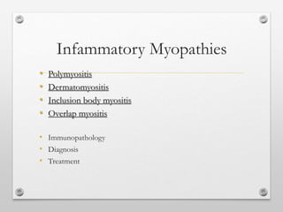
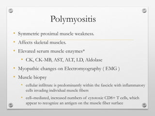
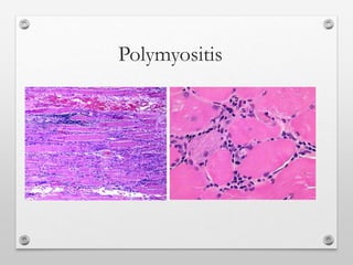
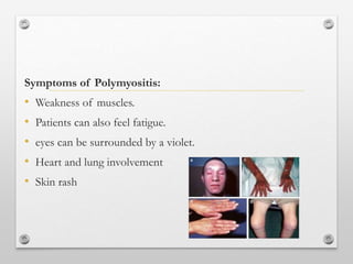
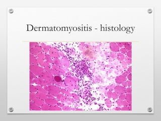
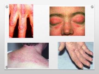
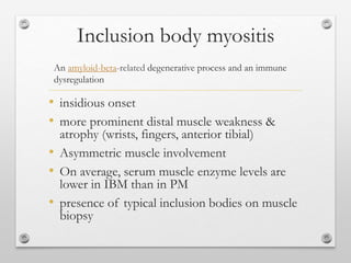
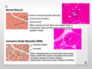
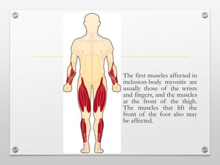
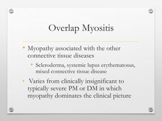
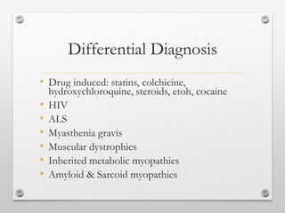
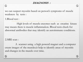
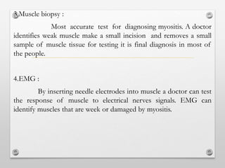
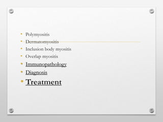
The document discusses various types of inflammatory myopathies (myositis):
1) Polymyositis causes symmetric proximal muscle weakness and inflammation within muscle fibers. Symptoms include fatigue and a skin rash.
2) Dermatomyositis causes similar muscle symptoms but also features a characteristic rash on the skin. It can also involve other organs.
3) Inclusion body myositis typically causes asymmetric weakness of wrist, finger, and thigh muscles. It involves degeneration in addition to immune dysfunction.
4) Overlap myositis combines myopathy with connective tissue diseases like lupus. Diagnosis involves blood tests, muscle enzyme levels, muscle biopsy and MRI or EMG. Treatment
2. “Myositis means muscle inflammation, and can be caused by
infection, injury, certain medicines, exercise, chronic disease,
or autoimmune disease”
Infammatory Myopathies…………?
NIH organization for research on Myositis-13 May 2014
3. The inflammatory myopathies are a group of diseases that involve
chronic muscle inflammation
which accompanied by :
6. Polymyositis
• Symmetric proximal muscle weakness.
• Affects skeletal muscles.
• Elevated serum muscle enzymes*
• CK, CK-MB, AST, ALT, LD, Aldolase
• Myopathic changes on Electromyography ( EMG )
• Muscle biopsy
• cellular infiltrate is predominantly within the fascicle with inflammatory
cells invading individual muscle fibers
• cell–mediated, increased numbers of cytotoxic CD8+ T cells, which
appear to recognize an antigen on the muscle fiber surface
7. Researchers are finding that each case of PM is quite different
from others. Sometimes, cases originally diagnosed as PM and
not responding to treatment are later found to be inclusion-
body myositis (IBM). Patients with certain types of PM may
have one or more other autoimmune diseases.
9. Symptoms of Polymyositis:
• Weakness of muscles.
• Patients can also feel fatigue.
• eyes can be surrounded by a violet.
• Heart and lung involvement
• Skin rash
11. Dermatomyositis
• Symmetric proximal muscle weakness
• Elevated serum muscle enzymes
• Myopathic changes on EMG
• Muscle biopsy
• humorally–mediated disorder (CD4 cells), cellular infiltrate,
located principally in perifascicular regions, focused around
blood vessels
• Rash
Dermatomyositis is an idiopathic inflammatory myopathy with characteristic
cutaneous findings that occur in children and adults. Systemic disorder most
frequently affects the skin and muscles but may also affect the joints; the
esophagus; the lungs; and, less commonly, the heart.
16. Both polymyositis and dermatomyositis can sometimes be
associated with cancers, including lymphoma, breast, lung,
ovarian, and colon cancer. The cancer risk is reported to be
much greater with dermatomyositis than polymyositis. (See
polymyositis).
18. Inclusion body myositis
• insidious onset
• more prominent distal muscle weakness &
atrophy (wrists, fingers, anterior tibial)
• Asymmetric muscle involvement
• On average, serum muscle enzyme levels are
lower in IBM than in PM
• presence of typical inclusion bodies on muscle
biopsy
An amyloid-beta-related degenerative process and an immune
dysregulation
19.
20. The first muscles affected in
inclusion-body myositis are
usually those of the wrists
and fingers, and the muscles
at the front of the thigh.
The muscles that lift the
front of the foot also may
be affected.
21. • Muscle weakness.
• Painless
• Heart and lungs are not affected in IBM.
Symptoms of Inclusion body myositis
23. Overlap Myositis
• Myopathy associated with the other
connective tissue diseases
• Scleroderma, systemic lupus erythematosus,
mixed connective tissue disease
• Varies from clinically insignificant to
typically severe PM or DM in which
myopathy dominates the clinical picture
25. Endothelium of the endomysial capillaries
DERMATOMYOSITIS
target
Attack mAc
Activation of cytokines
And chemokines
Migration of TGFb into endomysial
Or
Spaces lymphoid cells
26.
27. IMMUNOPATHOLOGY OF POLYMYOSITIS AND
INCLUSION BODY MYOSITIS :
In polymyositis and inclusion body myositis the
primary effector cells mediating muscle fiber injury
are CD8 cells that are surround and invade MHC-1
antigen expressing, non-necrotic, muscle fibers.
T- cell mediated cytotoxicity.
These cells have perforin and necrosis granules –
myoncerosis.
Antigen driven T- cell response.
Co- stimulatory molecules are up regulated.
28.
29.
30. IMMUNOPATHOGENESIS AND AUT0ANTIBODIES :
Polymyositis and dermatomyositis is an autoimmune disorder.
The impotance of these antibodies and their specificity in the
pathogenesis of polymysitis and dermatomyositis remains
unclear.
No specific target antigens have not been identified and the
agents initiating of disease/tissue remains unknown.
They occurs in less than 25% of patients.
They also occurs in patients with interstinal lung disease.
Without myositis.
34. DIAGNOSIS :
we can suspect myositis based on person’s symptoms of muscle
weakness by tests :
1.Blood test :
High levels of muscle enzymes such as creatine kinase
may means there is muscle inflammation. Blood tests check for
abnormal antibodies that may identify an autoimmune conditions.
2.MRI scan :
A scanner using a high powerd magnet and a computer
creats images of the muscles.it help to identify areas of myositis
and changes in the muscle over time.
35. 3.Muscle biopsy :
Most accurate test for diagnosing myositis. A doctor
identifies weak muscle make a small incision and removes a small
sample of muscle tissue for testing it is final diagnosis in most of
the people.
4.EMG :
By inserting needle electrodes into muscle a doctor can test
the response of muscle to electrical nerves signals. EMG can
identify muscles that are week or damaged by myositis.
37. TREATMENT
The goals of the therapy are to improve the ability to carry out activites
of daily living by increasing muscle strengh.
They are very few controlled clinical trails most on dermatomyositis
and inculsion body mysoitis. And inclusion body myositis is difficult to
treat. As there is no effective therapy.
The hypothesis that beta amyloid protein is key to IBM has been
supported in a mouse model using an Aβ vaccine that was found to be
effective against inclusion body myositis in mouse models.
The following agents are used in the treatment of polymyositis and
dermatomyositis :-
1. Corticosteroids :-
Prednisone is first line drug in treatment. Dose 80-100 mg per
day for 3-4 weeks single in morning dose (after breakfast).
if a patients with sever PM or DM are given IV route
methylprednisolone 1mg/day for 3-5 days. With oral prednisone as above.
38. Initation of therapy is due to :-
1. some patients are steroid resistance .
2. Increase dosage of steroids result in the worsening of muscle
strength.
3. Dose of prednisone for at least 2-3 month period become ineffective.
4. Disease is rapidly progressive with severe weakness and respiratory
failure.
So for this reasons we use immunosuppressive drugs.
Azathioprine :-
orally dose 2.5-3 mg/kg for 4-6 months.
Methotrexate :-
orally dose 25 mg weekly. This methotrexate act more quickly
than Azathioprine.
39. Cyclosporin :-
orally dose 100-150 mg twice daily may benefit for
childhood dermatomyositis. Acts faster than other drugs but its
efficancy has not been established with controlled studies.
Mycophenolate mofetil :-
2mg per day for 3 months is emerging as a promising and
well tolerated drug.
Cyclophosphamide :-
0.5-1.0 g/m2 intravenously has shown mixed resutls. It may
help for patients with interstitial lung disease.
Newer agents :-
Rituximab, Tancrolimus, Sirolimus (Rapamycin),TNFa
blockers(Infliximab).
40. The treatment of Overlap myositis is mainly based on the use
of corticosteroids and immunosuppressants. Biologic drugs,
i.e. anti-TNFα or anti-CD20 monoclonal antibodies, have
been recently introduced as alternative treatments in
refractory cases. There are some concerns with the use of
anti-TNF agents in patients with systemic autoimmune
diseases due to the risk of triggering disease exacerbations
Treatment for Overlap myositis