Definition: 
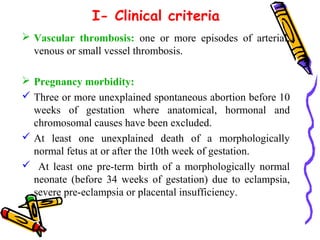
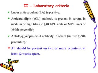
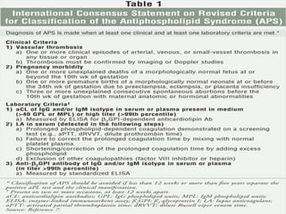
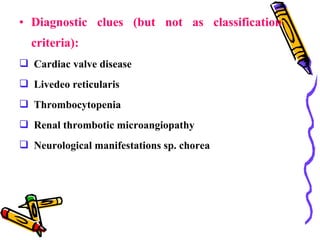
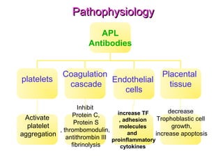
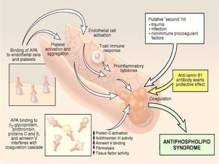
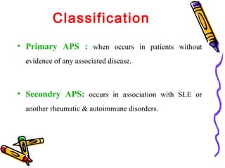
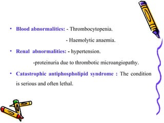
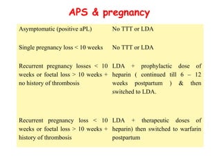
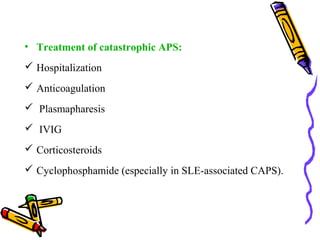
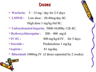
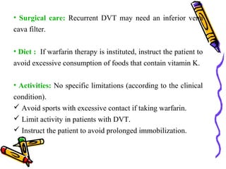
Antiphospholipid syndrome (APS) is an autoimmune 
disorder characterised by arterial and venous 
thrombosis, adverse pregnancy outcomes (for mother 
and fetus), and raised levels of antiphospholipid 
(aPL) antibodies.
Synonyms: 
• Anti-phospholipid syndrome. The immune system produces 
abnormal blood proteins called antiphospholipid antibodies. 
• lupus anti-coagulant syndrome: synonym can be 
confusing bec. patients with APS may not necessarily have 
SLE, LA is associated with thrombotic rather than 
hemorrhagic complications. 
• Anti-cardiolipin antibody syndrome
• Sticky blood syndrome. people with this condition are 
more likely to form clots in blood vessels 
• Hughes syndrome: named after Dr. Graham Hughes 
along with his team in London who described the disease 
between 1983 & 1985.
:History of APS 
 Anti-phospholipid antibodies were first noted in a 
group of people who had positive tests for syphilis 
without signs of infection (false-positive tests). 
 It was then noticed that some of these individuals 
developed systemic lupus erythematosus (SLE) and 
other rheumatic conditions.
 Later studies found a protein called the lupus 
anticoagulant in a number of individuals with SLE, 
provided further understanding of APS, including the 
testing for anticardiolipin antibodies.
Epidemiology : 
 1- 5% of healthy individuals have aPL antibodies. 
 Incidence of APS: about 5 cases per 100,000 persons 
per year. 
 50 % of APS cases : not associated with another 
rheumatic disease ( PAPS). 
 APL antibodies : found in about 30-40% of patients 
with SLE, but only about 10% have APS.
 APS is the cause of : - 14% of all strokes. 
- 11% of MI. 
- 10% of DVT. 
- 6% of pregnancy morbidity. 
- 9% of pregnancy losses.
 Catastrophic APS has mortality rate about 50% due to 
multi-organ infarctions over a period of days. 
 Sex : A female predominance specially for secondary 
APS. 
 Age : APS is common in young to middle-aged adults.
. • 
Diagnostic criteria ( Sapporo 
criteria): 
At least: 
 One of the clinical criteria 
 One of the laboratory criteria .
I- Clinical criteria 
 Vascular thrombosis: one or more episodes of arterial, 
venous or small vessel thrombosis. 
 Pregnancy morbidity: 
 Three or more unexplained spontaneous abortion before 10 
weeks of gestation where anatomical, hormonal and 
chromosomal causes have been excluded. 
 At least one unexplained death of a morphologically 
normal fetus at or after the 10th week of gestation. 
 At least one pre-term birth of a morphologically normal 
neonate (before 34 weeks of gestation) due to eclampsia, 
severe pre-eclampsia or placental insufficiency.
II - Laboratory criteria 
 Lupus anticoagulant (LA) is positive. 
 Anticardiolipin (aCL) antibody is present in serum, in 
medium or high titre (ie ≥40 GPL units or MPL units or 
≥99th percentile). 
 Anti-B2-glycoprotein-1 antibody in serum (in titre ≥99th 
percentile). 
All should be present on two or more occasions, at 
least 12 weeks apart.
• Diagnostic clues (but not as classification 
criteria): 
 Cardiac valve disease 
 Livedeo reticularis 
 Thrombocytopenia 
 Renal thrombotic microangiopathy 
 Neurological manifestations sp. chorea
:Pathophysiology 
 The homeostatic regulation of blood coagulation is altered. 
 Phospholipids are an integral part of platelet And endothelial 
cell surface membranes , it is expected that these antibodies 
have a effect on them.
PPaatthhoopphhyyssiioollooggyy 
APL 
Antibodies 
platelets Coagulation 
cascade Endothelial 
cells 
increase TF 
, adhesion 
molecules 
and 
proinflammatory 
cytokines 
Placental 
tissue 
decrease 
Trophoblastic cell 
growth, 
increase apoptosis 
Inhibit 
Protein C, 
Protein S 
, thrombomodulin, 
antithrombin III 
fibrinolysis 
Activate 
platelet 
aggregation
IInn pprreeggnnaannccyy
Classification 
• Primary APS : when occurs in patients without 
evidence of any associated disease. 
• Secondry APS: occurs in association with SLE or 
another rheumatic & autoimmune disorders.
• Catastrophic form : 
 A rapidely progressive lethal form of PAPS with 
widespread vascular occlusion ( in medium or small 
sized arteries) in multiple organs ( > 3 organs) in few 
days. 
 Mortality rate 50%
 Seronegative APS: 
 Clinical picture is highly suggestive for APS, while the 
laboratory tests fail to detect LAC or aCL. 
 These cases could be APS with other aPL which are not 
included in the criteria : e.g anti-cardiolipin IgA or other 
aPL( e.g: false positive test for syphilis, AMA). 
 It is also possible that during the acute event of thrombosis, 
aPL cannot be detected bec. they are consumed in the blood 
clot. 
 Repeated measurement of these autoantibodies several 
weeks later
Associated disorders ( secondary APS ) 
• SLE. 
• Rheumatoid arthritis. 
• Systemic sclerosis. 
• Behçet's disease. 
• Temporal arteritis. 
• Sjögren's syndrome. 
• Psoriatic arthropathy.
 Other clinical associations : 
 Infections : HIV, hepatitis C, syphilis, malaria. 
 Malignant lymphoma. 
 Drug exposure: phenothiazines, phenytoin, hydralazine. 
 Autoimmune thrombocytopenia. 
 Autoimmune haemolytic anaemia. 
 Sickle cell anaemia.
:Clinical presentation 
Skin disorders: - Livedo reticularis (most common). 
- Splinter haemorrhages . 
- Leg ulcers. 
-Superficial thrombophlebitis . 
- Vasculitis. 
Neurological defects: - Migraine headaches. 
- Seizures. 
- Dementia. 
Cardiac abnormalities: - MI. 
- Cardiac valve vegetations.
• Blood abnormalities: - Thrombocytopenia. 
- Haemolytic anaemia. 
• Renal abnormalities: - hypertension. 
-proteinuria due to thrombotic microangiopathy. 
• Catastrophic antiphospholipid syndrome : The condition 
is serious and often lethal.
Levideo reticularis
Levideo reticularis
Splinter haemorrhages
Vasculitis
Differential Diagnosis : 
 Disseminated Intravascular Coagulopathy(DIC). 
 Infective Endocarditis. 
 Thrombotic Thrombocytopenic Purpura(TTP).
 SO, Younger patients with a history of DVT, 
pulmonary embolism, MI , or CVA need to be 
investigated for antiphospholipid syndrome, particularly if 
no other risk factors for thrombosis are present.
MANAGEMENT 
OF APS
General roules: 
 Treatment regimens for APS must be according to the patient's 
clinical condition and history of thrombotic events. 
 Asymptomatic individuals (with positive blood tests) : 
no specific treatment. 
 Prophylactic therapy: Elimination of risk factors (e.g : oral 
contraceptives, smoking, hypertension, or hyperlipidemia.
 Prophylaxis is needed during surgery or hospitalization, as 
well as any associated autoimmune disease. 
 Low-dose aspirin is used widely in prophylaxis; however, 
the effectiveness of low-dose aspirin as primary 
prevention for APS remains unproven . 
 In patients with SLE, consider hydroxychloroquine, which 
may have intrinsic antithrombotic properties.
Thrombosis 
• Full anticoagulation with IV or SC heparin followed by 
warfarin therapy. 
• Our target for INR is 2 - 3 for venous thrombosis and 3 
for arterial thrombosis. 
• Patients with recurrent thrombotic events, may require an 
INR of 3 - 4. 
• Severe or refractory cases : a combination of warfarin 
and aspirin may be used. 
• Treatment for significant recurrent thrombotic events in 
patients with APS is generally lifelong.
 Rituximab can be considered for recurrent thrombosis 
despite adequate anticoagulation. A prospective study 
showed rituximab to be effective for non-criteria aPL 
manifestations (ie, thrombocytopenia and skin ulcers).
APS & pregnancy 
Asymptomatic (positive aPL) No TTT or LDA 
Single pregnancy loss < 10 weeks No TTT or LDA 
Recurrent pregnancy losses < 10 
weeks or foetal loss > 10 weeks + 
no history of thrombosis 
LDA + prophylactic dose of 
heparin ( continued till 6 – 12 
weeks postpartum ) & then 
switched to LDA. 
Recurrent pregnancy loss < 10 
weeks or foetal loss > 10 weeks + 
history of thrombosis 
LDA + therapeutic doses of 
heparin) then switched to warfarin 
postpartum
• Treatment of catastrophic APS: 
 Hospitalization 
 Anticoagulation 
 Plasmapharesis 
 IVIG 
 Corticosteroids 
 Cyclophosphamide (especially in SLE-associated CAPS).
DDoosseess 
• Warfarin: 5 – 15 mg / day for 2-5 days 
• LMWH : Low dose : 20-40mg/day SC. 
High dose 1 mg/kg bid SC. 
• Unfractionated heparin: 5000-10,000u /12h SC. 
• Hydroxychloroquine : 200 – 400 mg/d 
• IV IG : 400 mg/kg/d IV. for 5 days 
• Steroids : Prednisolone 1 mg/kg 
•Aspirin : 81 mg/day 
• Rituximab 1000mg IV (2 doses separated by 2 weeks).
• Surgical care: Recurrent DVT may need an inferior vena 
cava filter. 
• Diet : If warfarin therapy is instituted, instruct the patient to 
avoid excessive consumption of foods that contain vitamin K. 
• Activities: No specific limitations (according to the clinical 
condition). 
 Avoid sports with excessive contact if taking warfarin. 
 Limit activity in patients with DVT. 
 Instruct the patient to avoid prolonged immobilization.
Antiphospholipid Syndrome

Antiphospholipid Syndrome

  • 4.
    Definition: Antiphospholipid syndrome(APS) is an autoimmune disorder characterised by arterial and venous thrombosis, adverse pregnancy outcomes (for mother and fetus), and raised levels of antiphospholipid (aPL) antibodies.
  • 5.
    Synonyms: • Anti-phospholipidsyndrome. The immune system produces abnormal blood proteins called antiphospholipid antibodies. • lupus anti-coagulant syndrome: synonym can be confusing bec. patients with APS may not necessarily have SLE, LA is associated with thrombotic rather than hemorrhagic complications. • Anti-cardiolipin antibody syndrome
  • 6.
    • Sticky bloodsyndrome. people with this condition are more likely to form clots in blood vessels • Hughes syndrome: named after Dr. Graham Hughes along with his team in London who described the disease between 1983 & 1985.
  • 7.
    :History of APS  Anti-phospholipid antibodies were first noted in a group of people who had positive tests for syphilis without signs of infection (false-positive tests).  It was then noticed that some of these individuals developed systemic lupus erythematosus (SLE) and other rheumatic conditions.
  • 8.
     Later studiesfound a protein called the lupus anticoagulant in a number of individuals with SLE, provided further understanding of APS, including the testing for anticardiolipin antibodies.
  • 9.
    Epidemiology : 1- 5% of healthy individuals have aPL antibodies.  Incidence of APS: about 5 cases per 100,000 persons per year.  50 % of APS cases : not associated with another rheumatic disease ( PAPS).  APL antibodies : found in about 30-40% of patients with SLE, but only about 10% have APS.
  • 10.
     APS isthe cause of : - 14% of all strokes. - 11% of MI. - 10% of DVT. - 6% of pregnancy morbidity. - 9% of pregnancy losses.
  • 11.
     Catastrophic APShas mortality rate about 50% due to multi-organ infarctions over a period of days.  Sex : A female predominance specially for secondary APS.  Age : APS is common in young to middle-aged adults.
  • 12.
    . • Diagnosticcriteria ( Sapporo criteria): At least:  One of the clinical criteria  One of the laboratory criteria .
  • 13.
    I- Clinical criteria  Vascular thrombosis: one or more episodes of arterial, venous or small vessel thrombosis.  Pregnancy morbidity:  Three or more unexplained spontaneous abortion before 10 weeks of gestation where anatomical, hormonal and chromosomal causes have been excluded.  At least one unexplained death of a morphologically normal fetus at or after the 10th week of gestation.  At least one pre-term birth of a morphologically normal neonate (before 34 weeks of gestation) due to eclampsia, severe pre-eclampsia or placental insufficiency.
  • 14.
    II - Laboratorycriteria  Lupus anticoagulant (LA) is positive.  Anticardiolipin (aCL) antibody is present in serum, in medium or high titre (ie ≥40 GPL units or MPL units or ≥99th percentile).  Anti-B2-glycoprotein-1 antibody in serum (in titre ≥99th percentile). All should be present on two or more occasions, at least 12 weeks apart.
  • 16.
    • Diagnostic clues(but not as classification criteria):  Cardiac valve disease  Livedeo reticularis  Thrombocytopenia  Renal thrombotic microangiopathy  Neurological manifestations sp. chorea
  • 17.
    :Pathophysiology  Thehomeostatic regulation of blood coagulation is altered.  Phospholipids are an integral part of platelet And endothelial cell surface membranes , it is expected that these antibodies have a effect on them.
  • 18.
    PPaatthhoopphhyyssiioollooggyy APL Antibodies platelets Coagulation cascade Endothelial cells increase TF , adhesion molecules and proinflammatory cytokines Placental tissue decrease Trophoblastic cell growth, increase apoptosis Inhibit Protein C, Protein S , thrombomodulin, antithrombin III fibrinolysis Activate platelet aggregation
  • 20.
  • 21.
    Classification • PrimaryAPS : when occurs in patients without evidence of any associated disease. • Secondry APS: occurs in association with SLE or another rheumatic & autoimmune disorders.
  • 22.
    • Catastrophic form:  A rapidely progressive lethal form of PAPS with widespread vascular occlusion ( in medium or small sized arteries) in multiple organs ( > 3 organs) in few days.  Mortality rate 50%
  • 23.
     Seronegative APS:  Clinical picture is highly suggestive for APS, while the laboratory tests fail to detect LAC or aCL.  These cases could be APS with other aPL which are not included in the criteria : e.g anti-cardiolipin IgA or other aPL( e.g: false positive test for syphilis, AMA).  It is also possible that during the acute event of thrombosis, aPL cannot be detected bec. they are consumed in the blood clot.  Repeated measurement of these autoantibodies several weeks later
  • 24.
    Associated disorders (secondary APS ) • SLE. • Rheumatoid arthritis. • Systemic sclerosis. • Behçet's disease. • Temporal arteritis. • Sjögren's syndrome. • Psoriatic arthropathy.
  • 25.
     Other clinicalassociations :  Infections : HIV, hepatitis C, syphilis, malaria.  Malignant lymphoma.  Drug exposure: phenothiazines, phenytoin, hydralazine.  Autoimmune thrombocytopenia.  Autoimmune haemolytic anaemia.  Sickle cell anaemia.
  • 26.
    :Clinical presentation Skindisorders: - Livedo reticularis (most common). - Splinter haemorrhages . - Leg ulcers. -Superficial thrombophlebitis . - Vasculitis. Neurological defects: - Migraine headaches. - Seizures. - Dementia. Cardiac abnormalities: - MI. - Cardiac valve vegetations.
  • 27.
    • Blood abnormalities:- Thrombocytopenia. - Haemolytic anaemia. • Renal abnormalities: - hypertension. -proteinuria due to thrombotic microangiopathy. • Catastrophic antiphospholipid syndrome : The condition is serious and often lethal.
  • 28.
  • 29.
  • 30.
  • 31.
  • 32.
    Differential Diagnosis :  Disseminated Intravascular Coagulopathy(DIC).  Infective Endocarditis.  Thrombotic Thrombocytopenic Purpura(TTP).
  • 33.
     SO, Youngerpatients with a history of DVT, pulmonary embolism, MI , or CVA need to be investigated for antiphospholipid syndrome, particularly if no other risk factors for thrombosis are present.
  • 34.
  • 35.
    General roules: Treatment regimens for APS must be according to the patient's clinical condition and history of thrombotic events.  Asymptomatic individuals (with positive blood tests) : no specific treatment.  Prophylactic therapy: Elimination of risk factors (e.g : oral contraceptives, smoking, hypertension, or hyperlipidemia.
  • 36.
     Prophylaxis isneeded during surgery or hospitalization, as well as any associated autoimmune disease.  Low-dose aspirin is used widely in prophylaxis; however, the effectiveness of low-dose aspirin as primary prevention for APS remains unproven .  In patients with SLE, consider hydroxychloroquine, which may have intrinsic antithrombotic properties.
  • 37.
    Thrombosis • Fullanticoagulation with IV or SC heparin followed by warfarin therapy. • Our target for INR is 2 - 3 for venous thrombosis and 3 for arterial thrombosis. • Patients with recurrent thrombotic events, may require an INR of 3 - 4. • Severe or refractory cases : a combination of warfarin and aspirin may be used. • Treatment for significant recurrent thrombotic events in patients with APS is generally lifelong.
  • 38.
     Rituximab canbe considered for recurrent thrombosis despite adequate anticoagulation. A prospective study showed rituximab to be effective for non-criteria aPL manifestations (ie, thrombocytopenia and skin ulcers).
  • 39.
    APS & pregnancy Asymptomatic (positive aPL) No TTT or LDA Single pregnancy loss < 10 weeks No TTT or LDA Recurrent pregnancy losses < 10 weeks or foetal loss > 10 weeks + no history of thrombosis LDA + prophylactic dose of heparin ( continued till 6 – 12 weeks postpartum ) & then switched to LDA. Recurrent pregnancy loss < 10 weeks or foetal loss > 10 weeks + history of thrombosis LDA + therapeutic doses of heparin) then switched to warfarin postpartum
  • 40.
    • Treatment ofcatastrophic APS:  Hospitalization  Anticoagulation  Plasmapharesis  IVIG  Corticosteroids  Cyclophosphamide (especially in SLE-associated CAPS).
  • 41.
    DDoosseess • Warfarin:5 – 15 mg / day for 2-5 days • LMWH : Low dose : 20-40mg/day SC. High dose 1 mg/kg bid SC. • Unfractionated heparin: 5000-10,000u /12h SC. • Hydroxychloroquine : 200 – 400 mg/d • IV IG : 400 mg/kg/d IV. for 5 days • Steroids : Prednisolone 1 mg/kg •Aspirin : 81 mg/day • Rituximab 1000mg IV (2 doses separated by 2 weeks).
  • 42.
    • Surgical care:Recurrent DVT may need an inferior vena cava filter. • Diet : If warfarin therapy is instituted, instruct the patient to avoid excessive consumption of foods that contain vitamin K. • Activities: No specific limitations (according to the clinical condition).  Avoid sports with excessive contact if taking warfarin.  Limit activity in patients with DVT.  Instruct the patient to avoid prolonged immobilization.

Editor's Notes

  • #9 A history of a biologic false positive serologic test for syphilis (BFPTS) may be a clue to the presence of any type of aPL: aCL, anti-beta2-GPI antibodies, or an LA. This phenomenon occurs because the syphilis antigen used in the Venereal Disease Research Laboratory (VDRL) and rapid plasma reagin (RPR) tests is cardiolipin mixed with cephaline and cholesterol. Examples of BFPTS are positive RPR or VDRL tests that are not confirmed by specific treponemal assays. However, because of the nonspecific nature of the BFPTS, the presence of one or more aPL should be confirmed with one of the tests indicated below
  • #36 Clopidogrel has anecdotally been reported to be helpful in persons with APS and may be useful in patients allergic to aspirin.
  • #39 No data exist regarding clopidogrel &amp; new oral anticoagulants (ie, direct thrombin inhibitors and factor Xa inhibitors) in APS patients.
  • #42 Warfarin---5-15mg/day qd for 2-5 days INR 2.5-3.5 LMWH—low dose 20-40mg/day SC,1 mg/kg SC bid –high dose. Unfractionated—SC 5000-10,000u q12h