The document provides an overview of microscopy including:
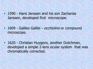
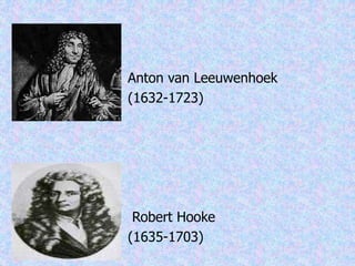
1. It discusses the historical development of the microscope from the 16th century to present day. Key figures mentioned include Hans Janssen, Galileo Galilei, Christian Huygens, Anton van Leeuwenhoek, and Robert Hooke.
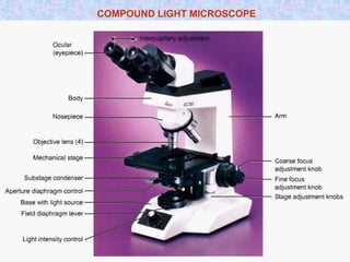
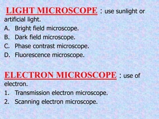
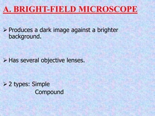
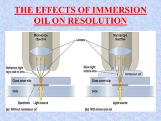
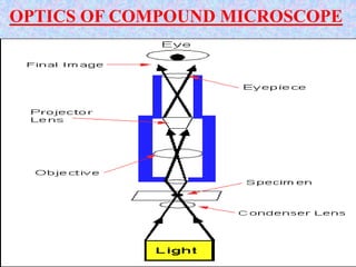
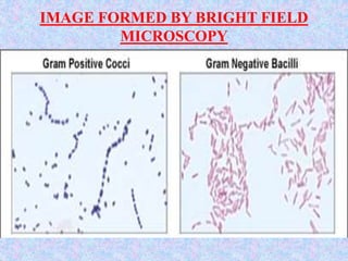
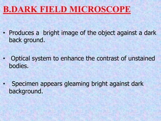
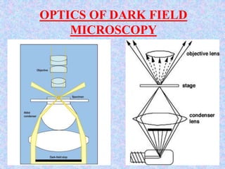
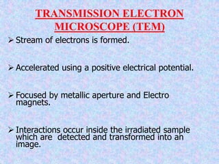
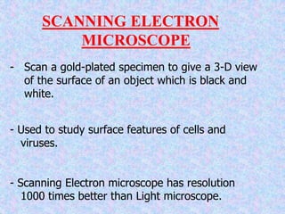
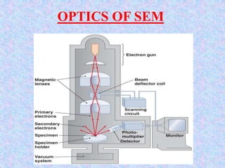
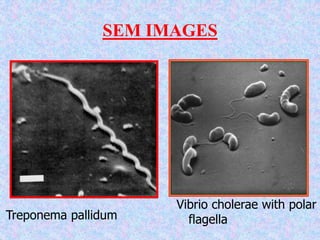
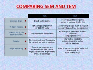
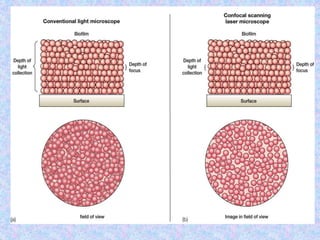
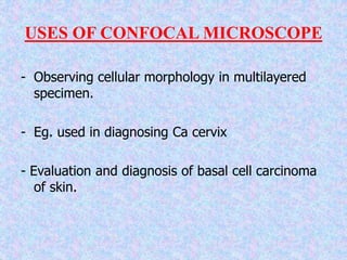
2. It describes different types of microscopes like brightfield, darkfield, phase contrast, fluorescence, electron (TEM and SEM), confocal, and scanning probe microscopes.
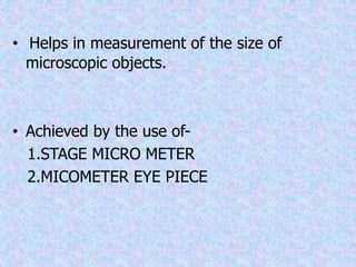
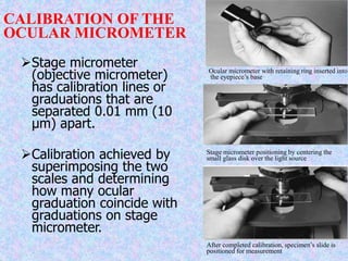
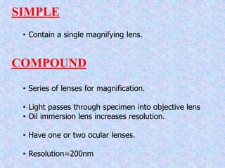
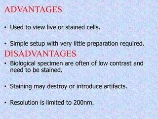
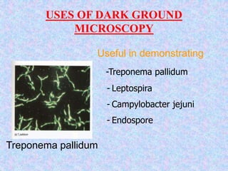
3. It explains various optical and imaging principles of different microscope types as well as their applications, advantages, and limitations. Microscopy techniques like micrometry, staining, and immunostaining are also covered.