Cytoskeleton structure. Microfilaments, microtubules and intermediate filaments.
The cytoskeleton is a network of filaments and tubules in all cells that provides structure, shape, and organization for organelles, composed mainly of microfilaments, microtubules, and intermediate filaments. Microfilaments, made of actin, aid in cell movement and division, while microtubules, constructed from tubulin, play critical roles in cell division and structure formation. Intermediate filaments provide structural support and maintain cell shape, contributing to overall cellular function.
Cytoskeleton
The cytoskeleton isa network of filaments and tubules that extends throughout a cell,
through the cytoplasm. It is found in all cells, though the proteins that is made of vary
between organisms.
• It supports the cell
• Gives it shape
• Organizes the organelles
(::)
4.
Structure of the
Cytoskeleton
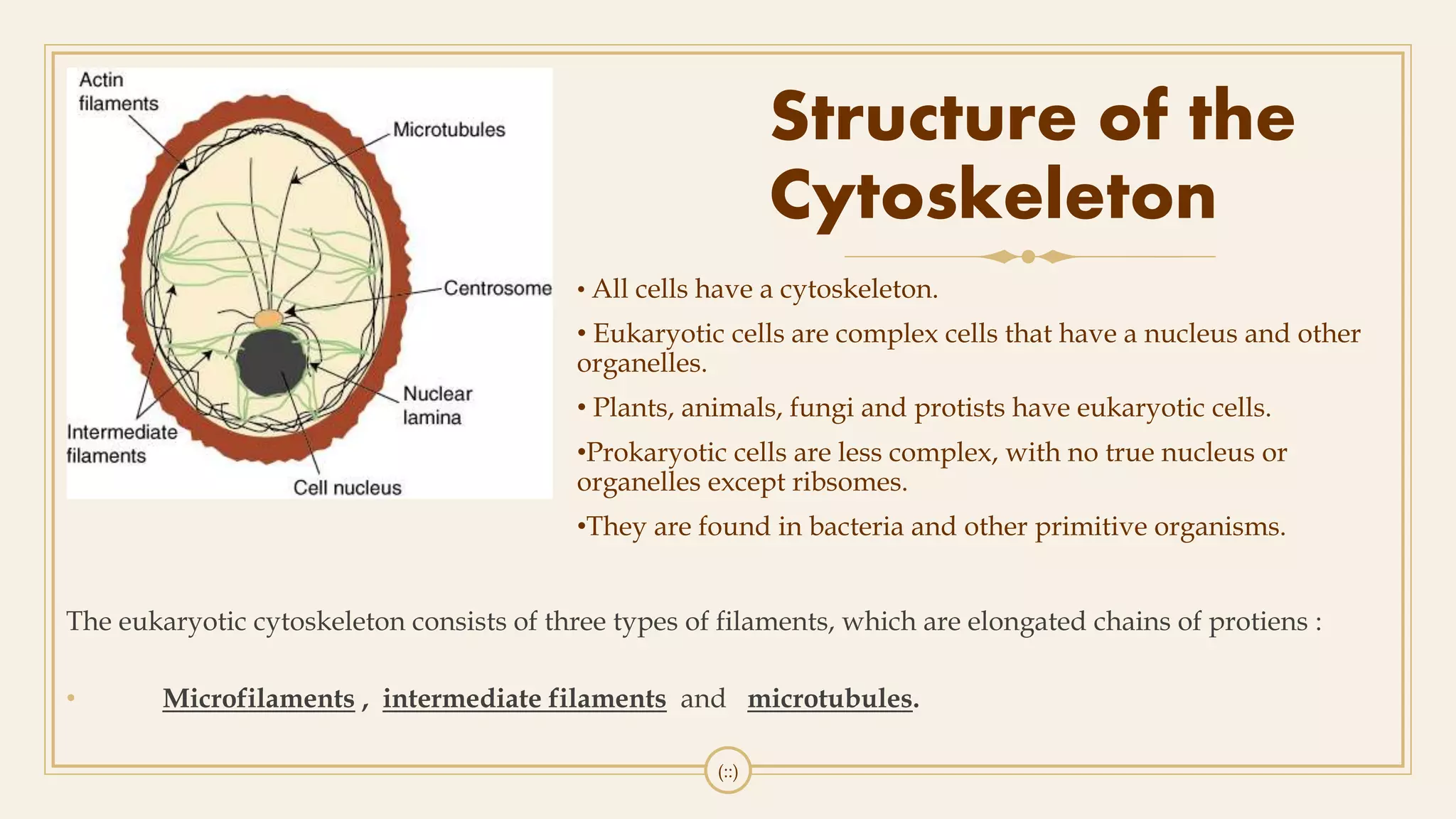
Theeukaryotic cytoskeleton consists of three types of filaments, which are elongated chains of protiens :
• Microfilaments , intermediate filaments and microtubules.
(::)
• All cells have a cytoskeleton.
• Eukaryotic cells are complex cells that have a nucleus and other
organelles.
• Plants, animals, fungi and protists have eukaryotic cells.
•Prokaryotic cells are less complex, with no true nucleus or
organelles except ribsomes.
•They are found in bacteria and other primitive organisms.
5.
Microfilaments
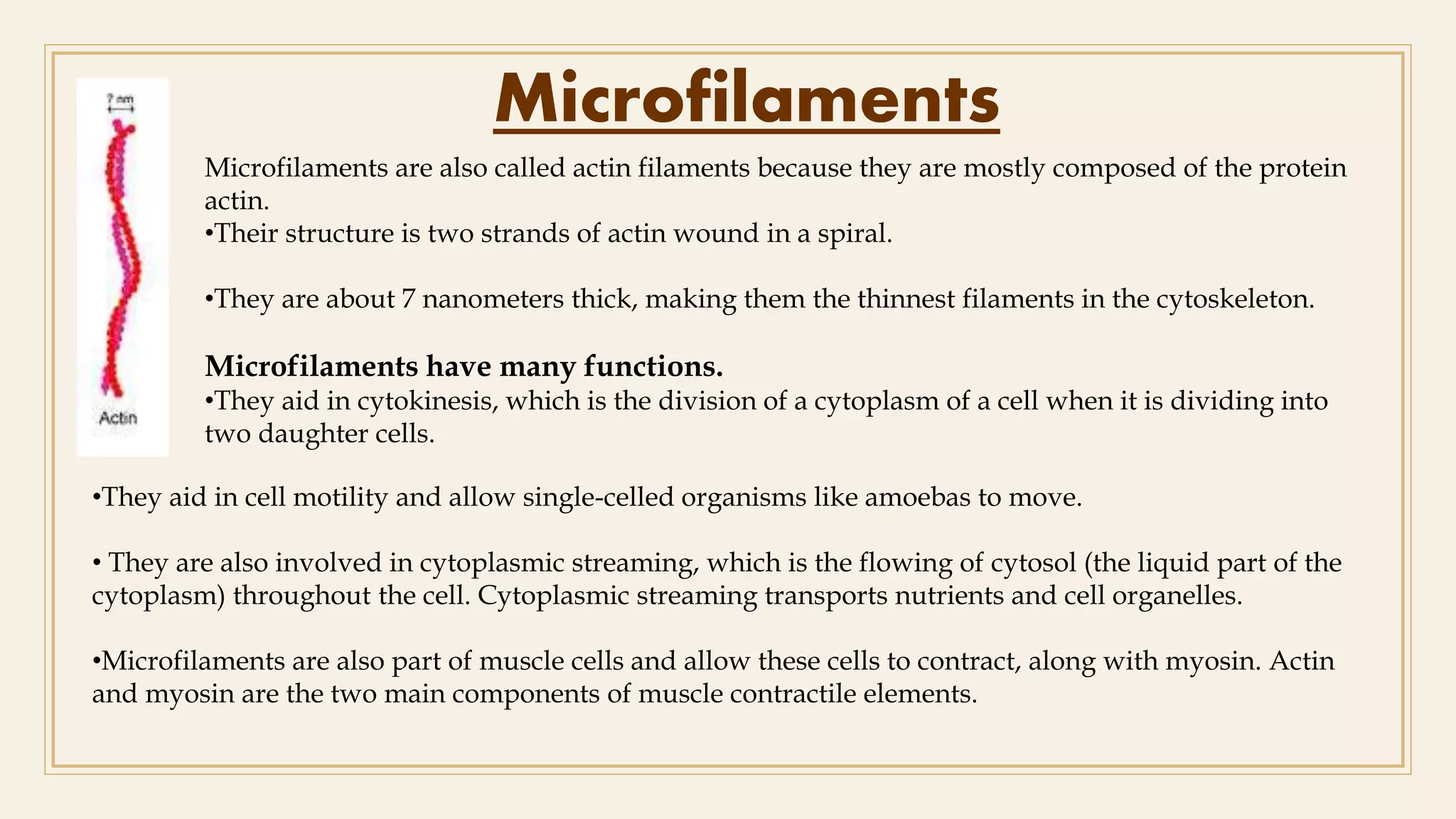
Microfilaments are alsocalled actin filaments because they are mostly composed of the protein
actin.
•Their structure is two strands of actin wound in a spiral.
•They are about 7 nanometers thick, making them the thinnest filaments in the cytoskeleton.
Microfilaments have many functions.
•They aid in cytokinesis, which is the division of a cytoplasm of a cell when it is dividing into
two daughter cells.
•They aid in cell motility and allow single-celled organisms like amoebas to move.
• They are also involved in cytoplasmic streaming, which is the flowing of cytosol (the liquid part of the
cytoplasm) throughout the cell. Cytoplasmic streaming transports nutrients and cell organelles.
•Microfilaments are also part of muscle cells and allow these cells to contract, along with myosin. Actin
and myosin are the two main components of muscle contractile elements.
6.
Microfilament Structure
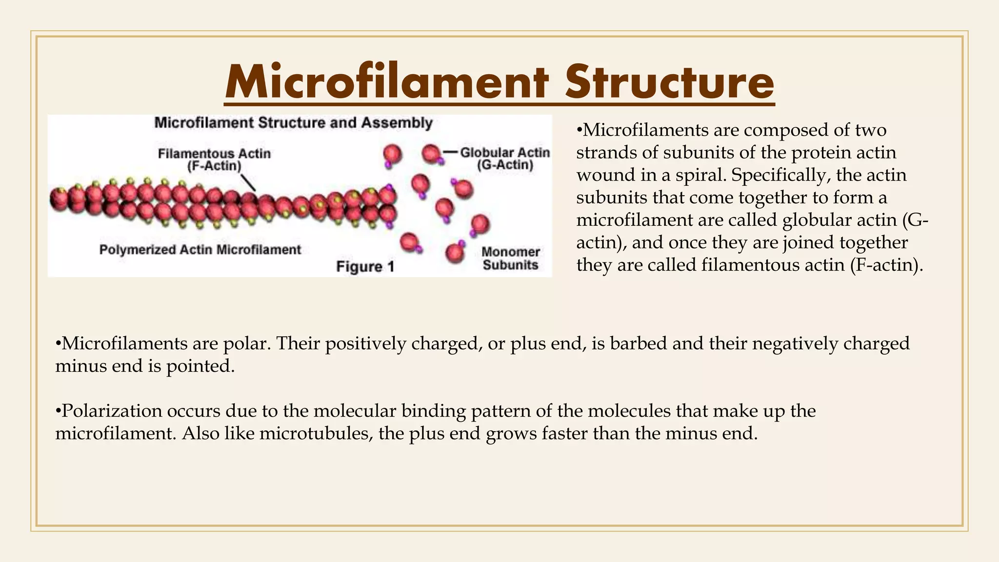
•Microfilaments arepolar. Their positively charged, or plus end, is barbed and their negatively charged
minus end is pointed.
•Polarization occurs due to the molecular binding pattern of the molecules that make up the
microfilament. Also like microtubules, the plus end grows faster than the minus end.
•Microfilaments are composed of two
strands of subunits of the protein actin
wound in a spiral. Specifically, the actin
subunits that come together to form a
microfilament are called globular actin (G-
actin), and once they are joined together
they are called filamentous actin (F-actin).
7.
Functions of Microfilaments
•Microfilamentsare the thinnest filaments of the cytoskeleton, with a diameter of about 6 to 7
nanometers.
•A microfilament begins to form when three G-actin proteins come together by themselves to form a
trimer. Then, more actin binds to the barbed end. The process of self-assembly is aided by autoclampin
proteins, which act as motors to help assemble the long strands that make up microfilaments.
•Two long strands of actin arrange in a spiral in order to form a microfilament.
•Muscle Contraction
•Cell Movement
•Cell Division
8.
Microtubules
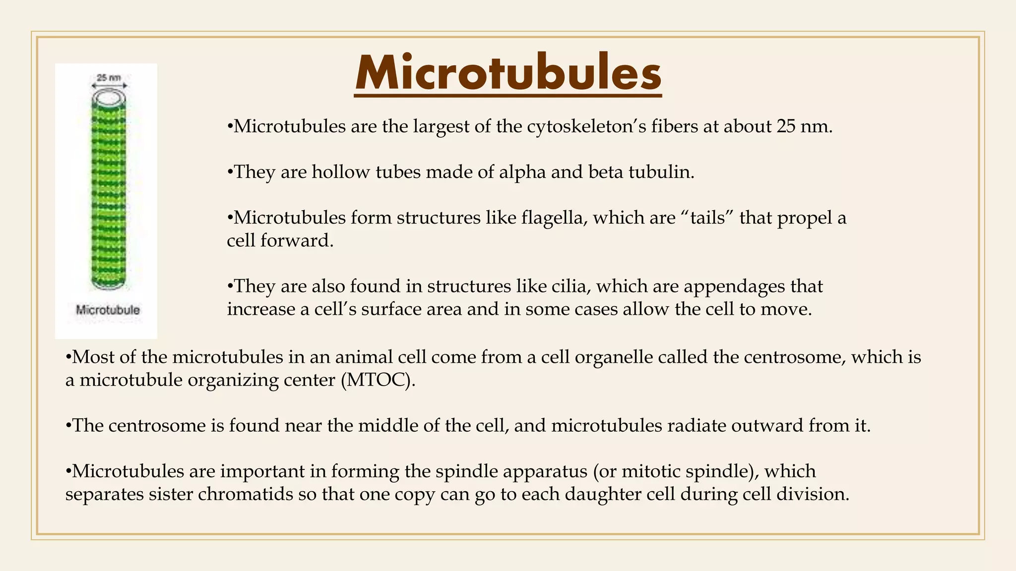
•Microtubules are thelargest of the cytoskeleton’s fibers at about 25 nm.
•They are hollow tubes made of alpha and beta tubulin.
•Microtubules form structures like flagella, which are “tails” that propel a
cell forward.
•They are also found in structures like cilia, which are appendages that
increase a cell’s surface area and in some cases allow the cell to move.
•Most of the microtubules in an animal cell come from a cell organelle called the centrosome, which is
a microtubule organizing center (MTOC).
•The centrosome is found near the middle of the cell, and microtubules radiate outward from it.
•Microtubules are important in forming the spindle apparatus (or mitotic spindle), which
separates sister chromatids so that one copy can go to each daughter cell during cell division.
9.
Microtubule Structure
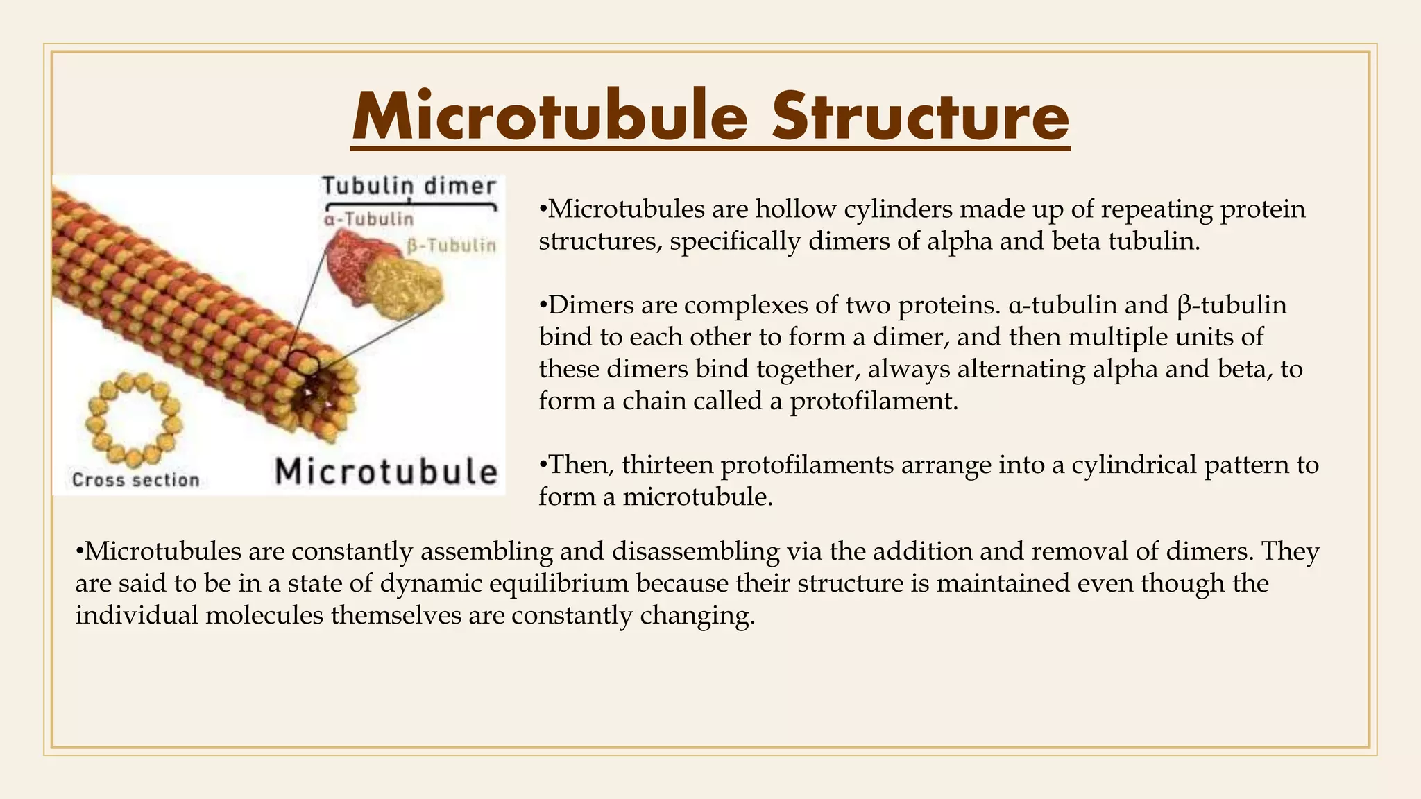
•Microtubules arehollow cylinders made up of repeating protein
structures, specifically dimers of alpha and beta tubulin.
•Dimers are complexes of two proteins. ɑ-tubulin and β-tubulin
bind to each other to form a dimer, and then multiple units of
these dimers bind together, always alternating alpha and beta, to
form a chain called a protofilament.
•Then, thirteen protofilaments arrange into a cylindrical pattern to
form a microtubule.
•Microtubules are constantly assembling and disassembling via the addition and removal of dimers. They
are said to be in a state of dynamic equilibrium because their structure is maintained even though the
individual molecules themselves are constantly changing.
10.
Function of Microtubules
•Microtubulesare polar molecules, with a positively charged end that grows relatively fast and a
negatively charged end that grows relatively slow.
• Protofilaments arrange themselves parallel to each other in a microtubule, so the positive end of the
microtubule always has beta subunits exposed, while the negative end has alpha subunits exposed.
Having polarity allows the microtubule to assemble in a specific way and function correctly.
•Cell Movement
•Cell Division
•Cell Transport
11.
Intermediate Filaments
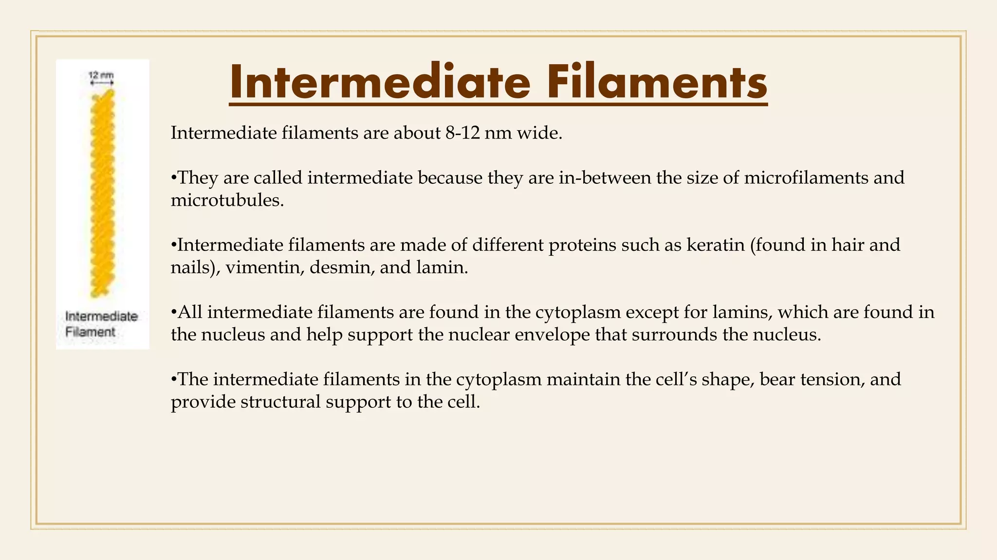
Intermediate filamentsare about 8-12 nm wide.
•They are called intermediate because they are in-between the size of microfilaments and
microtubules.
•Intermediate filaments are made of different proteins such as keratin (found in hair and
nails), vimentin, desmin, and lamin.
•All intermediate filaments are found in the cytoplasm except for lamins, which are found in
the nucleus and help support the nuclear envelope that surrounds the nucleus.
•The intermediate filaments in the cytoplasm maintain the cell’s shape, bear tension, and
provide structural support to the cell.
12.
Features of IntermediateFilaments
•The intermediate filaments are the type of cytoskeleton whose diameter is intermediate of the other two
types.
•Its diameter is about 10 nm (or ranges from 8 to 12 nm).
•An intermediate filament is comprised of two anti-parallel helices or dimers of varying protein sub-units.
It may be composed of any of a number of different proteins and form a ring around the cell nucleus.
•Intermediate filaments are stretchable. They can be extended from their initial length.
•The intermediate filaments are cytoplasmic.
•Unlike microfilaments and microtubules, the intermediate filaments do not exhibit polarity. This means
that they do not have a minus (-) end and a (+) end.
13.
Function of theCytoskeleton
As described above, the cytoskeleton has several functions :
•It gives the cell shape. This is especially important in cells without cell walls, such as animal cells, that
do not get their shape from a thick outer layer.
•It can also give the cell movement.
•The microfilaments and microtubules can disassemble, reassemble, and contract, allowing cells to crawl
and migrate .
•Microtubules help form structures like cilia and flagella that allow for cell movement.
•The cytoskeleton organizes the cell and keeps the cell’s organelles in place, but it also aids in the
movement of organelles throughout the cell. For example, during endocytosis, when a cell engulfs a
molecule, microfilaments pull the vesicle containing the engulfed particles into the cell. Similarly, the
cytoskeleton helps move chromosomes during cell division.
• The cytoskeleton is the “frame” of the cell, keeping structures in place, providing support, and giving
the cell a definite shape.
14.
Reference
1. The Cell:A Molecular Approach. (Cooper, Geoffrey M)
2. Journal of Cell Science. (Geli MI, Riezman H)
3. Cell Motility and the Cytoskeleton. (Frixione E)