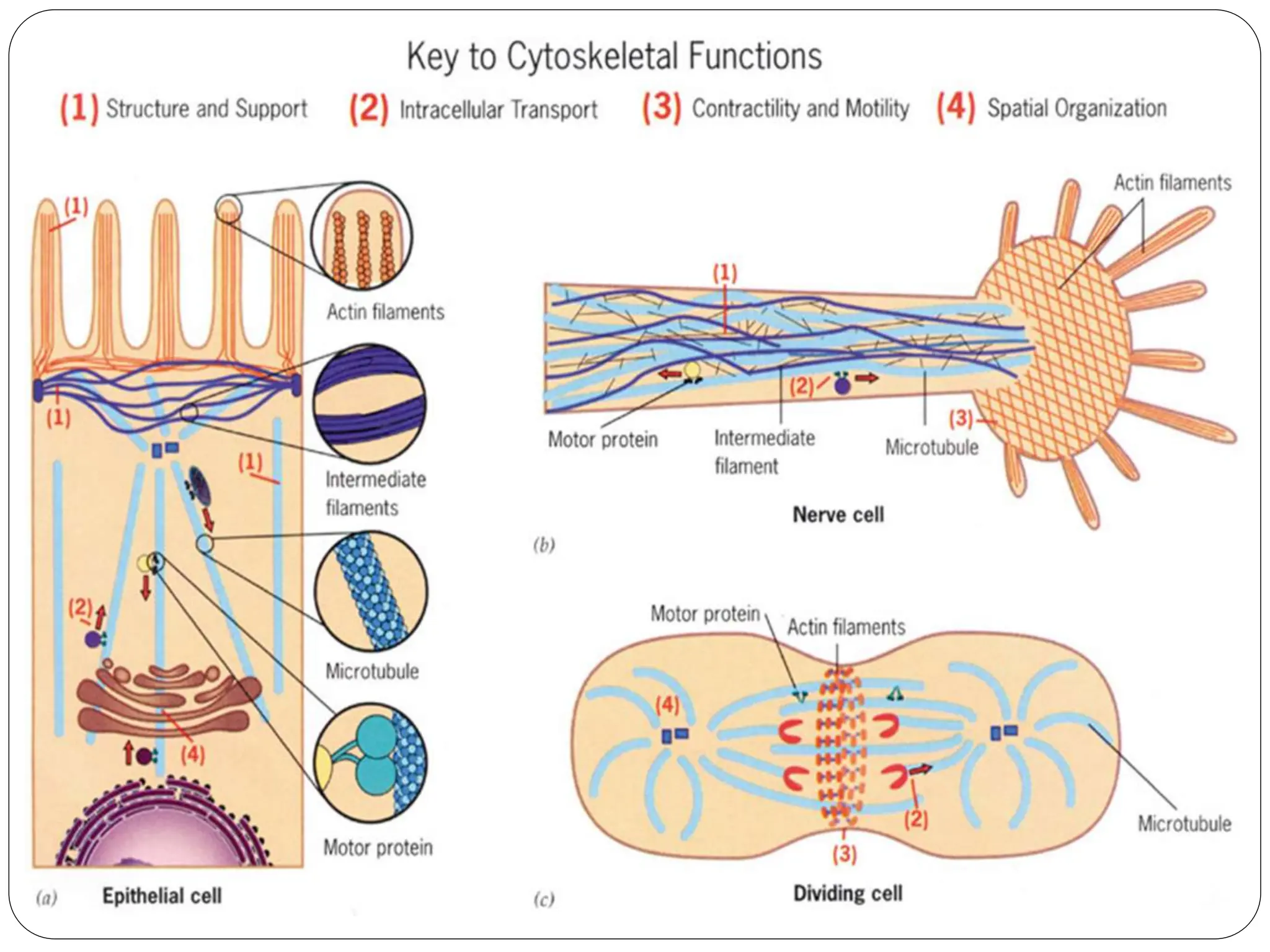
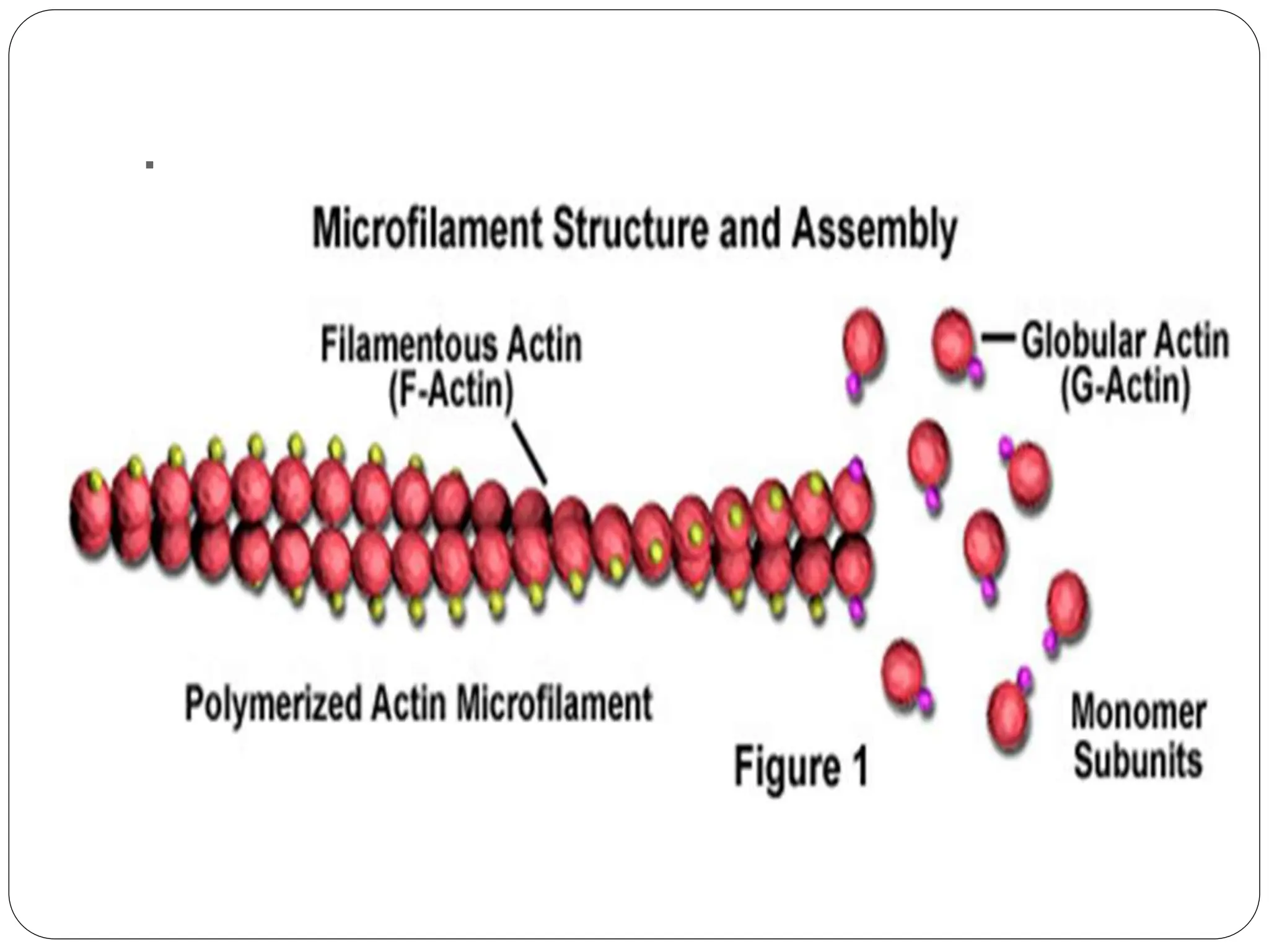
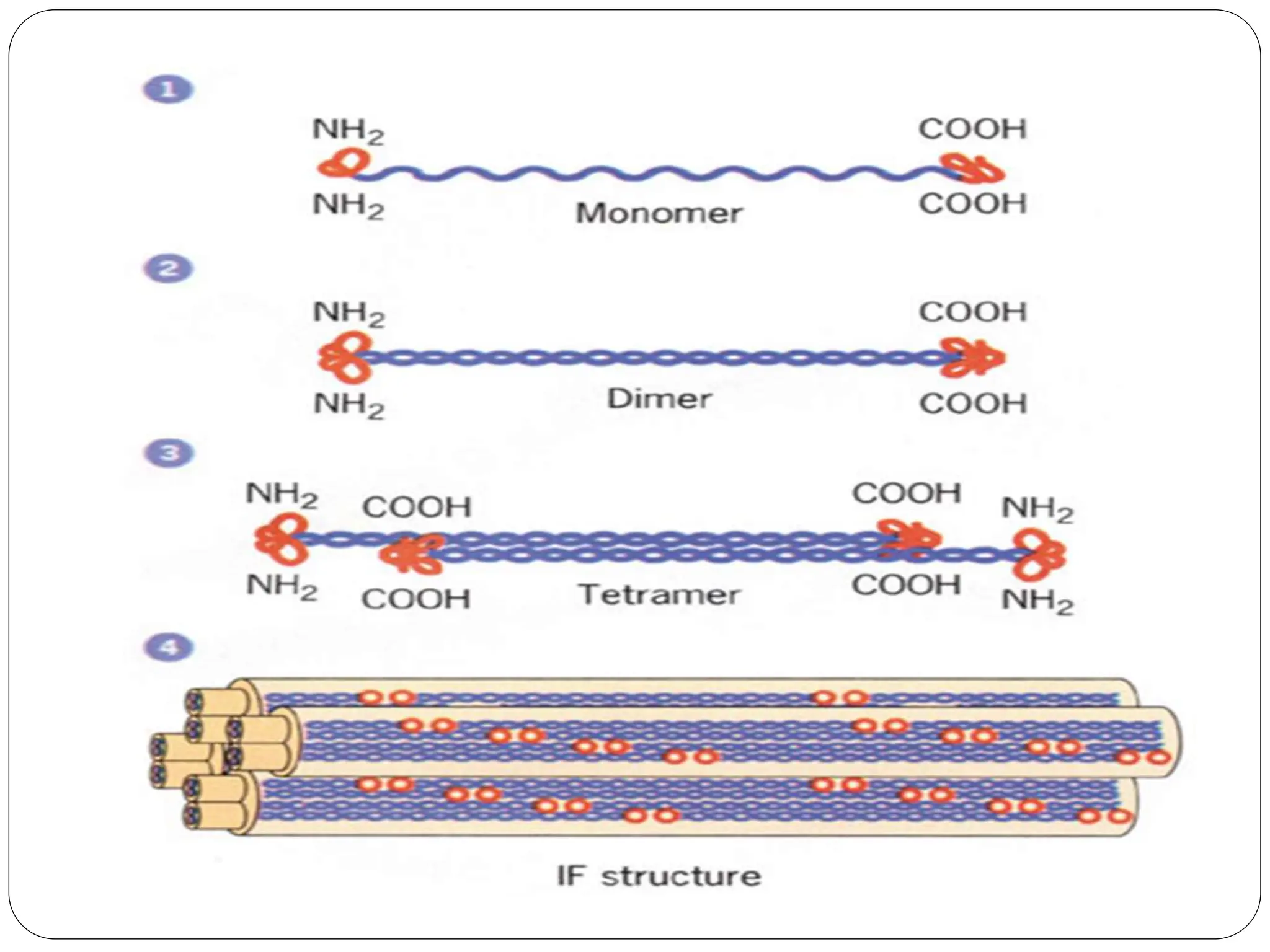
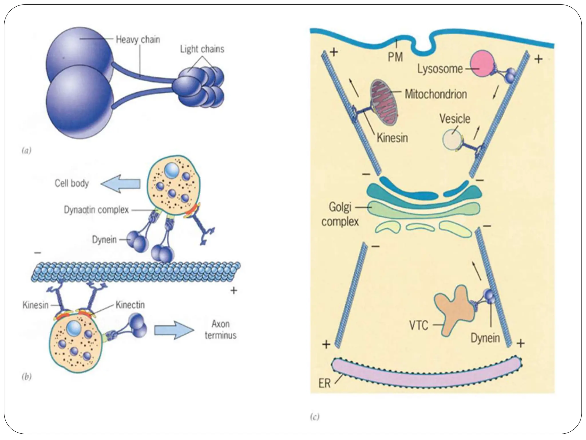
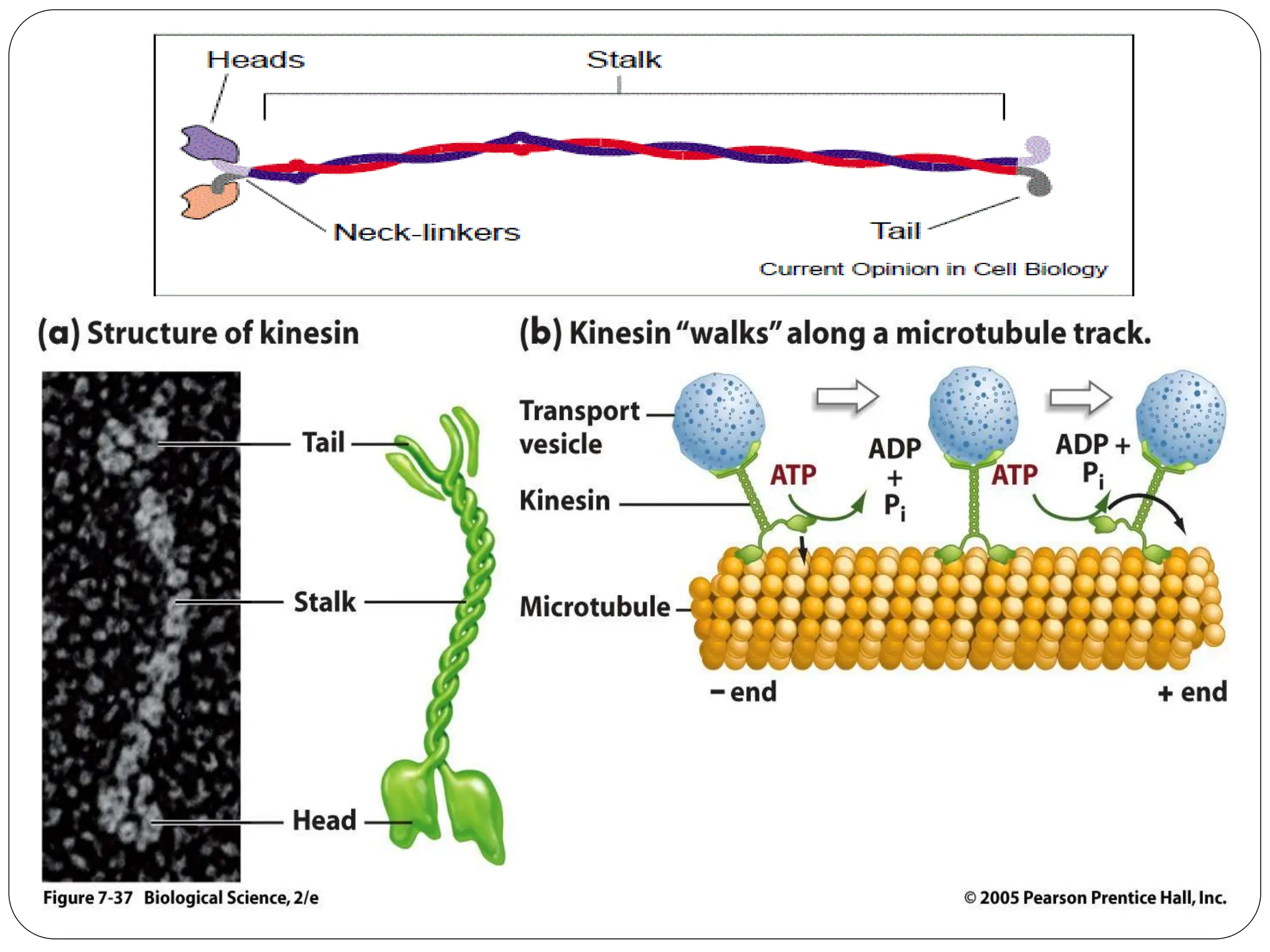
Cytoskeleton of the cell
Asma Saleh Alilesh
Cytoskeleton
 The cytoskeleton is composed of three
well-defined filamentous structures:
1. Microtubules,
2. microfilaments
3. intermediate filaments
 Each of the three types of cytoskeletal
filaments is a polymer of protein subunits
held together by weak, noncovalent bonds.
 They together form an elaborate interactive
network.
Cytoskeletal Functions
1- structural support that can determine the shape of
the cell and resist forces that tend to deform it.
2- responsible for positioning the various organelles
within the interior of the cell.
3- direct the movement of materials and organelles
within cells.
4- The force-generating apparatus that moves cells
from one
place to another
5- An essential component of the cell’s division
machinery. Cytoskeletal elements make up the
apparatus responsible for separating the
Microtubules
 Hollow, relatively rigid, tubular structures.
 They found in nearly every eukaryotic cell.
 Microtubules have an outer diameter of 25 nm and
a wall thickness of approximately 4 nm.
 The wall of a microtubule is composed of globular
proteins (tubulin) arranged in rows, termed
protofilaments, that are aligned parallel to the
long axis of the tubule.
 Each protofilament is assembled from dimeric
building blocks consisting of one α-tubulin and one
β-tubulin subunit.
 Has polarity.One end of a microtubule is known as
the plus end and is terminated by a row of β -
tubulin subunits The opposite end is the minus end
and is terminated by a row of α -tubulin subunits
 Microtubules function as structural supports and
organizers: (structure of cilia and flagella and the
axons of nerve cells.)
 Microtubules as agents of intracellular motility.
Microfilaments
 Flexible helical filament composed of globular
subunits of the protein actin In the presence of ATP,
actin monomers polymerize to form.
 They also called actin filament, F-actin.
 8 nm diameters
 Actin filaments have polarity, which is denoted as a
plus and minus end.
 Actin is an ATPase.
 They found in nearly every eukaryotic cell.
 functioning in cytokinesis, amoeboid movement,
and changes in cell shape and involved in
intracellular motile processes.
.
Intermediate filaments (IFs)
 Intermediate filaments are strong, flexible ropelike
filaments.
 Have only been identified in animal cells.
 10 nm diameter
 provide mechanical strength to cells that are
subjected to physical stress, including neurons,
muscle cells, and the epithelial cells that line the
body’s cavities.
 IFs are a chemically heterogeneous polypeptide
(Keratin and Keratin- like proteins), in humans, are
encoded by approximately 70 different genes.
 Have no enzymatic activity or polarity
Motor proteins
 The motor proteins of a cell are proteins convert
chemical energy (stored in ATP) into mechanical energy,
which is used to generate force to move cellular cargo
attached to the motor.
 Types of cellular cargo transported by these proteins
include vesicles, mitochondria, lysosomes,
chromosomes.
 A single cell may contain a hundred different motor
proteins, specialized for a different activity.
 motor proteins can be grouped into three broad
superfamilies: kinesins, dyneins, and myosins.
 Kinesins and dyneins move along microtubules.
 Myosins move along microfilaments.
Kinesins :
 Kinesin is a tetramer constructed from two
identical heavy chains and two identical light
chains.
 Heads that bind a microtubule and act as ATP-
hydrolyzing unit (engines).
 Move along microtubules toward their plus end.
Kinesin is therefore said to be a plus end-directed
microtubular motor.
 kinesins tend to move vesicles and organelles
(e.g., peroxisomes and mitochondria) in an
outward direction toward the cell’s plasma
membrane.
Dynein:
 Cytoplasmic dynein is a huge protein (molecular
mass of approximately 1.5 million daltons) composed
of two identical heavy chains and a variety of
intermediate and light chains.
 Dynein moves along a microtubule toward the minus
end—opposite that of kinesin.
- Function as:
1. As a force-generating agent in positioning the
spindle and moving chromosomes during mitosis
2. As a minus end–directed microtubular motor with a
role in positioning the centrosome and Golgi complex
and moving organelles, vesicles, and particles through
the cytoplasm.

cytoskeleton (Microtubules, microfilaments intermediate filaments)

  • 1.
    Cytoskeleton of thecell Asma Saleh Alilesh
  • 2.
    Cytoskeleton  The cytoskeletonis composed of three well-defined filamentous structures: 1. Microtubules, 2. microfilaments 3. intermediate filaments  Each of the three types of cytoskeletal filaments is a polymer of protein subunits held together by weak, noncovalent bonds.  They together form an elaborate interactive network.
  • 4.
    Cytoskeletal Functions 1- structuralsupport that can determine the shape of the cell and resist forces that tend to deform it. 2- responsible for positioning the various organelles within the interior of the cell. 3- direct the movement of materials and organelles within cells. 4- The force-generating apparatus that moves cells from one place to another 5- An essential component of the cell’s division machinery. Cytoskeletal elements make up the apparatus responsible for separating the
  • 5.
    Microtubules  Hollow, relativelyrigid, tubular structures.  They found in nearly every eukaryotic cell.  Microtubules have an outer diameter of 25 nm and a wall thickness of approximately 4 nm.  The wall of a microtubule is composed of globular proteins (tubulin) arranged in rows, termed protofilaments, that are aligned parallel to the long axis of the tubule.  Each protofilament is assembled from dimeric building blocks consisting of one α-tubulin and one β-tubulin subunit.
  • 7.
     Has polarity.Oneend of a microtubule is known as the plus end and is terminated by a row of β - tubulin subunits The opposite end is the minus end and is terminated by a row of α -tubulin subunits  Microtubules function as structural supports and organizers: (structure of cilia and flagella and the axons of nerve cells.)  Microtubules as agents of intracellular motility.
  • 8.
    Microfilaments  Flexible helicalfilament composed of globular subunits of the protein actin In the presence of ATP, actin monomers polymerize to form.  They also called actin filament, F-actin.  8 nm diameters  Actin filaments have polarity, which is denoted as a plus and minus end.  Actin is an ATPase.  They found in nearly every eukaryotic cell.  functioning in cytokinesis, amoeboid movement, and changes in cell shape and involved in intracellular motile processes.
  • 9.
  • 10.
    Intermediate filaments (IFs) Intermediate filaments are strong, flexible ropelike filaments.  Have only been identified in animal cells.  10 nm diameter  provide mechanical strength to cells that are subjected to physical stress, including neurons, muscle cells, and the epithelial cells that line the body’s cavities.  IFs are a chemically heterogeneous polypeptide (Keratin and Keratin- like proteins), in humans, are encoded by approximately 70 different genes.  Have no enzymatic activity or polarity
  • 12.
    Motor proteins  Themotor proteins of a cell are proteins convert chemical energy (stored in ATP) into mechanical energy, which is used to generate force to move cellular cargo attached to the motor.  Types of cellular cargo transported by these proteins include vesicles, mitochondria, lysosomes, chromosomes.  A single cell may contain a hundred different motor proteins, specialized for a different activity.  motor proteins can be grouped into three broad superfamilies: kinesins, dyneins, and myosins.  Kinesins and dyneins move along microtubules.  Myosins move along microfilaments.
  • 14.
    Kinesins :  Kinesinis a tetramer constructed from two identical heavy chains and two identical light chains.  Heads that bind a microtubule and act as ATP- hydrolyzing unit (engines).  Move along microtubules toward their plus end. Kinesin is therefore said to be a plus end-directed microtubular motor.  kinesins tend to move vesicles and organelles (e.g., peroxisomes and mitochondria) in an outward direction toward the cell’s plasma membrane.
  • 16.
    Dynein:  Cytoplasmic dyneinis a huge protein (molecular mass of approximately 1.5 million daltons) composed of two identical heavy chains and a variety of intermediate and light chains.  Dynein moves along a microtubule toward the minus end—opposite that of kinesin. - Function as: 1. As a force-generating agent in positioning the spindle and moving chromosomes during mitosis 2. As a minus end–directed microtubular motor with a role in positioning the centrosome and Golgi complex and moving organelles, vesicles, and particles through the cytoplasm.