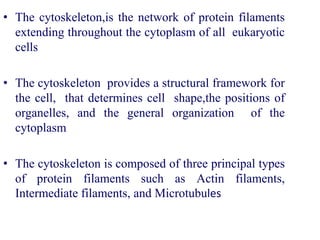
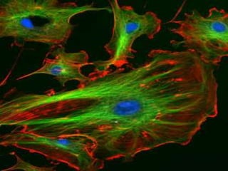
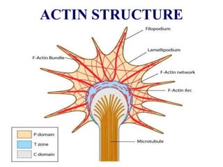
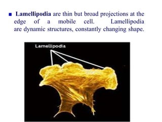
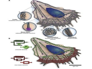
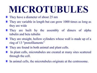
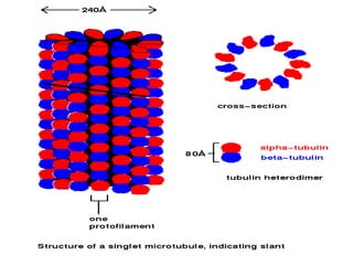
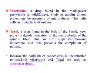
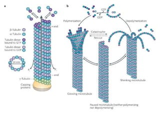
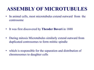
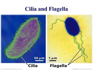
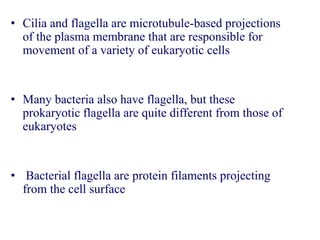
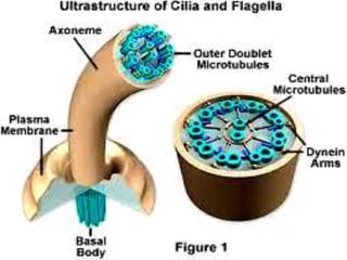
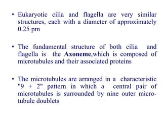
SlideShare a Scribd company logo
CYTOSKELETON
• The cytoskeleton,is the network of protein filaments
extending throughout the cytoplasm of all eukaryotic
cells
• The cytoskeleton provides a structural framework for
the cell, that determines cell shape,the positions of
organelles, and the general organization of the
cytoplasm
• The cytoskeleton is composed of three principal types
of protein filaments such as Actin filaments,
Intermediate filaments, and Microtubules
FUNCTIONS OF CYTOSKELETON
• Establishing cell shape
• Providing mechanical strength
• Locomotion
• Chromosome separation in mitosis and meiosis
• Intracellular transport of organelles
• The major cytoskeletal protein of most cells is Actin
• which polymerizes to form actin filaments- thin,
flexible fibers approximately 7 nm in diameter and
up to several micrometers in length
• Being the thinnest of the cytoskeletal filaments,This
are also called Microfilaments
(Actin cytoskeleton stained with phalloidin
• In skeletal muscle fibers they are called Thin
filaments
• Generate Cytoplasmic streaming in some cells
• Generate Locomotion in cells such as white blood
cells
• Interact with myosin ("thick") filaments in skeletal
muscle fibres to provide the force of muscular
contraction
ACTIN STRUCTURE
• Filopodia (also called Microspikes) are long, thin and
transient structures that extend out from the cell surface.
• Bundles of parallel actin filaments, with their plus ends
oriented toward the filopodial tip
• Microvilli are shorter and more numerous protrusions of
the cell surface found in some cells.
• Tightly bundled actin filaments within these structures
their plus ends oriented toward the tip
• Small cross-linking proteins such as fimbrin and
villin bind actin filaments together within microvilli
provide stiffness
Lamellipodia are thin but broad projections at the
edge of a mobile cell. Lamellipodia
are dynamic structures, constantly changing shape.
• Stress fibers form when a cell makes stable
connections to a substrate
• Bundles of actin filaments extend from the cell
surface through the cytosol. The actin filaments,
whose plus ends are oriented toward the cell
surface
• Myosin mediates sliding of anti-parallel actin
filaments during contraction of stress fibers
• -Actinin may cross-link actin filaments within
stress fibers
• Some cells have a cytoskeletal network just inside the
plasma membrane that includes actin along with various
other proteins such as spectrin
• This cytoskeleton has a role in maintaining cell shape.
An example is found in erythrocytes
• Spectrin is an actin-binding protein that forms an
elongated tetrameric complex having an actin-binding
domain at each end
• With short actin filaments, spectrin forms a cytoskeletal
network on the cytosolic surface of the plasma
membrane of erythrocytes and some other cells
Spectrin - actin-binding
ORGANISATION OF ACTIN FILAMENTS
• Individual actin filaments are assembled into two general types
of structures called Actin bundles and Actin networks,
which play different roles in the cell.
• The first type of bundle, containing closely spaced actin
filaments aligned in parallel supports projections of the plasma
membrane, such as microvilli
• In these bundles, all the filaments have the same polarity, with
their barbed ends adjacent to the plasma membrane
• An example of a bundling protein involved in the formation of
these structures is fimbrin
• which was first isolated from intestinal microvilli and later
found in surface projections of a wide variety of cell types
• The second type of actin bundle is composed of
filaments that are more widely spaced, allowing the
bundle to contract
• In contrast to fimbrin, actin binds to actin as a
dimer, each subunit of which is a 102 kd protein
containing a single actin-binding site
• The actin filaments in networks are held together by large actin-
binding proteins, such as filamin.
• Filamin binds actin as a dimer of two 280 kd subunits. The
actin-binding domains and dimerization domains are at opposite
ends of each subunit
• so the filamin dimer is a flexible V-shaped molecule with actin-
binding domains at the ends of each arm.
• As a result, filamin forms cross-links between orthogonal actin
filaments, creating a loose three-dimensional mesh .
They have a diameter of about 25 nm
They are variable in length but can grow 1000 times as long as
they are wide
They are built by the assembly of dimers of alpha
tubulin and beta tubulin
They are straight, hollow cylinders whose wall is made up of a
ring of 13 "protofilaments”
They are found in both animal and plant cells.
In plant cells, microtubules are created at many sites scattered
through the cell.
In animal cells, the microtubules originate at the centrosome.
• The attached end is called the minus end; the other
end is the plus end
• grow at the plus end by the polymerization of tubulin
dimers (powered by the hydrolysis of GTP)
 Microtubules participate in a wide variety of cell
activities
• Most involve motion. The motion is provided by
protein "motors" that use the energy of ATP to move
along micotubule
Microtubule motors
• There are two major groups of microtubule motors:
• kinesins most of these move toward the plus end of the
microtubules
• dyneins which move toward the minus end
Some examples:
• The migration of chromosomes in mitosis and
meiosis takes place on microtubules that make up the
spindle fibres
• Both Kinesins and Dyneins are used as motors.
• The rapid transport of organelles, like vesicles and
mitochondria, along the axons of neurons takes
place along microtubules
• The motors used are kinesins
Vincristine, a drug found in the Madagascar
periwinkle (a wildflower), binds to tubulin dimers
preventing the assembly of microtubules. This halts
cells in metaphase of mitosis.
Taxol, a drug found in the bark of the Pacific yew,
prevents depolymerization of the microtubules of the
spindle fiber. This, in turn, stops chromosome
movement, and thus prevents the completion of
mitosis.
• Because the hallmark of cancer cells is uncontrolled
mitosis,both vincristine and Taxol are used as
anticancer drugs .
Madagascar periwinkle
Vincristine
Taxol
Pacific yew
STRUCTURE OF MICROTUBULE
• The building blocks of microtubules are tubulin dimers
consisting of two closely related 55 kd polypeptides: α-
tubulin and β-tubulin
• Like actin, both α and β tubulin are encoded by small
families of related genes
• In addition, a third type of tubulin (y-tubulin) is concentrated
in the centrosome where it plays a critical role in initiating
microtubule assembly
• Tubulin dimers can depolymerize as well as polymerize, and
micro-tubules can undergo rapid cycles of assembly and
disassembly.
ASSEMBLY OF MICROTUBULES
• In animal cells, most microtubules extend outward from the
centrosome
• It was first discovered by Theoder Boveri in 1888
• During mitosis Microtubules similarly extend outward from
duplicated centrosomes to form mititic spindle
• which is responsible for the separation and distribution of
chromosomes to daughter cells
• Microtubules grow by the addition of tubulin to
their plus ends
• which extend outward from the centrosome
toward the cell periphery, so the role of the
centrosome is to initiate microtubule growth.
• The centrosomes of most animal cells contain a
pair of centrioles, oriented perpendicular to each
other
• The centrioles are cylindrical structures based on
nine triplets of microtubules, similar to the basal
bodies of cilia and flagella.
Cilia and Flagella
• Cilia and flagella are microtubule-based projections
of the plasma membrane that are responsible for
movement of a variety of eukaryotic cells
• Many bacteria also have flagella, but these
prokaryotic flagella are quite different from those of
eukaryotes
• Bacterial flagella are protein filaments projecting
from the cell surface
• Eukaryotic cilia and flagella are very similar
structures, each with a diameter of approximately
0.25 pm
• The fundamental structure of both cilia and
flagella is the Axoneme,which is composed of
microtubules and their associated proteins
• The microtubules are arranged in a characteristic
"9 + 2" pattern in which a central pair of
microtubules is surrounded by nine outer micro-
tubule doublets
THE CYTOSKELETON

More Related Content

What's hot

Intermediate filaments
Intermediate filamentsIntermediate filaments
Intermediate filaments
Sarita Nanda
 
The Cytoskeleton- An overview
The Cytoskeleton- An overviewThe Cytoskeleton- An overview
The Cytoskeleton- An overview
Dariyus Kabraji
 
Golgi complex structure and functions
Golgi complex structure and functionsGolgi complex structure and functions
Golgi complex structure and functions
bikashsingh2510
 
Cyclin dependent kinases
Cyclin dependent kinasesCyclin dependent kinases
Cyclin dependent kinases
Mandira bhosale
 
Peroxisome
PeroxisomePeroxisome
Peroxisome
Dilip Pandya
 
Cytoskeleton - microtubules ,microfilaments and intermediate filaments
Cytoskeleton - microtubules ,microfilaments and intermediate filamentsCytoskeleton - microtubules ,microfilaments and intermediate filaments
Cytoskeleton - microtubules ,microfilaments and intermediate filaments
BIOTECH SIMPLIFIED
 
ribosomes
ribosomesribosomes
ribosomes
Dilip Pandya
 
Microtubules
MicrotubulesMicrotubules
Microtubules
Sarita Nanda
 
Structure of model membrane
Structure of model membrane Structure of model membrane
Structure of model membrane
gohil sanjay bhagvanji
 
Vacuole
VacuoleVacuole
Vacuole
Dilip Pandya
 
Nucleus Structure
Nucleus StructureNucleus Structure
Nucleus Structure
Morgan Stanley
 
Cell motility cilia and flagella
Cell motility  cilia and flagellaCell motility  cilia and flagella
Cell motility cilia and flagella
ALLIENU
 
Cell cell interaction
Cell cell interactionCell cell interaction
Cell cell interaction
Praveen Garg
 
Plasma membrane
Plasma membranePlasma membrane
Plasma membrane
Rajpal Choudhary
 
Structure and functions of endoplasmic reticulum
Structure and functions of endoplasmic reticulumStructure and functions of endoplasmic reticulum
Structure and functions of endoplasmic reticulum
ICHHA PURAK
 
Peroxisomes
PeroxisomesPeroxisomes
Peroxisomes
Sam Shaikh
 
Structure and function of plasma membrane 2
Structure and function of plasma membrane 2Structure and function of plasma membrane 2
Structure and function of plasma membrane 2
ICHHA PURAK
 
Biological membrane & its chemical composition
Biological membrane & its chemical compositionBiological membrane & its chemical composition
Biological membrane & its chemical composition
Pradeep Singh Narwat
 
Nucleus
NucleusNucleus
Nucleus
Hema Mallika
 

What's hot (20)

Intermediate filaments
Intermediate filamentsIntermediate filaments
Intermediate filaments
 
The Cytoskeleton- An overview
The Cytoskeleton- An overviewThe Cytoskeleton- An overview
The Cytoskeleton- An overview
 
Golgi complex structure and functions
Golgi complex structure and functionsGolgi complex structure and functions
Golgi complex structure and functions
 
Cyclin dependent kinases
Cyclin dependent kinasesCyclin dependent kinases
Cyclin dependent kinases
 
Peroxisome
PeroxisomePeroxisome
Peroxisome
 
Cytoskeleton - microtubules ,microfilaments and intermediate filaments
Cytoskeleton - microtubules ,microfilaments and intermediate filamentsCytoskeleton - microtubules ,microfilaments and intermediate filaments
Cytoskeleton - microtubules ,microfilaments and intermediate filaments
 
Nuclear pores
Nuclear poresNuclear pores
Nuclear pores
 
ribosomes
ribosomesribosomes
ribosomes
 
Microtubules
MicrotubulesMicrotubules
Microtubules
 
Structure of model membrane
Structure of model membrane Structure of model membrane
Structure of model membrane
 
Vacuole
VacuoleVacuole
Vacuole
 
Nucleus Structure
Nucleus StructureNucleus Structure
Nucleus Structure
 
Cell motility cilia and flagella
Cell motility  cilia and flagellaCell motility  cilia and flagella
Cell motility cilia and flagella
 
Cell cell interaction
Cell cell interactionCell cell interaction
Cell cell interaction
 
Plasma membrane
Plasma membranePlasma membrane
Plasma membrane
 
Structure and functions of endoplasmic reticulum
Structure and functions of endoplasmic reticulumStructure and functions of endoplasmic reticulum
Structure and functions of endoplasmic reticulum
 
Peroxisomes
PeroxisomesPeroxisomes
Peroxisomes
 
Structure and function of plasma membrane 2
Structure and function of plasma membrane 2Structure and function of plasma membrane 2
Structure and function of plasma membrane 2
 
Biological membrane & its chemical composition
Biological membrane & its chemical compositionBiological membrane & its chemical composition
Biological membrane & its chemical composition
 
Nucleus
NucleusNucleus
Nucleus
 

Similar to THE CYTOSKELETON

Cytoskeleton structure. Microfilaments, microtubules and intermediate filaments.
Cytoskeleton structure. Microfilaments, microtubules and intermediate filaments.Cytoskeleton structure. Microfilaments, microtubules and intermediate filaments.
Cytoskeleton structure. Microfilaments, microtubules and intermediate filaments.
GURUNath18
 
Group 6 presentation(cytoskeleton).pptx
Group 6 presentation(cytoskeleton).pptxGroup 6 presentation(cytoskeleton).pptx
Group 6 presentation(cytoskeleton).pptx
RoyBisenti
 
Cytoskeleton
CytoskeletonCytoskeleton
Cytoskeleton
Gauri Haval
 
Cytoskeleton
CytoskeletonCytoskeleton
Cytoskeleton
Ali Zia Kamboh
 
CYTOSKELETON STRUCTURE.ppt
CYTOSKELETON STRUCTURE.pptCYTOSKELETON STRUCTURE.ppt
CYTOSKELETON STRUCTURE.ppt
EstherShobhaR
 
CMB_L28_2008s.ppt
CMB_L28_2008s.pptCMB_L28_2008s.ppt
CMB_L28_2008s.ppt
GazmendBojaj1
 
Microtubules by jimmy
Microtubules by jimmyMicrotubules by jimmy
Microtubules by jimmy
Ahmad Ali
 
Cell movement
Cell movementCell movement
Cell movement
GOPAL KUMBHANI
 
Cytoskeleton
Cytoskeleton Cytoskeleton
Cytoskeleton
Muhammadasif909
 
CYTOSKELETON presentation by (Aneeq) (1).pptx
CYTOSKELETON presentation  by (Aneeq) (1).pptxCYTOSKELETON presentation  by (Aneeq) (1).pptx
CYTOSKELETON presentation by (Aneeq) (1).pptx
AneeqJaved
 
Cytoskeleton
CytoskeletonCytoskeleton
Cytoskeleton
Muzna Kashaf
 
Microtubules & filaments.pptx 2
Microtubules & filaments.pptx 2Microtubules & filaments.pptx 2
Microtubules & filaments.pptx 2
Arv080
 
Cytoskeleton
CytoskeletonCytoskeleton
Cytoskeleton
Geonyzl Alviola
 
Cytoskeleton
Cytoskeleton  Cytoskeleton
Cytoskeleton
zooomaniazooostory
 
Cytoskeleton[1]
Cytoskeleton[1]Cytoskeleton[1]
Cytoskeleton[1]
Dilip Pandya
 
structure and functions of cytoskeletons
structure and functions of cytoskeletonsstructure and functions of cytoskeletons
structure and functions of cytoskeletons
zaferkaragozlu
 
HUMAN Cell _RDP
HUMAN Cell _RDPHUMAN Cell _RDP
HUMAN Cell _RDP
rishi2789
 
Introduction to Anatomy (Lec 4) 2021-2022.pdf
Introduction to Anatomy (Lec 4) 2021-2022.pdfIntroduction to Anatomy (Lec 4) 2021-2022.pdf
Introduction to Anatomy (Lec 4) 2021-2022.pdf
M SHAKEEL
 
Cytoskeleton
CytoskeletonCytoskeleton
Cytoskeleton
Bharat Lodha
 

Similar to THE CYTOSKELETON (20)

Cytoskeleton structure. Microfilaments, microtubules and intermediate filaments.
Cytoskeleton structure. Microfilaments, microtubules and intermediate filaments.Cytoskeleton structure. Microfilaments, microtubules and intermediate filaments.
Cytoskeleton structure. Microfilaments, microtubules and intermediate filaments.
 
Group 6 presentation(cytoskeleton).pptx
Group 6 presentation(cytoskeleton).pptxGroup 6 presentation(cytoskeleton).pptx
Group 6 presentation(cytoskeleton).pptx
 
Cytoskeleton
CytoskeletonCytoskeleton
Cytoskeleton
 
Cytoskeleton
CytoskeletonCytoskeleton
Cytoskeleton
 
CYTOSKELETON STRUCTURE.ppt
CYTOSKELETON STRUCTURE.pptCYTOSKELETON STRUCTURE.ppt
CYTOSKELETON STRUCTURE.ppt
 
CMB_L28_2008s.ppt
CMB_L28_2008s.pptCMB_L28_2008s.ppt
CMB_L28_2008s.ppt
 
Microtubules by jimmy
Microtubules by jimmyMicrotubules by jimmy
Microtubules by jimmy
 
Cell movement
Cell movementCell movement
Cell movement
 
Cytoskeleton
CytoskeletonCytoskeleton
Cytoskeleton
 
Cytoskeleton
Cytoskeleton Cytoskeleton
Cytoskeleton
 
CYTOSKELETON presentation by (Aneeq) (1).pptx
CYTOSKELETON presentation  by (Aneeq) (1).pptxCYTOSKELETON presentation  by (Aneeq) (1).pptx
CYTOSKELETON presentation by (Aneeq) (1).pptx
 
Cytoskeleton
CytoskeletonCytoskeleton
Cytoskeleton
 
Microtubules & filaments.pptx 2
Microtubules & filaments.pptx 2Microtubules & filaments.pptx 2
Microtubules & filaments.pptx 2
 
Cytoskeleton
CytoskeletonCytoskeleton
Cytoskeleton
 
Cytoskeleton
Cytoskeleton  Cytoskeleton
Cytoskeleton
 
Cytoskeleton[1]
Cytoskeleton[1]Cytoskeleton[1]
Cytoskeleton[1]
 
structure and functions of cytoskeletons
structure and functions of cytoskeletonsstructure and functions of cytoskeletons
structure and functions of cytoskeletons
 
HUMAN Cell _RDP
HUMAN Cell _RDPHUMAN Cell _RDP
HUMAN Cell _RDP
 
Introduction to Anatomy (Lec 4) 2021-2022.pdf
Introduction to Anatomy (Lec 4) 2021-2022.pdfIntroduction to Anatomy (Lec 4) 2021-2022.pdf
Introduction to Anatomy (Lec 4) 2021-2022.pdf
 
Cytoskeleton
CytoskeletonCytoskeleton
Cytoskeleton
 

More from Mariya Raju

MANUFACTURE OF BREAD
MANUFACTURE OF BREADMANUFACTURE OF BREAD
MANUFACTURE OF BREAD
Mariya Raju
 
FOOD QUALITY CONTROL
FOOD QUALITY CONTROLFOOD QUALITY CONTROL
FOOD QUALITY CONTROL
Mariya Raju
 
EXTREMOPHILES
EXTREMOPHILESEXTREMOPHILES
EXTREMOPHILES
Mariya Raju
 
EMBRYO CULTURE AND EMBRYO RESCUE
EMBRYO CULTURE AND EMBRYO RESCUEEMBRYO CULTURE AND EMBRYO RESCUE
EMBRYO CULTURE AND EMBRYO RESCUE
Mariya Raju
 
COVALENT MODIFICATION AND ZYMOGEN ACTIVATION
COVALENT MODIFICATION AND ZYMOGEN ACTIVATIONCOVALENT MODIFICATION AND ZYMOGEN ACTIVATION
COVALENT MODIFICATION AND ZYMOGEN ACTIVATION
Mariya Raju
 
ELECTRON SPIN RESONANCE SPECTROSCOPY
ELECTRON SPIN RESONANCE SPECTROSCOPYELECTRON SPIN RESONANCE SPECTROSCOPY
ELECTRON SPIN RESONANCE SPECTROSCOPY
Mariya Raju
 
MULTIPLE SEQUENCE ALIGNMENT
MULTIPLE  SEQUENCE  ALIGNMENTMULTIPLE  SEQUENCE  ALIGNMENT
MULTIPLE SEQUENCE ALIGNMENT
Mariya Raju
 
HIGH FRUCTOSE CORN SYRUP
HIGH FRUCTOSE CORN SYRUPHIGH FRUCTOSE CORN SYRUP
HIGH FRUCTOSE CORN SYRUP
Mariya Raju
 
NEURODEGENERATIVE DISORDERS
NEURODEGENERATIVE DISORDERSNEURODEGENERATIVE DISORDERS
NEURODEGENERATIVE DISORDERS
Mariya Raju
 
THE ARCHAEA
THE ARCHAEATHE ARCHAEA
THE ARCHAEA
Mariya Raju
 
MYCOPROTEINS
MYCOPROTEINSMYCOPROTEINS
MYCOPROTEINS
Mariya Raju
 
FERMENTATIVE PRODUCTION OF BEER,WINE AND OTHER DISTILLED BEVARAGES
FERMENTATIVE PRODUCTION OF BEER,WINE AND OTHER DISTILLED BEVARAGESFERMENTATIVE PRODUCTION OF BEER,WINE AND OTHER DISTILLED BEVARAGES
FERMENTATIVE PRODUCTION OF BEER,WINE AND OTHER DISTILLED BEVARAGES
Mariya Raju
 
Hypersensitivity
HypersensitivityHypersensitivity
Hypersensitivity
Mariya Raju
 
VITAMINS
VITAMINSVITAMINS
VITAMINS
Mariya Raju
 
Bacteriological analysis of drinking water
Bacteriological analysis of drinking waterBacteriological analysis of drinking water
Bacteriological analysis of drinking water
Mariya Raju
 

More from Mariya Raju (15)

MANUFACTURE OF BREAD
MANUFACTURE OF BREADMANUFACTURE OF BREAD
MANUFACTURE OF BREAD
 
FOOD QUALITY CONTROL
FOOD QUALITY CONTROLFOOD QUALITY CONTROL
FOOD QUALITY CONTROL
 
EXTREMOPHILES
EXTREMOPHILESEXTREMOPHILES
EXTREMOPHILES
 
EMBRYO CULTURE AND EMBRYO RESCUE
EMBRYO CULTURE AND EMBRYO RESCUEEMBRYO CULTURE AND EMBRYO RESCUE
EMBRYO CULTURE AND EMBRYO RESCUE
 
COVALENT MODIFICATION AND ZYMOGEN ACTIVATION
COVALENT MODIFICATION AND ZYMOGEN ACTIVATIONCOVALENT MODIFICATION AND ZYMOGEN ACTIVATION
COVALENT MODIFICATION AND ZYMOGEN ACTIVATION
 
ELECTRON SPIN RESONANCE SPECTROSCOPY
ELECTRON SPIN RESONANCE SPECTROSCOPYELECTRON SPIN RESONANCE SPECTROSCOPY
ELECTRON SPIN RESONANCE SPECTROSCOPY
 
MULTIPLE SEQUENCE ALIGNMENT
MULTIPLE  SEQUENCE  ALIGNMENTMULTIPLE  SEQUENCE  ALIGNMENT
MULTIPLE SEQUENCE ALIGNMENT
 
HIGH FRUCTOSE CORN SYRUP
HIGH FRUCTOSE CORN SYRUPHIGH FRUCTOSE CORN SYRUP
HIGH FRUCTOSE CORN SYRUP
 
NEURODEGENERATIVE DISORDERS
NEURODEGENERATIVE DISORDERSNEURODEGENERATIVE DISORDERS
NEURODEGENERATIVE DISORDERS
 
THE ARCHAEA
THE ARCHAEATHE ARCHAEA
THE ARCHAEA
 
MYCOPROTEINS
MYCOPROTEINSMYCOPROTEINS
MYCOPROTEINS
 
FERMENTATIVE PRODUCTION OF BEER,WINE AND OTHER DISTILLED BEVARAGES
FERMENTATIVE PRODUCTION OF BEER,WINE AND OTHER DISTILLED BEVARAGESFERMENTATIVE PRODUCTION OF BEER,WINE AND OTHER DISTILLED BEVARAGES
FERMENTATIVE PRODUCTION OF BEER,WINE AND OTHER DISTILLED BEVARAGES
 
Hypersensitivity
HypersensitivityHypersensitivity
Hypersensitivity
 
VITAMINS
VITAMINSVITAMINS
VITAMINS
 
Bacteriological analysis of drinking water
Bacteriological analysis of drinking waterBacteriological analysis of drinking water
Bacteriological analysis of drinking water
 

Recently uploaded

Earliest Galaxies in the JADES Origins Field: Luminosity Function and Cosmic ...
Earliest Galaxies in the JADES Origins Field: Luminosity Function and Cosmic ...Earliest Galaxies in the JADES Origins Field: Luminosity Function and Cosmic ...
Earliest Galaxies in the JADES Origins Field: Luminosity Function and Cosmic ...
Sérgio Sacani
 
platelets- lifespan -Clot retraction-disorders.pptx
platelets- lifespan -Clot retraction-disorders.pptxplatelets- lifespan -Clot retraction-disorders.pptx
platelets- lifespan -Clot retraction-disorders.pptx
muralinath2
 
Comparing Evolved Extractive Text Summary Scores of Bidirectional Encoder Rep...
Comparing Evolved Extractive Text Summary Scores of Bidirectional Encoder Rep...Comparing Evolved Extractive Text Summary Scores of Bidirectional Encoder Rep...
Comparing Evolved Extractive Text Summary Scores of Bidirectional Encoder Rep...
University of Maribor
 
In silico drugs analogue design: novobiocin analogues.pptx
In silico drugs analogue design: novobiocin analogues.pptxIn silico drugs analogue design: novobiocin analogues.pptx
In silico drugs analogue design: novobiocin analogues.pptx
AlaminAfendy1
 
Unveiling the Energy Potential of Marshmallow Deposits.pdf
Unveiling the Energy Potential of Marshmallow Deposits.pdfUnveiling the Energy Potential of Marshmallow Deposits.pdf
Unveiling the Energy Potential of Marshmallow Deposits.pdf
Erdal Coalmaker
 
Seminar of U.V. Spectroscopy by SAMIR PANDA
 Seminar of U.V. Spectroscopy by SAMIR PANDA Seminar of U.V. Spectroscopy by SAMIR PANDA
Seminar of U.V. Spectroscopy by SAMIR PANDA
SAMIR PANDA
 
Body fluids_tonicity_dehydration_hypovolemia_hypervolemia.pptx
Body fluids_tonicity_dehydration_hypovolemia_hypervolemia.pptxBody fluids_tonicity_dehydration_hypovolemia_hypervolemia.pptx
Body fluids_tonicity_dehydration_hypovolemia_hypervolemia.pptx
muralinath2
 
Lab report on liquid viscosity of glycerin
Lab report on liquid viscosity of glycerinLab report on liquid viscosity of glycerin
Lab report on liquid viscosity of glycerin
ossaicprecious19
 
NuGOweek 2024 Ghent - programme - final version
NuGOweek 2024 Ghent - programme - final versionNuGOweek 2024 Ghent - programme - final version
NuGOweek 2024 Ghent - programme - final version
pablovgd
 
Structures and textures of metamorphic rocks
Structures and textures of metamorphic rocksStructures and textures of metamorphic rocks
Structures and textures of metamorphic rocks
kumarmathi863
 
What is greenhouse gasses and how many gasses are there to affect the Earth.
What is greenhouse gasses and how many gasses are there to affect the Earth.What is greenhouse gasses and how many gasses are there to affect the Earth.
What is greenhouse gasses and how many gasses are there to affect the Earth.
moosaasad1975
 
Astronomy Update- Curiosity’s exploration of Mars _ Local Briefs _ leadertele...
Astronomy Update- Curiosity’s exploration of Mars _ Local Briefs _ leadertele...Astronomy Update- Curiosity’s exploration of Mars _ Local Briefs _ leadertele...
Astronomy Update- Curiosity’s exploration of Mars _ Local Briefs _ leadertele...
NathanBaughman3
 
Structural Classification Of Protein (SCOP)
Structural Classification Of Protein  (SCOP)Structural Classification Of Protein  (SCOP)
Structural Classification Of Protein (SCOP)
aishnasrivastava
 
Richard's aventures in two entangled wonderlands
Richard's aventures in two entangled wonderlandsRichard's aventures in two entangled wonderlands
Richard's aventures in two entangled wonderlands
Richard Gill
 
Comparative structure of adrenal gland in vertebrates
Comparative structure of adrenal gland in vertebratesComparative structure of adrenal gland in vertebrates
Comparative structure of adrenal gland in vertebrates
sachin783648
 
Nutraceutical market, scope and growth: Herbal drug technology
Nutraceutical market, scope and growth: Herbal drug technologyNutraceutical market, scope and growth: Herbal drug technology
Nutraceutical market, scope and growth: Herbal drug technology
Lokesh Patil
 
Mammalian Pineal Body Structure and Also Functions
Mammalian Pineal Body Structure and Also FunctionsMammalian Pineal Body Structure and Also Functions
Mammalian Pineal Body Structure and Also Functions
YOGESH DOGRA
 
Cancer cell metabolism: special Reference to Lactate Pathway
Cancer cell metabolism: special Reference to Lactate PathwayCancer cell metabolism: special Reference to Lactate Pathway
Cancer cell metabolism: special Reference to Lactate Pathway
AADYARAJPANDEY1
 
Circulatory system_ Laplace law. Ohms law.reynaults law,baro-chemo-receptors-...
Circulatory system_ Laplace law. Ohms law.reynaults law,baro-chemo-receptors-...Circulatory system_ Laplace law. Ohms law.reynaults law,baro-chemo-receptors-...
Circulatory system_ Laplace law. Ohms law.reynaults law,baro-chemo-receptors-...
muralinath2
 
extra-chromosomal-inheritance[1].pptx.pdfpdf
extra-chromosomal-inheritance[1].pptx.pdfpdfextra-chromosomal-inheritance[1].pptx.pdfpdf
extra-chromosomal-inheritance[1].pptx.pdfpdf
DiyaBiswas10
 

Recently uploaded (20)

Earliest Galaxies in the JADES Origins Field: Luminosity Function and Cosmic ...
Earliest Galaxies in the JADES Origins Field: Luminosity Function and Cosmic ...Earliest Galaxies in the JADES Origins Field: Luminosity Function and Cosmic ...
Earliest Galaxies in the JADES Origins Field: Luminosity Function and Cosmic ...
 
platelets- lifespan -Clot retraction-disorders.pptx
platelets- lifespan -Clot retraction-disorders.pptxplatelets- lifespan -Clot retraction-disorders.pptx
platelets- lifespan -Clot retraction-disorders.pptx
 
Comparing Evolved Extractive Text Summary Scores of Bidirectional Encoder Rep...
Comparing Evolved Extractive Text Summary Scores of Bidirectional Encoder Rep...Comparing Evolved Extractive Text Summary Scores of Bidirectional Encoder Rep...
Comparing Evolved Extractive Text Summary Scores of Bidirectional Encoder Rep...
 
In silico drugs analogue design: novobiocin analogues.pptx
In silico drugs analogue design: novobiocin analogues.pptxIn silico drugs analogue design: novobiocin analogues.pptx
In silico drugs analogue design: novobiocin analogues.pptx
 
Unveiling the Energy Potential of Marshmallow Deposits.pdf
Unveiling the Energy Potential of Marshmallow Deposits.pdfUnveiling the Energy Potential of Marshmallow Deposits.pdf
Unveiling the Energy Potential of Marshmallow Deposits.pdf
 
Seminar of U.V. Spectroscopy by SAMIR PANDA
 Seminar of U.V. Spectroscopy by SAMIR PANDA Seminar of U.V. Spectroscopy by SAMIR PANDA
Seminar of U.V. Spectroscopy by SAMIR PANDA
 
Body fluids_tonicity_dehydration_hypovolemia_hypervolemia.pptx
Body fluids_tonicity_dehydration_hypovolemia_hypervolemia.pptxBody fluids_tonicity_dehydration_hypovolemia_hypervolemia.pptx
Body fluids_tonicity_dehydration_hypovolemia_hypervolemia.pptx
 
Lab report on liquid viscosity of glycerin
Lab report on liquid viscosity of glycerinLab report on liquid viscosity of glycerin
Lab report on liquid viscosity of glycerin
 
NuGOweek 2024 Ghent - programme - final version
NuGOweek 2024 Ghent - programme - final versionNuGOweek 2024 Ghent - programme - final version
NuGOweek 2024 Ghent - programme - final version
 
Structures and textures of metamorphic rocks
Structures and textures of metamorphic rocksStructures and textures of metamorphic rocks
Structures and textures of metamorphic rocks
 
What is greenhouse gasses and how many gasses are there to affect the Earth.
What is greenhouse gasses and how many gasses are there to affect the Earth.What is greenhouse gasses and how many gasses are there to affect the Earth.
What is greenhouse gasses and how many gasses are there to affect the Earth.
 
Astronomy Update- Curiosity’s exploration of Mars _ Local Briefs _ leadertele...
Astronomy Update- Curiosity’s exploration of Mars _ Local Briefs _ leadertele...Astronomy Update- Curiosity’s exploration of Mars _ Local Briefs _ leadertele...
Astronomy Update- Curiosity’s exploration of Mars _ Local Briefs _ leadertele...
 
Structural Classification Of Protein (SCOP)
Structural Classification Of Protein  (SCOP)Structural Classification Of Protein  (SCOP)
Structural Classification Of Protein (SCOP)
 
Richard's aventures in two entangled wonderlands
Richard's aventures in two entangled wonderlandsRichard's aventures in two entangled wonderlands
Richard's aventures in two entangled wonderlands
 
Comparative structure of adrenal gland in vertebrates
Comparative structure of adrenal gland in vertebratesComparative structure of adrenal gland in vertebrates
Comparative structure of adrenal gland in vertebrates
 
Nutraceutical market, scope and growth: Herbal drug technology
Nutraceutical market, scope and growth: Herbal drug technologyNutraceutical market, scope and growth: Herbal drug technology
Nutraceutical market, scope and growth: Herbal drug technology
 
Mammalian Pineal Body Structure and Also Functions
Mammalian Pineal Body Structure and Also FunctionsMammalian Pineal Body Structure and Also Functions
Mammalian Pineal Body Structure and Also Functions
 
Cancer cell metabolism: special Reference to Lactate Pathway
Cancer cell metabolism: special Reference to Lactate PathwayCancer cell metabolism: special Reference to Lactate Pathway
Cancer cell metabolism: special Reference to Lactate Pathway
 
Circulatory system_ Laplace law. Ohms law.reynaults law,baro-chemo-receptors-...
Circulatory system_ Laplace law. Ohms law.reynaults law,baro-chemo-receptors-...Circulatory system_ Laplace law. Ohms law.reynaults law,baro-chemo-receptors-...
Circulatory system_ Laplace law. Ohms law.reynaults law,baro-chemo-receptors-...
 
extra-chromosomal-inheritance[1].pptx.pdfpdf
extra-chromosomal-inheritance[1].pptx.pdfpdfextra-chromosomal-inheritance[1].pptx.pdfpdf
extra-chromosomal-inheritance[1].pptx.pdfpdf
 

THE CYTOSKELETON

  • 1.
  • 3. • The cytoskeleton,is the network of protein filaments extending throughout the cytoplasm of all eukaryotic cells • The cytoskeleton provides a structural framework for the cell, that determines cell shape,the positions of organelles, and the general organization of the cytoplasm • The cytoskeleton is composed of three principal types of protein filaments such as Actin filaments, Intermediate filaments, and Microtubules
  • 4.
  • 5. FUNCTIONS OF CYTOSKELETON • Establishing cell shape • Providing mechanical strength • Locomotion • Chromosome separation in mitosis and meiosis • Intracellular transport of organelles
  • 6. • The major cytoskeletal protein of most cells is Actin • which polymerizes to form actin filaments- thin, flexible fibers approximately 7 nm in diameter and up to several micrometers in length • Being the thinnest of the cytoskeletal filaments,This are also called Microfilaments
  • 7. (Actin cytoskeleton stained with phalloidin
  • 8. • In skeletal muscle fibers they are called Thin filaments • Generate Cytoplasmic streaming in some cells • Generate Locomotion in cells such as white blood cells • Interact with myosin ("thick") filaments in skeletal muscle fibres to provide the force of muscular contraction
  • 10. • Filopodia (also called Microspikes) are long, thin and transient structures that extend out from the cell surface. • Bundles of parallel actin filaments, with their plus ends oriented toward the filopodial tip • Microvilli are shorter and more numerous protrusions of the cell surface found in some cells. • Tightly bundled actin filaments within these structures their plus ends oriented toward the tip • Small cross-linking proteins such as fimbrin and villin bind actin filaments together within microvilli provide stiffness
  • 11.
  • 12. Lamellipodia are thin but broad projections at the edge of a mobile cell. Lamellipodia are dynamic structures, constantly changing shape.
  • 13. • Stress fibers form when a cell makes stable connections to a substrate • Bundles of actin filaments extend from the cell surface through the cytosol. The actin filaments, whose plus ends are oriented toward the cell surface • Myosin mediates sliding of anti-parallel actin filaments during contraction of stress fibers • -Actinin may cross-link actin filaments within stress fibers
  • 14.
  • 15. • Some cells have a cytoskeletal network just inside the plasma membrane that includes actin along with various other proteins such as spectrin • This cytoskeleton has a role in maintaining cell shape. An example is found in erythrocytes • Spectrin is an actin-binding protein that forms an elongated tetrameric complex having an actin-binding domain at each end • With short actin filaments, spectrin forms a cytoskeletal network on the cytosolic surface of the plasma membrane of erythrocytes and some other cells
  • 17. ORGANISATION OF ACTIN FILAMENTS • Individual actin filaments are assembled into two general types of structures called Actin bundles and Actin networks, which play different roles in the cell. • The first type of bundle, containing closely spaced actin filaments aligned in parallel supports projections of the plasma membrane, such as microvilli • In these bundles, all the filaments have the same polarity, with their barbed ends adjacent to the plasma membrane • An example of a bundling protein involved in the formation of these structures is fimbrin • which was first isolated from intestinal microvilli and later found in surface projections of a wide variety of cell types
  • 18.
  • 19. • The second type of actin bundle is composed of filaments that are more widely spaced, allowing the bundle to contract • In contrast to fimbrin, actin binds to actin as a dimer, each subunit of which is a 102 kd protein containing a single actin-binding site
  • 20. • The actin filaments in networks are held together by large actin- binding proteins, such as filamin. • Filamin binds actin as a dimer of two 280 kd subunits. The actin-binding domains and dimerization domains are at opposite ends of each subunit • so the filamin dimer is a flexible V-shaped molecule with actin- binding domains at the ends of each arm. • As a result, filamin forms cross-links between orthogonal actin filaments, creating a loose three-dimensional mesh .
  • 21.
  • 22. They have a diameter of about 25 nm They are variable in length but can grow 1000 times as long as they are wide They are built by the assembly of dimers of alpha tubulin and beta tubulin They are straight, hollow cylinders whose wall is made up of a ring of 13 "protofilaments” They are found in both animal and plant cells. In plant cells, microtubules are created at many sites scattered through the cell. In animal cells, the microtubules originate at the centrosome.
  • 23.
  • 24. • The attached end is called the minus end; the other end is the plus end • grow at the plus end by the polymerization of tubulin dimers (powered by the hydrolysis of GTP)  Microtubules participate in a wide variety of cell activities • Most involve motion. The motion is provided by protein "motors" that use the energy of ATP to move along micotubule
  • 25.
  • 26. Microtubule motors • There are two major groups of microtubule motors: • kinesins most of these move toward the plus end of the microtubules • dyneins which move toward the minus end
  • 27.
  • 28. Some examples: • The migration of chromosomes in mitosis and meiosis takes place on microtubules that make up the spindle fibres • Both Kinesins and Dyneins are used as motors. • The rapid transport of organelles, like vesicles and mitochondria, along the axons of neurons takes place along microtubules • The motors used are kinesins
  • 29. Vincristine, a drug found in the Madagascar periwinkle (a wildflower), binds to tubulin dimers preventing the assembly of microtubules. This halts cells in metaphase of mitosis. Taxol, a drug found in the bark of the Pacific yew, prevents depolymerization of the microtubules of the spindle fiber. This, in turn, stops chromosome movement, and thus prevents the completion of mitosis. • Because the hallmark of cancer cells is uncontrolled mitosis,both vincristine and Taxol are used as anticancer drugs .
  • 30.
  • 32. STRUCTURE OF MICROTUBULE • The building blocks of microtubules are tubulin dimers consisting of two closely related 55 kd polypeptides: α- tubulin and β-tubulin • Like actin, both α and β tubulin are encoded by small families of related genes • In addition, a third type of tubulin (y-tubulin) is concentrated in the centrosome where it plays a critical role in initiating microtubule assembly • Tubulin dimers can depolymerize as well as polymerize, and micro-tubules can undergo rapid cycles of assembly and disassembly.
  • 33.
  • 34. ASSEMBLY OF MICROTUBULES • In animal cells, most microtubules extend outward from the centrosome • It was first discovered by Theoder Boveri in 1888 • During mitosis Microtubules similarly extend outward from duplicated centrosomes to form mititic spindle • which is responsible for the separation and distribution of chromosomes to daughter cells
  • 35. • Microtubules grow by the addition of tubulin to their plus ends • which extend outward from the centrosome toward the cell periphery, so the role of the centrosome is to initiate microtubule growth. • The centrosomes of most animal cells contain a pair of centrioles, oriented perpendicular to each other • The centrioles are cylindrical structures based on nine triplets of microtubules, similar to the basal bodies of cilia and flagella.
  • 37. • Cilia and flagella are microtubule-based projections of the plasma membrane that are responsible for movement of a variety of eukaryotic cells • Many bacteria also have flagella, but these prokaryotic flagella are quite different from those of eukaryotes • Bacterial flagella are protein filaments projecting from the cell surface
  • 38.
  • 39. • Eukaryotic cilia and flagella are very similar structures, each with a diameter of approximately 0.25 pm • The fundamental structure of both cilia and flagella is the Axoneme,which is composed of microtubules and their associated proteins • The microtubules are arranged in a characteristic "9 + 2" pattern in which a central pair of microtubules is surrounded by nine outer micro- tubule doublets